Loading video…


 29 Concepts
 135.5K views

Video Summary: Extrinsic and Intrinsic Pathways of Hemostasis Explained
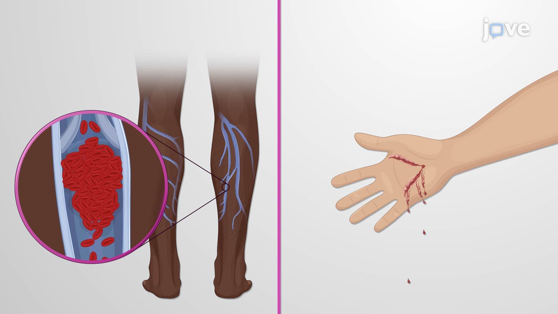
Did you know that when you cut your finger, two completely different molecular pathways spring into action within seconds? The extrinsic intrinsic hemostasis pathways work like a sophisticated emergency response system, with the extrinsic pathway responding to external tissue damage while the intrinsic pathway handles internal blood vessel injuries. Consider how quickly bleeding stops after a paper cut versus internal bruising from a sports injury at your local high school-these scenarios activate different coagulation mechanisms. Watch the full video on JoVE Coach to master this concept with expert-led visuals and step-by-step explanations.
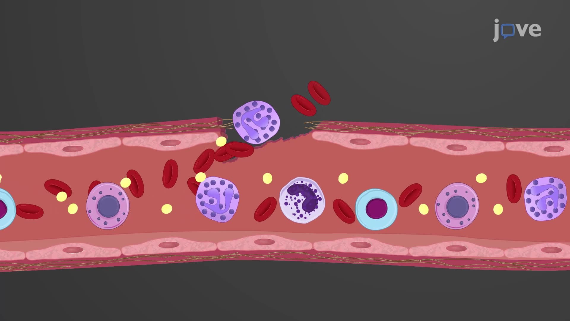
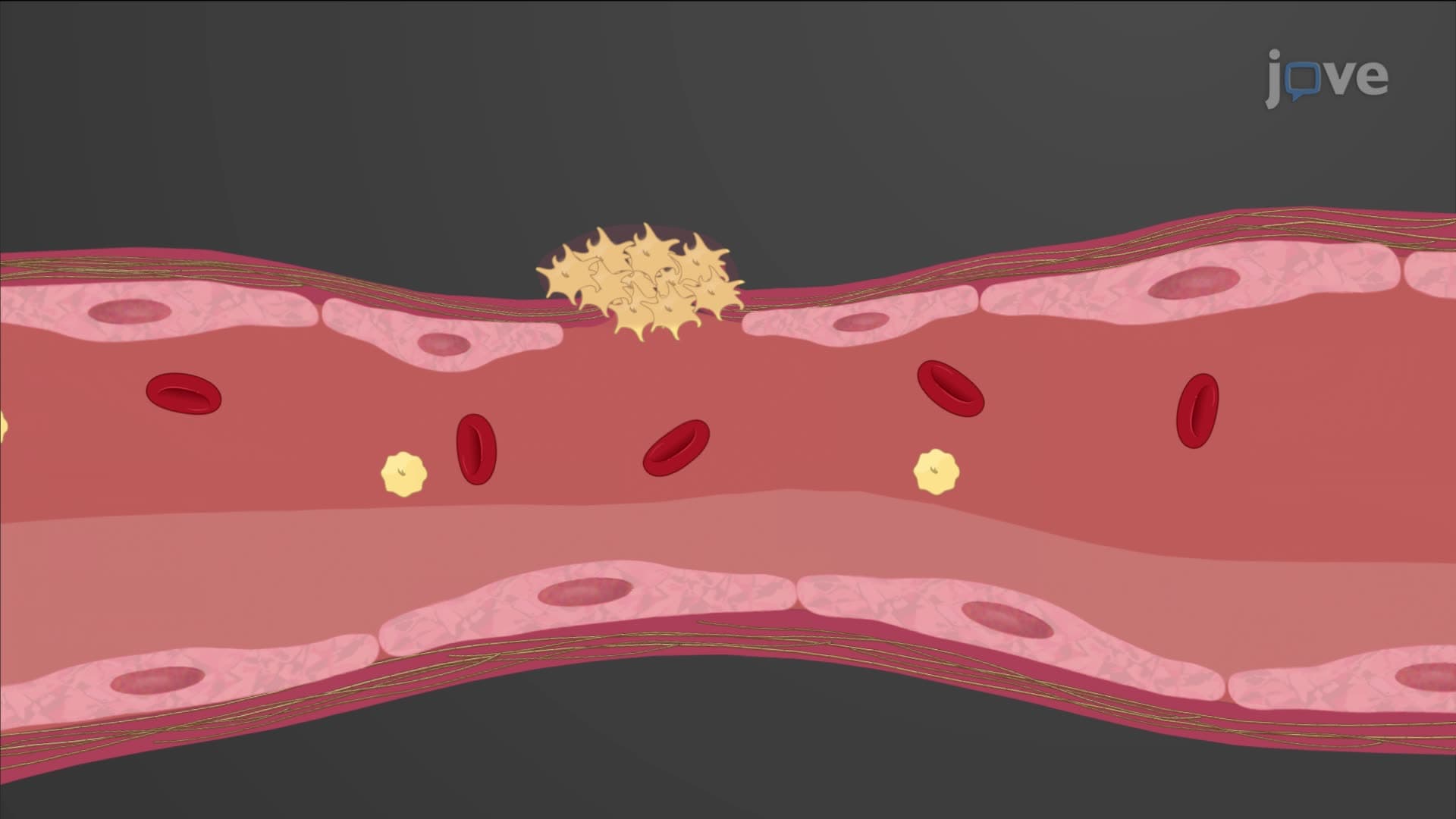
The human body's ability to stop bleeding represents one of biology's most elegant emergency systems. When blood vessels suffer damage, two distinct molecular cascades-the extrinsic intrinsic hemostasis pathways-activate to prevent life-threatening blood loss. These pathways demonstrate how cellular injury triggers precise biochemical responses that ultimately save lives.
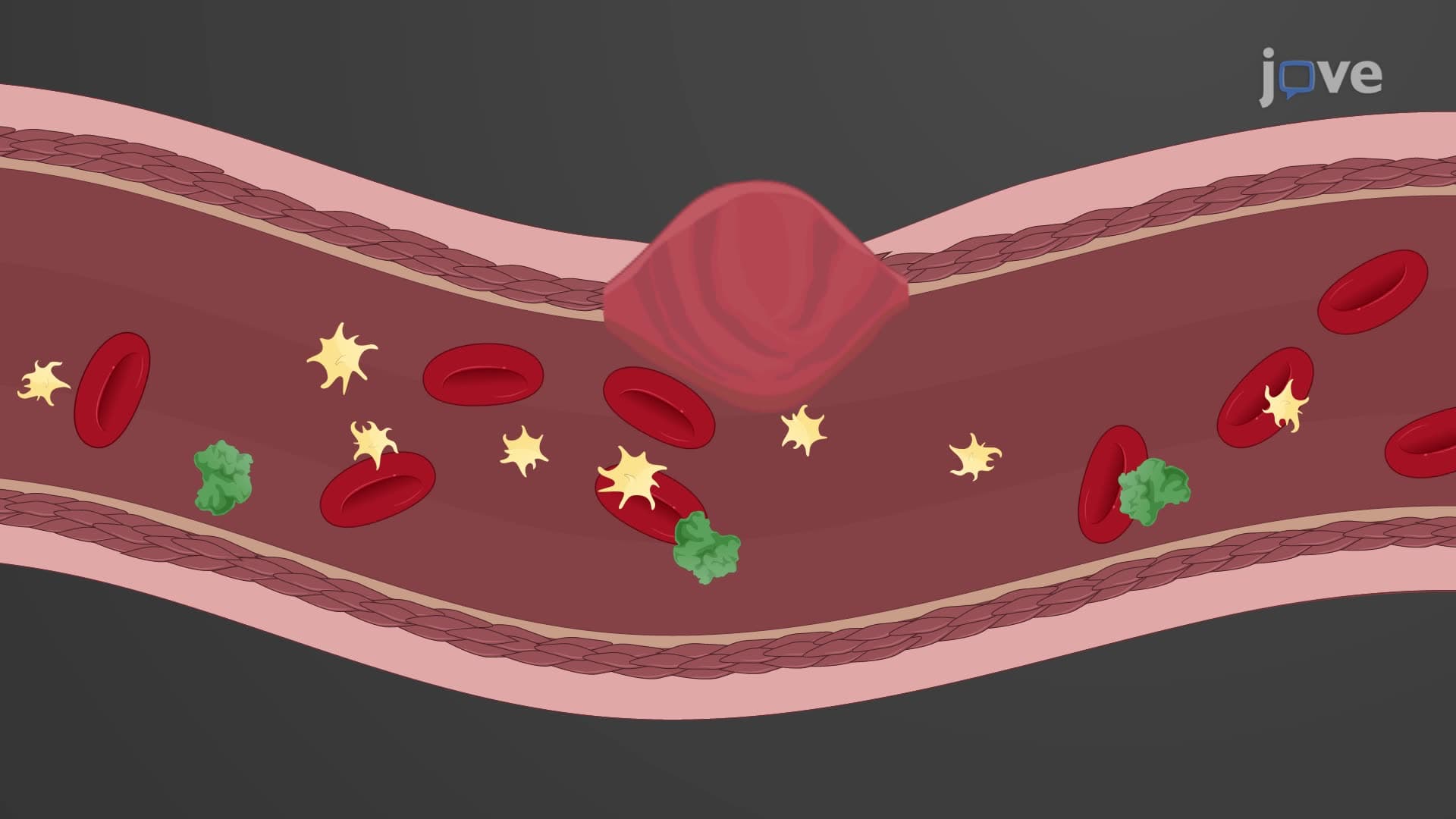
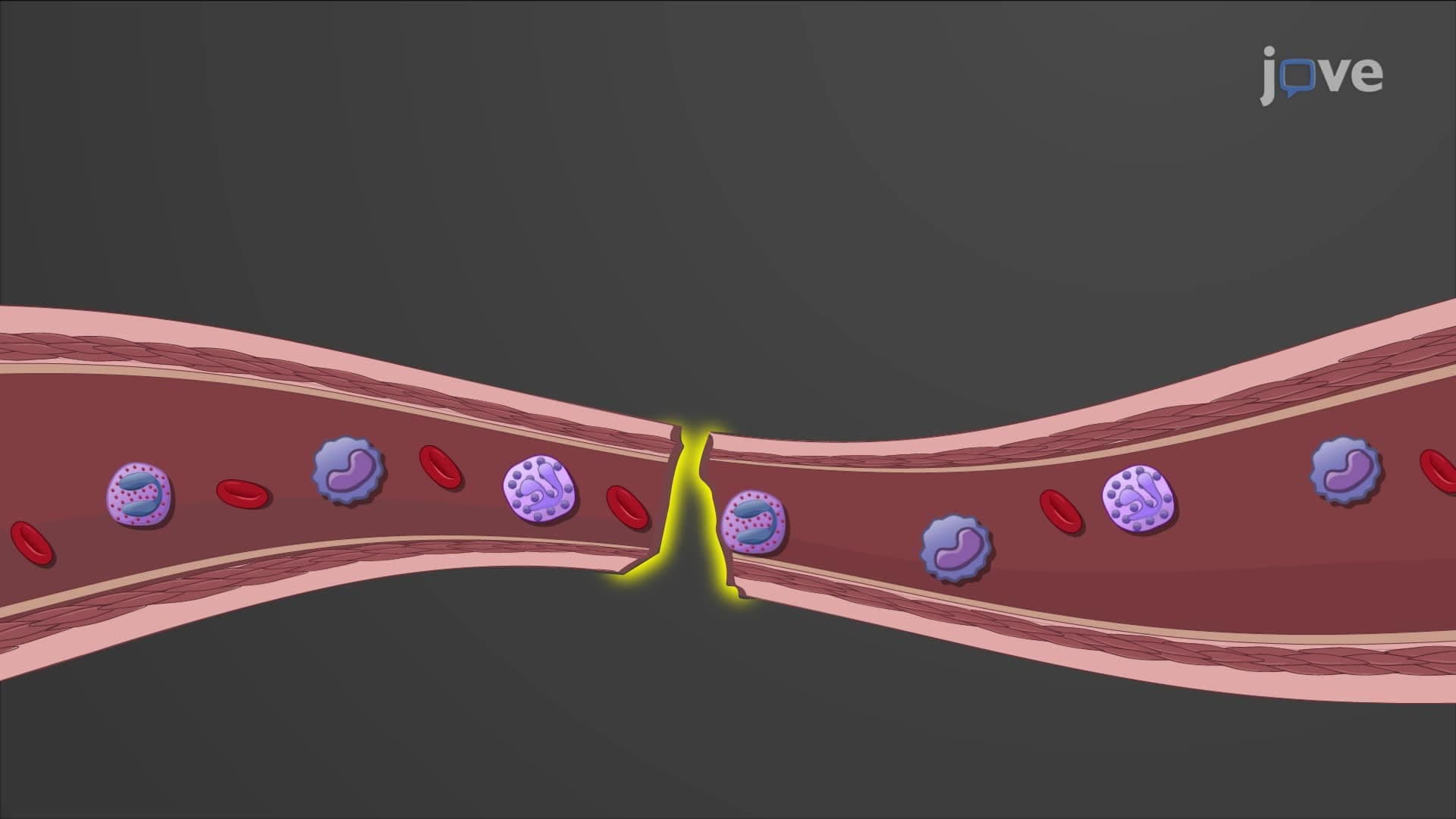
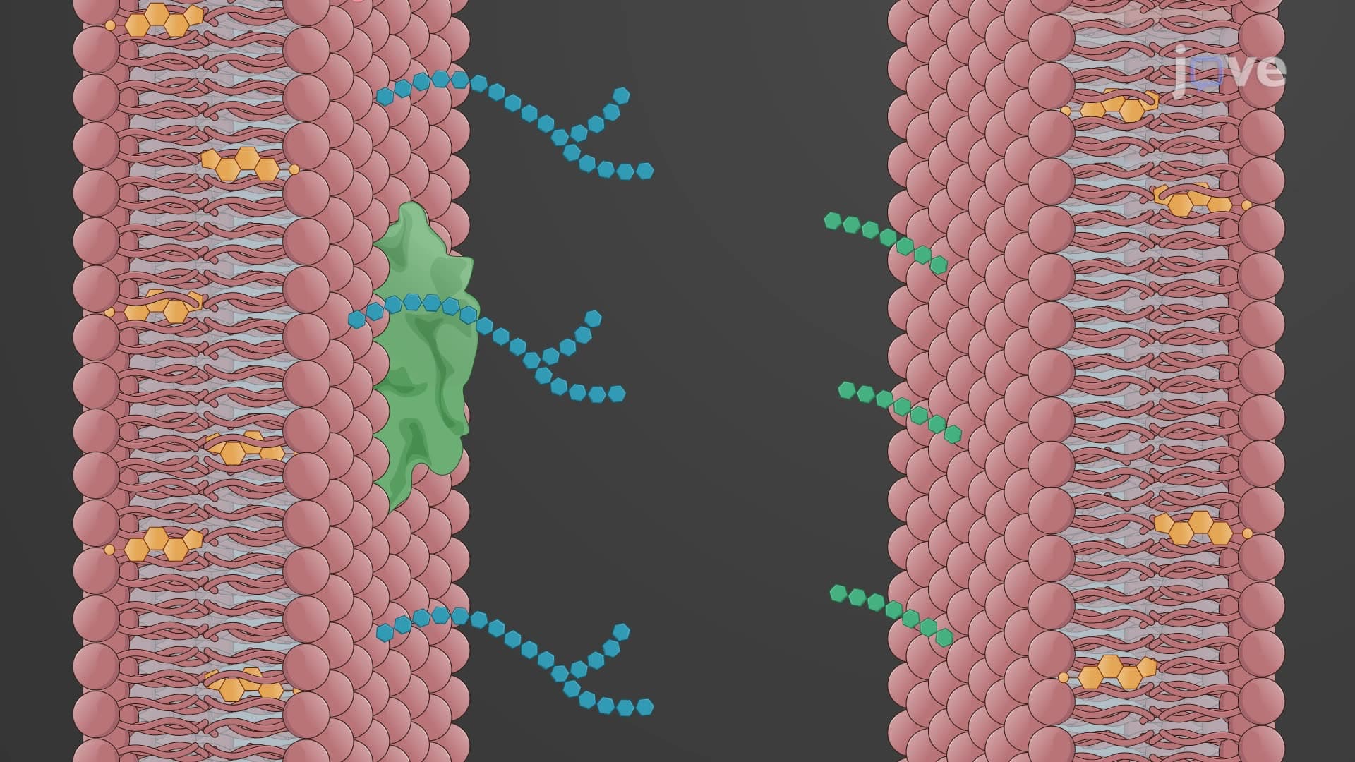
The extrinsic pathway activates when trauma breaches blood vessel walls, exposing circulating blood to surrounding tissues. This external damage releases tissue factor (thromboplastin), a powerful lipoprotein complex that serves as the pathway's primary trigger. In clinical settings like emergency departments at major US hospitals, physicians see this pathway activate in cases ranging from surgical incisions to automobile accident injuries.
The molecular sequence begins when tissue factor combines with circulating Factor VII in the presence of calcium ions, forming the tissue factor-Factor VII complex. This complex directly activates Factor X, bypassing multiple steps seen in other coagulation routes. The efficiency of this "shortcut" explains why external wounds often clot faster than internal bleeding-a phenomenon AP Biology students frequently encounter in laboratory exercises involving controlled tissue injury.
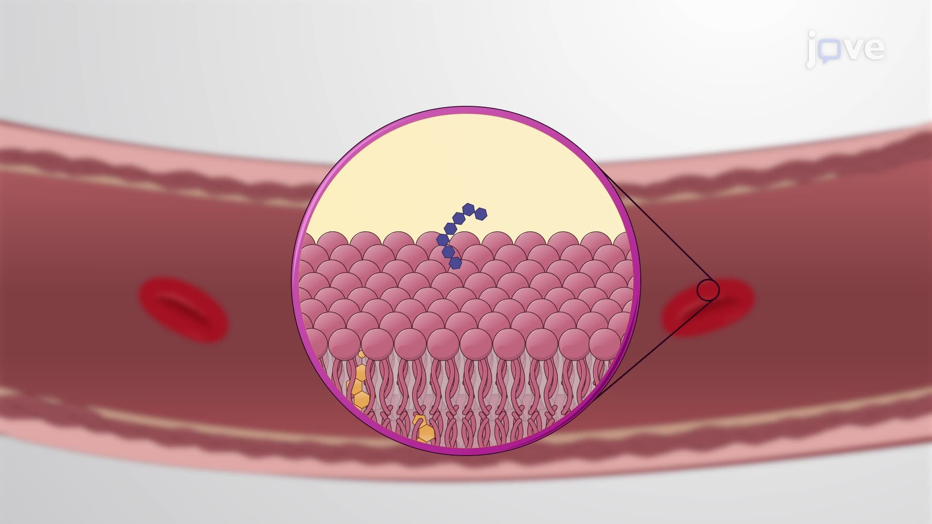
Unlike its extrinsic counterpart, the intrinsic pathway responds to internal blood vessel damage, particularly injury to the endothelial lining. This pathway activates entirely within the bloodstream, utilizing a more complex cascade of reactions. Contact activation occurs when blood encounters damaged vessel surfaces, triggering a series of factor activations including Factors XII, XI, IX, and VIII.
The intrinsic pathway requires platelet factor 3 and calcium ions to function effectively. Factors VIII and IX combine to form an enzyme complex that ultimately activates Factor X-the same convergence point reached by the extrinsic pathway. This redundancy ensures clot formation even when one pathway fails, explaining why patients with single factor deficiencies (like hemophilia A affecting Factor VIII) may still achieve hemostasis through alternative routes.
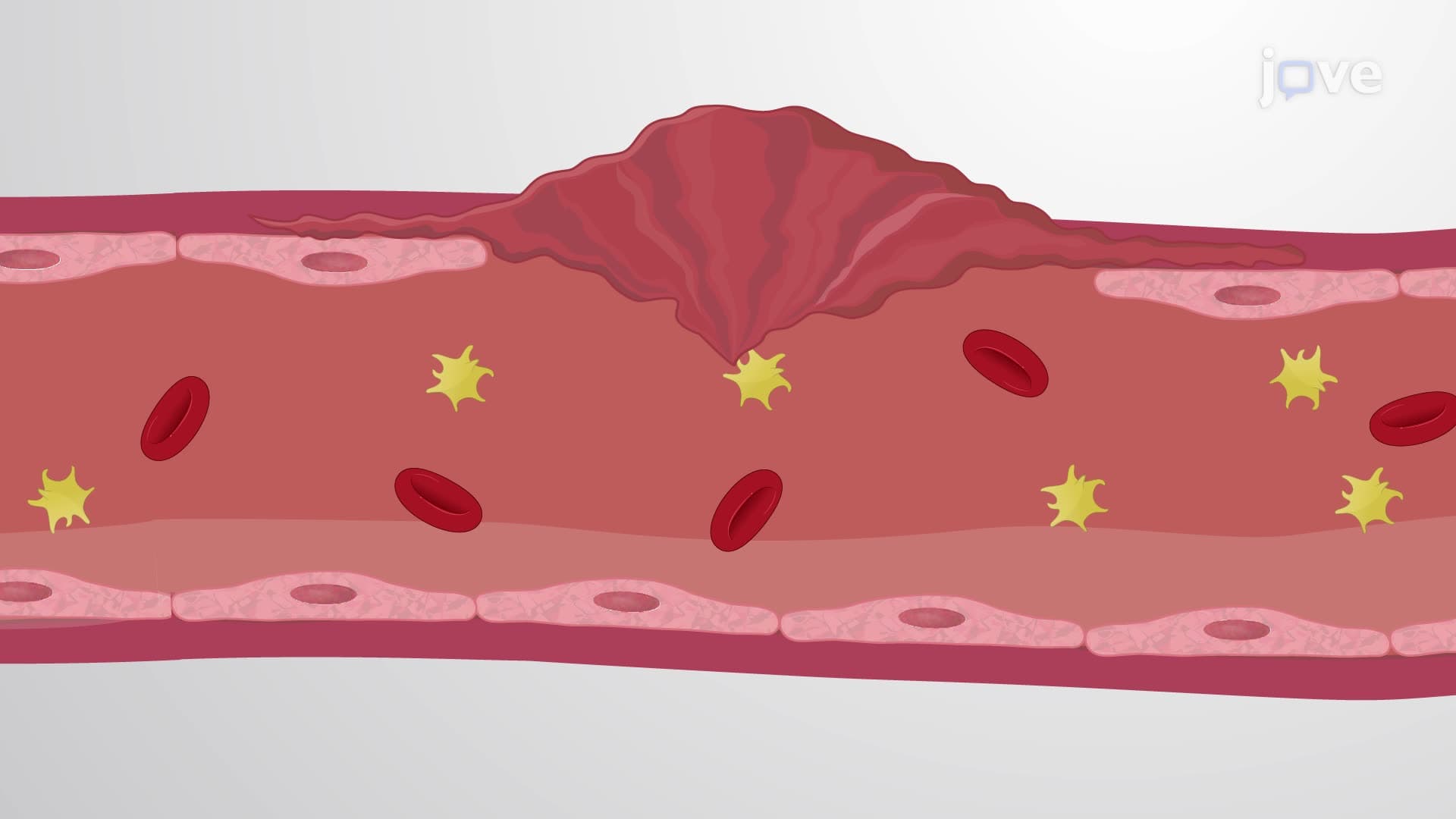
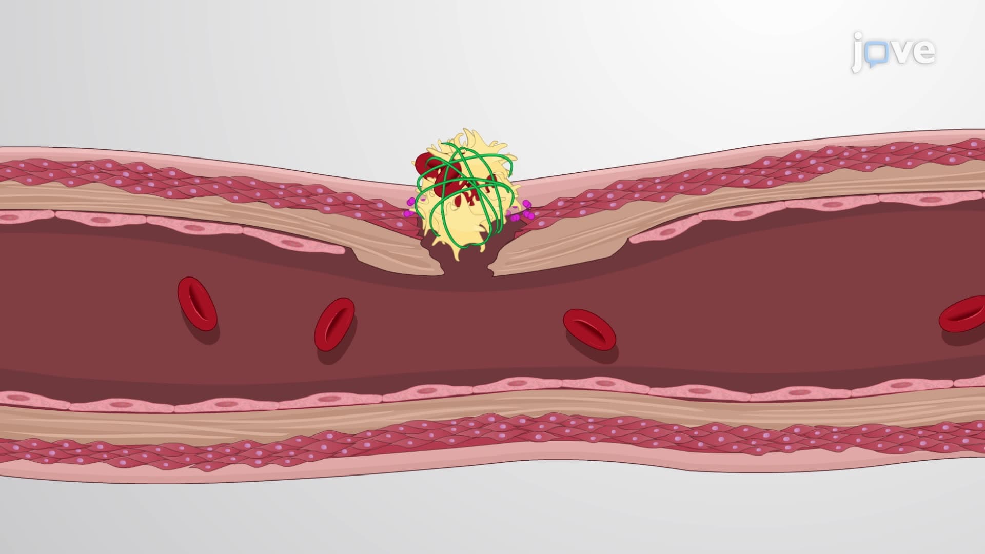
Both pathways merge at Factor X activation, initiating the common pathway that converts prothrombin to thrombin. Thrombin then converts soluble fibrinogen into insoluble fibrin strands, creating the structural framework of blood clots. Medical professionals use this convergence point for diagnostic testing-PT (Prothrombin Time) tests evaluate extrinsic pathway function, while PTT (Partial Thromboplastin Time) assesses intrinsic pathway integrity.
Pre-med students preparing for the MCAT must understand these pathways' clinical relevance, particularly how anticoagulant medications like warfarin (targeting extrinsic pathway) versus heparin (affecting intrinsic pathway) produce different therapeutic effects. Understanding these mechanisms proves essential for healthcare career preparation and explains why bleeding disorders require specific diagnostic approaches.
Related Micro-courses