Loading subject page…


135,525 views

Video Summary: What Is Hemostasis
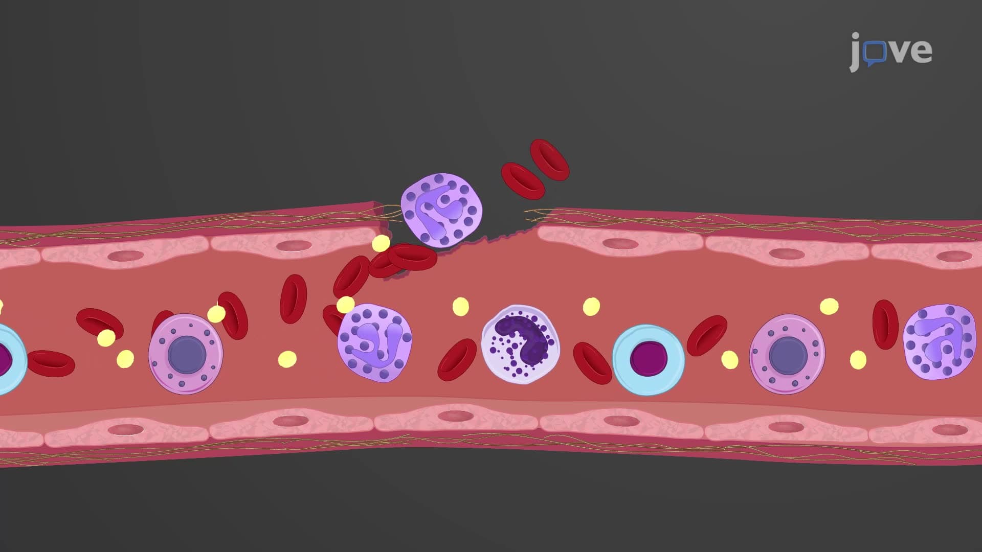
When you cut your finger, why doesn't it bleed forever? Hemostasis biology explained reveals how your body orchestrates a remarkable three-phase rescue mission to stop bleeding within minutes. Consider emergency room physicians at Johns Hopkins Hospital treating trauma patients-they rely on understanding what is hemostasis to save lives daily. This critical physiological process involves vascular constriction, platelet plug formation, and blood coagulation working together seamlessly. Watch the full video on JoVE Coach to master this concept with expert-led visuals and step-by-step explanations.
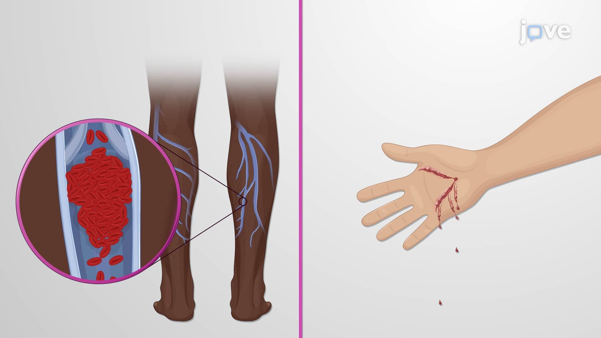
Hemostasis biology explained encompasses far more than simple "blood clotting." This sophisticated physiological mechanism represents one of medicine's most elegant examples of biological engineering. When trauma surgeons at Mayo Clinic treat accident victims, they witness hemostasis in action-a coordinated cellular and molecular response that can mean the difference between life and death.
The blood clotting hemostasis process begins within seconds of vascular injury. Unlike generalized inflammatory responses, hemostasis operates with surgical precision, activating only at damage sites while preserving normal circulation elsewhere. This localized specificity prevents unnecessary clot formation that could cause strokes or heart attacks.
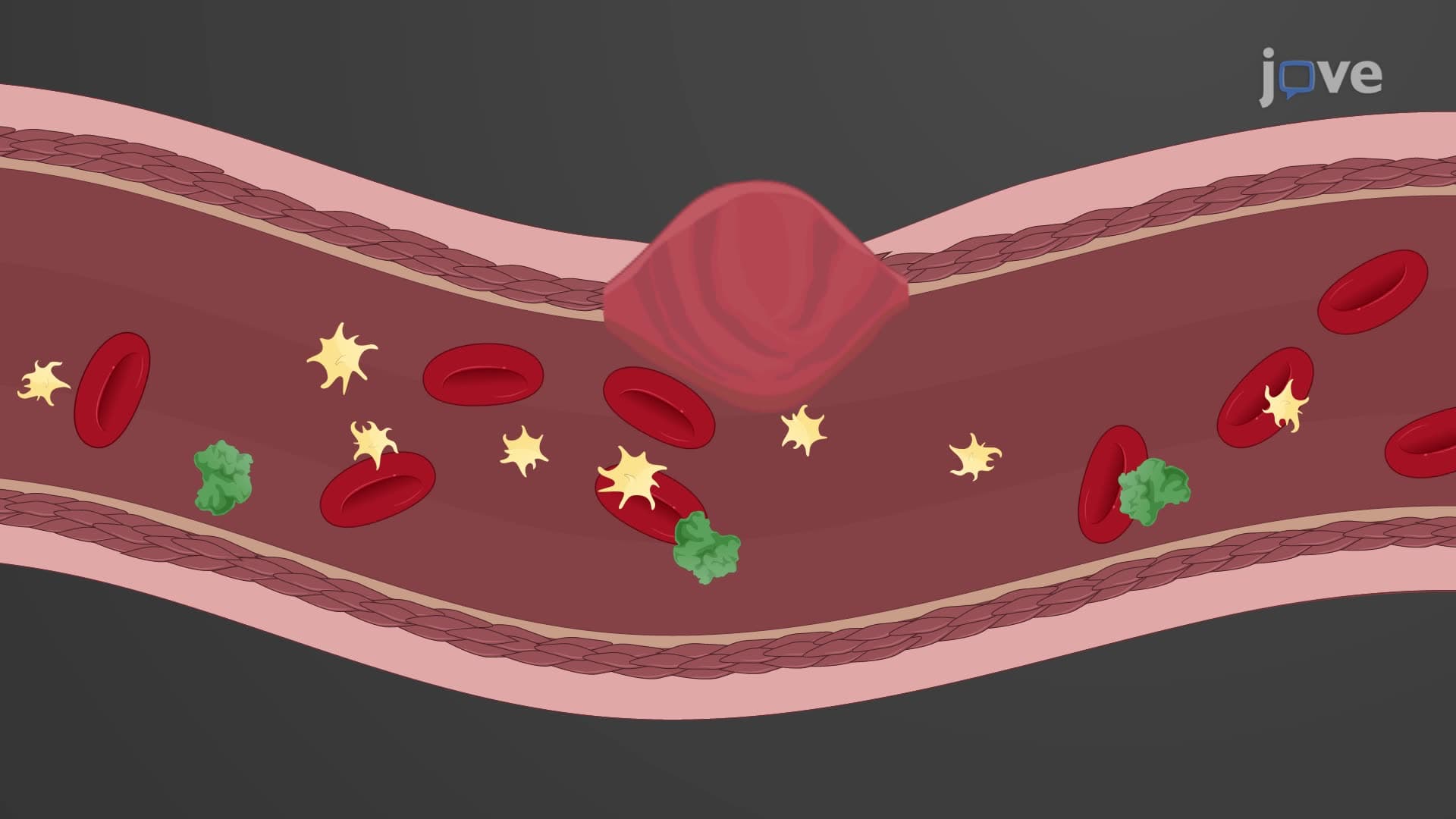
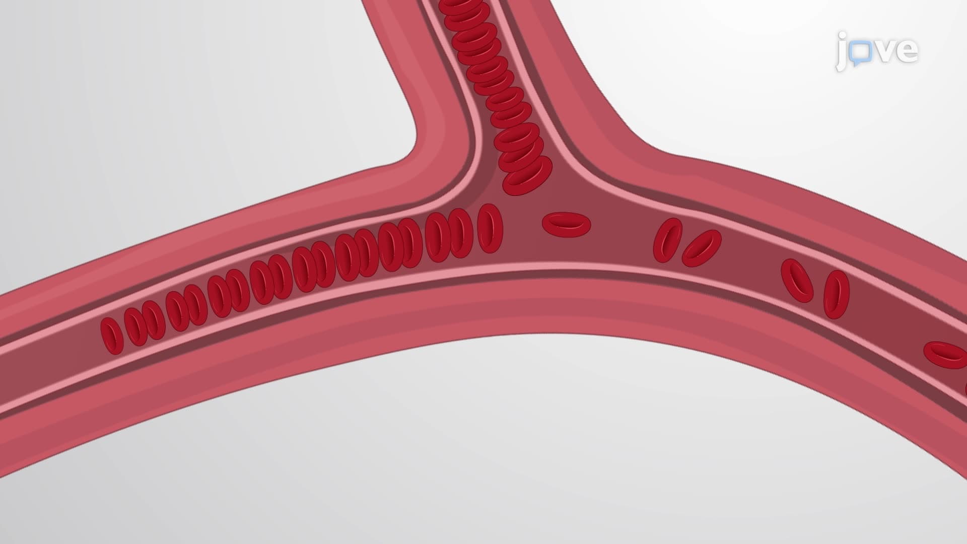
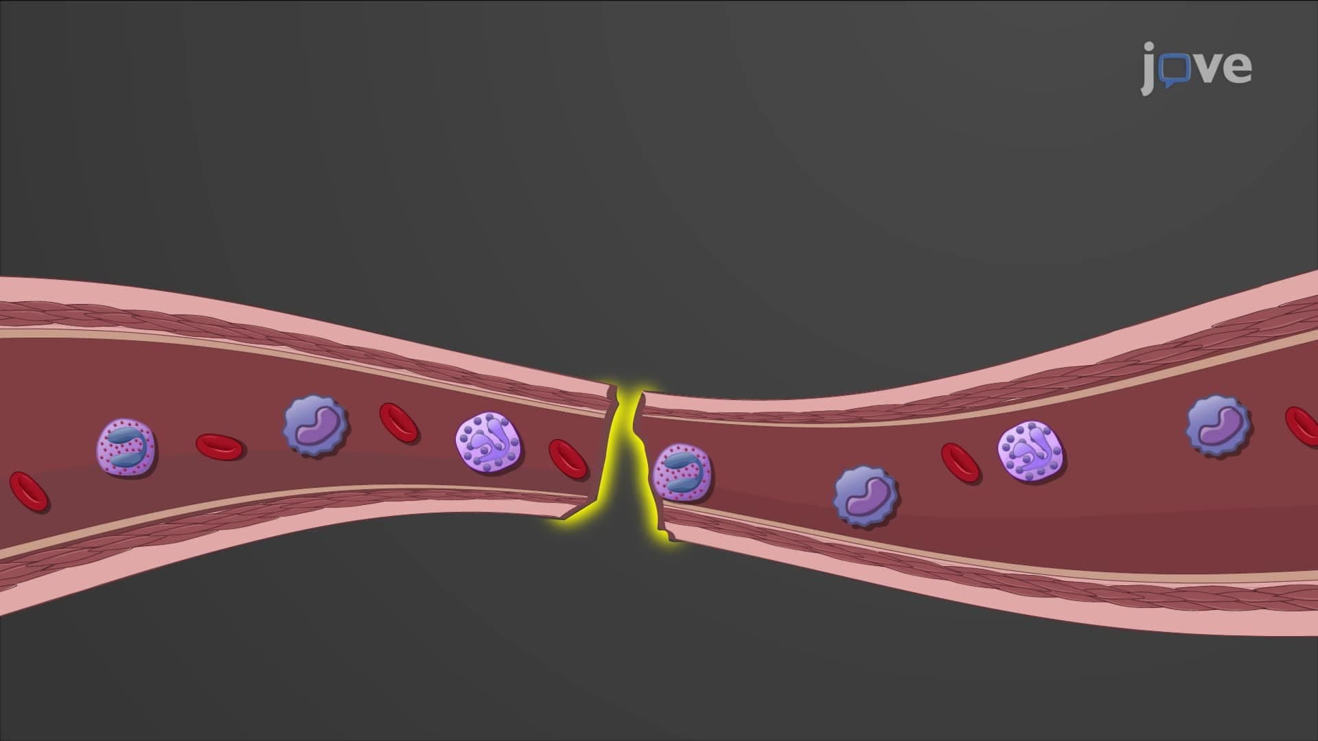
Vascular spasm hemostasis initiates immediately upon injury through smooth muscle contraction in blood vessel walls. This vasoconstriction can reduce blood flow by up to 30%, buying precious time for subsequent phases. Emergency medical technicians observe this phenomenon when treating patients-initial bleeding often slows before any medical intervention.
The spasm results from direct trauma to vascular smooth muscle, local reflexes, and chemical mediators released by damaged tissues. Smaller vessels can completely close through this mechanism alone, explaining why paper cuts stop bleeding quickly without forming visible clots.
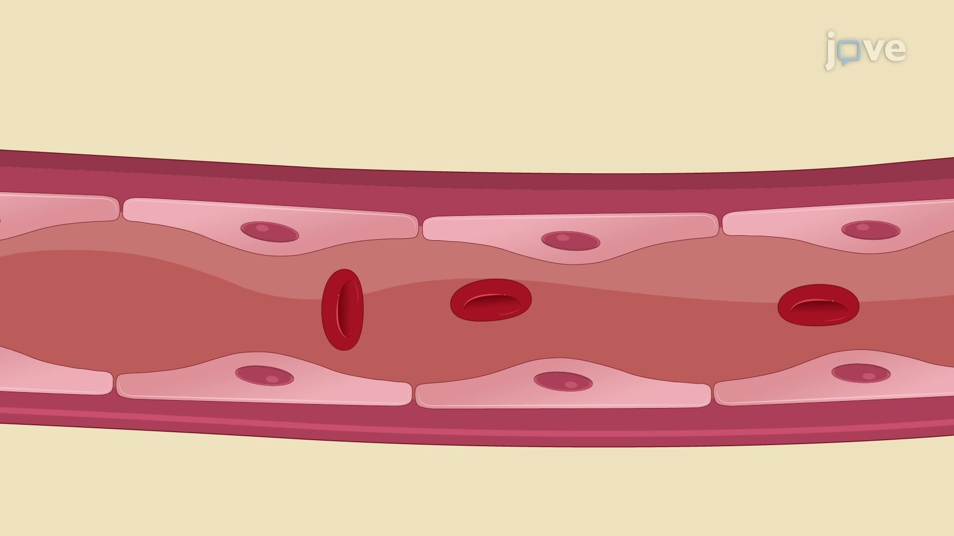
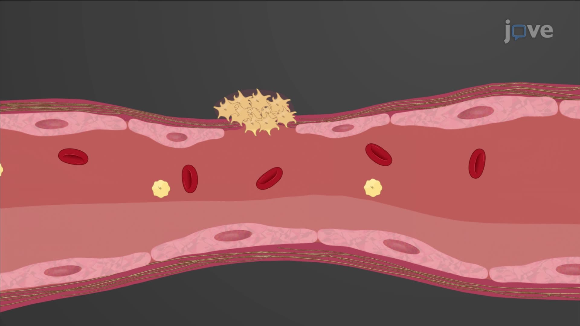
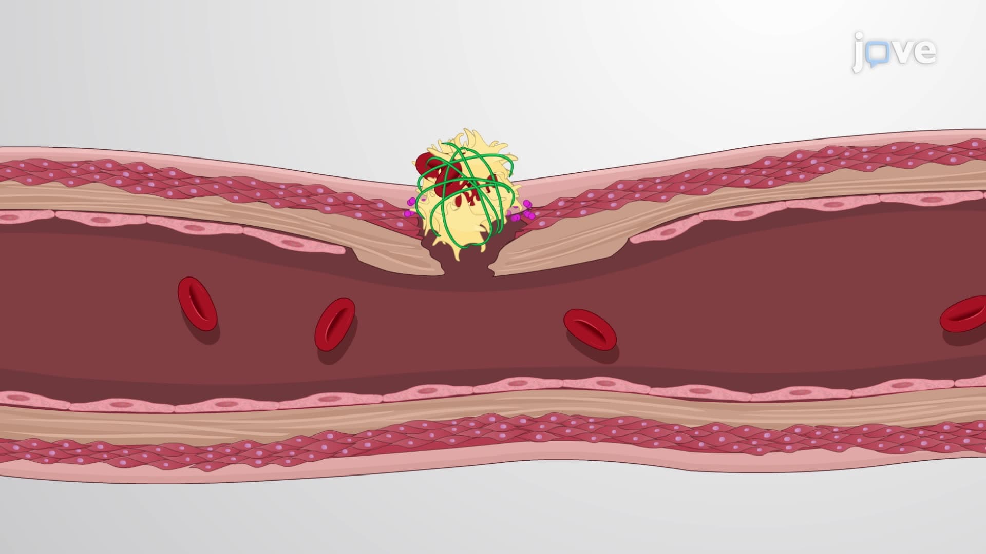
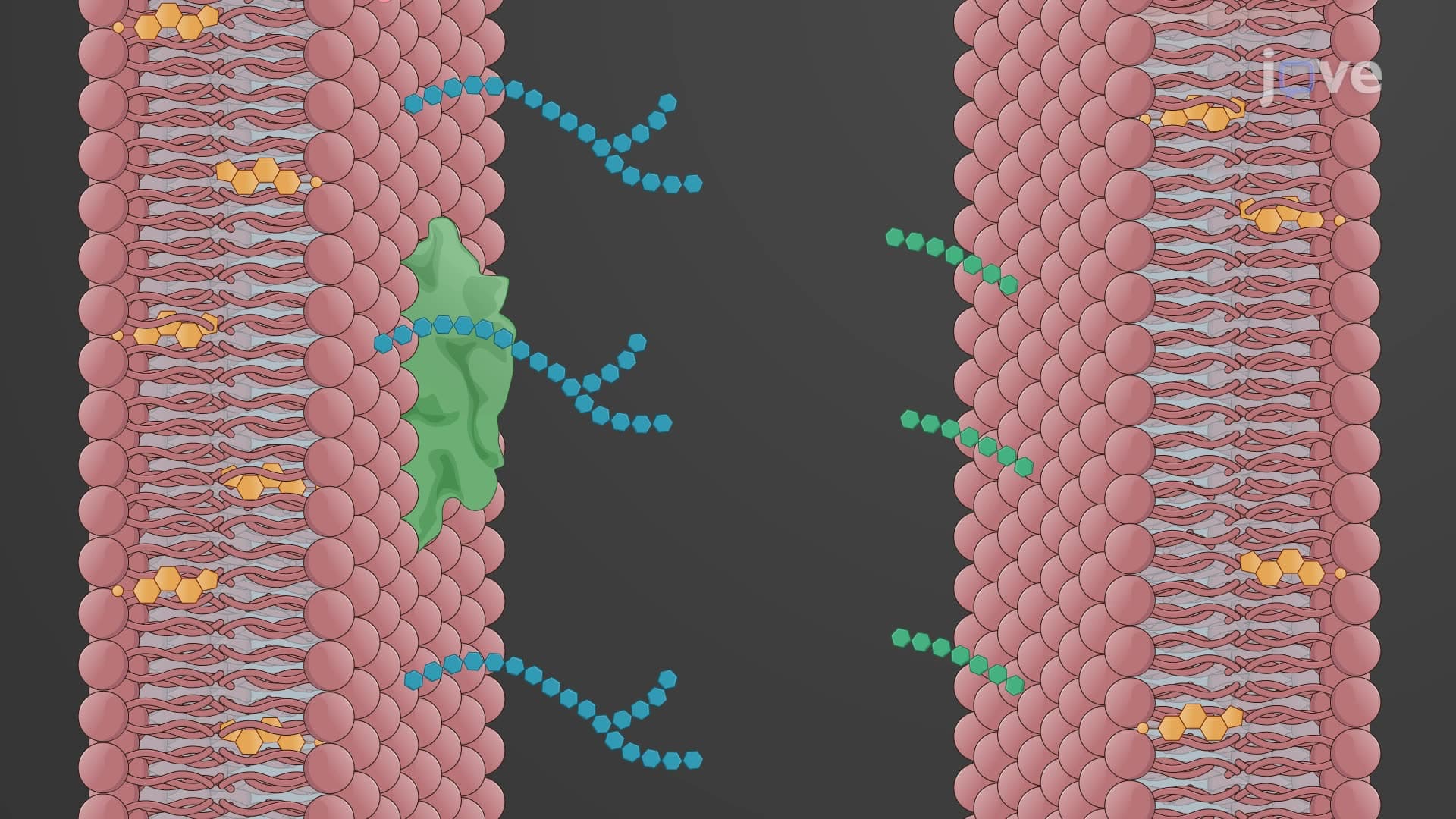
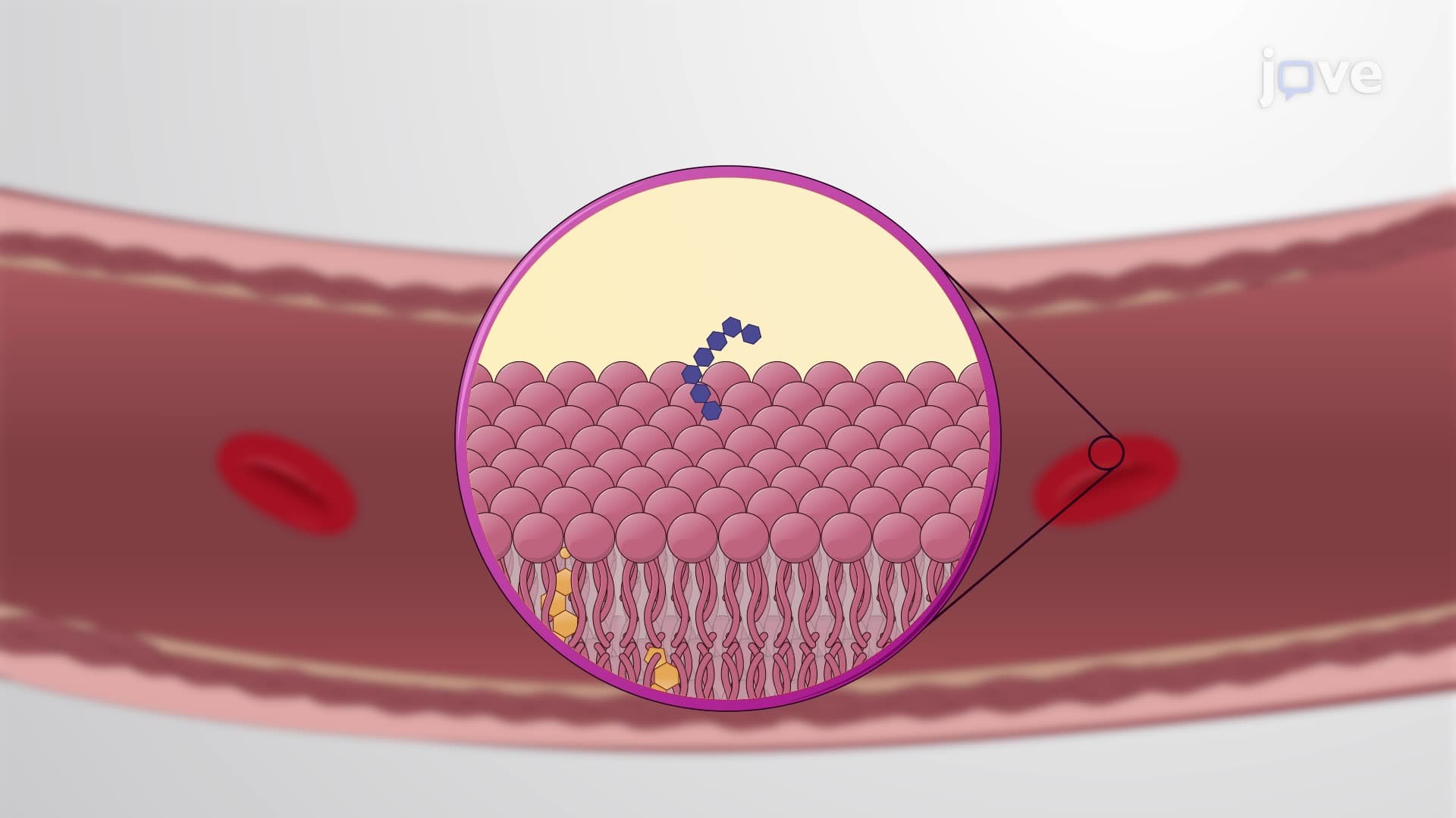
Platelet plug primary hemostasis transforms circulating platelets into a cohesive barrier. When endothelial cells lining blood vessels suffer damage, they expose underlying collagen fibers. Platelets possess specific receptors that bind to collagen with remarkable affinity, like molecular velcro.
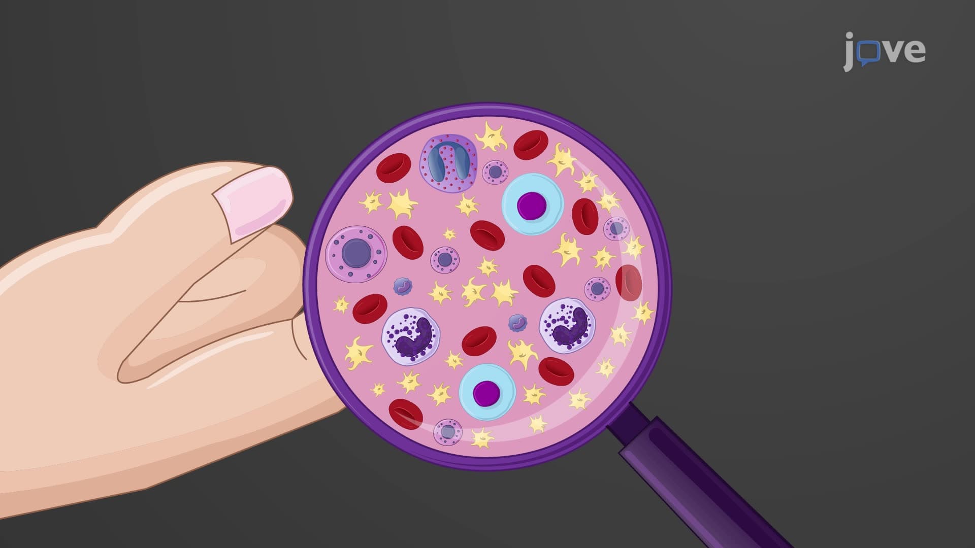
Activated platelets undergo dramatic shape changes, extending pseudopodia that help them adhere to injury sites and each other. They simultaneously release adenosine diphosphate (ADP), thromboxane A2, and other chemical signals that attract additional platelets-a process called aggregation. This creates a loose but effective plug within 1-3 minutes.
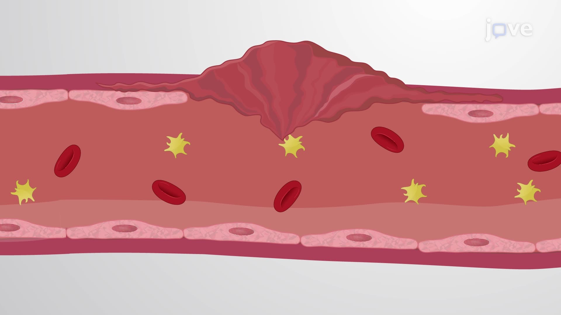
Coagulation secondary hemostasis represents biochemistry in action, involving over a dozen clotting factors in two interconnected pathways. The intrinsic pathway activates when blood contacts damaged vessel surfaces, while the extrinsic pathway responds to tissue factor released by injured cells.
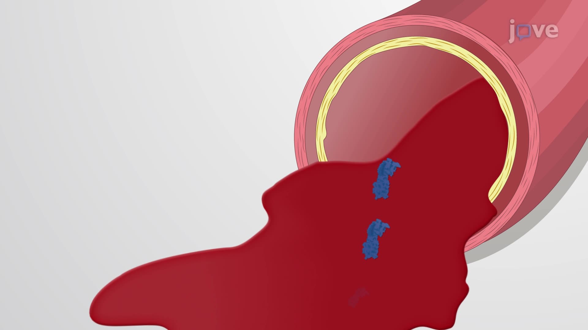
Both pathways converge at Factor X activation, leading to thrombin generation. Thrombin converts soluble fibrinogen into insoluble fibrin strands that weave through the platelet plug like reinforcing cables in concrete. This transforms a temporary platelet barrier into a permanent, stable clot.
Students preparing for the MCAT or AP Biology exams should understand that hemostasis three stages work synergistically, not sequentially. All phases can occur simultaneously, with positive feedback loops amplifying the response until bleeding stops completely.
Related Micro-courses