Loading video…


 29 Concepts
 135.5K views

Video Summary: What Is Lifecycle of Erythrocytes
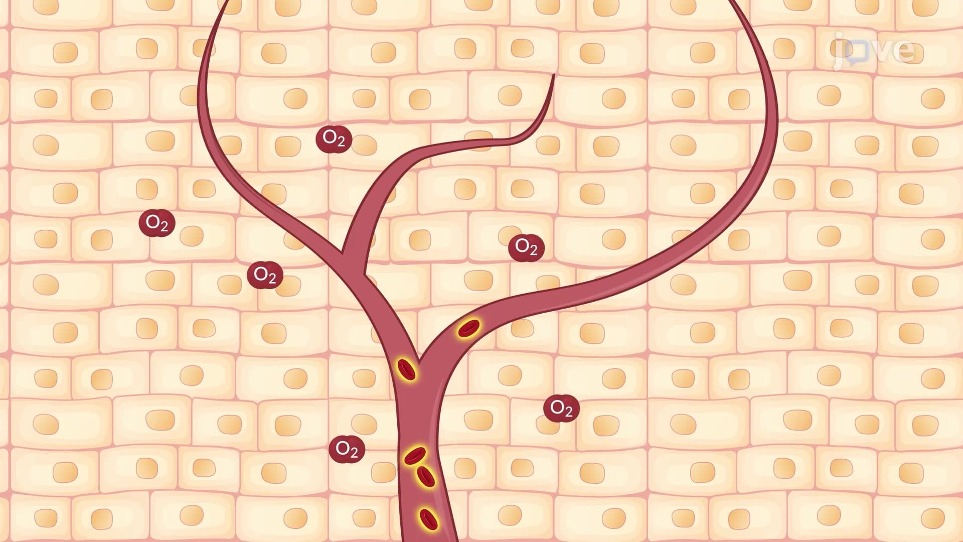
Every second, your body destroys approximately 2.4 million red blood cells while simultaneously creating new ones-a perfectly balanced cellular recycling system. The erythrocyte lifecycle biology demonstrates how these oxygen-carrying cells complete their 120-day journey from bone marrow production to splenic breakdown. Consider how patients with sickle cell disease at Johns Hopkins Hospital experience disrupted red blood cell lifespans, requiring frequent monitoring of this critical process. Watch the full video on JoVE Coach to master this concept with expert-led visuals and step-by-step explanations.
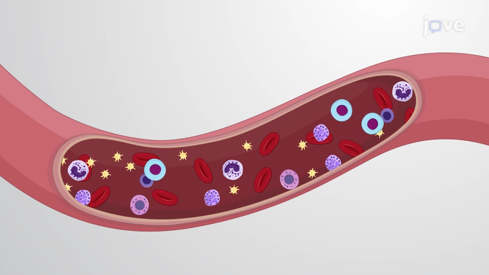
The erythrocyte lifecycle biology represents one of the most efficient recycling systems in human physiology, ensuring continuous oxygen transport while conserving valuable cellular components. This 120-day cellular journey involves precise coordination between bone marrow production, circulatory function, and systematic breakdown.
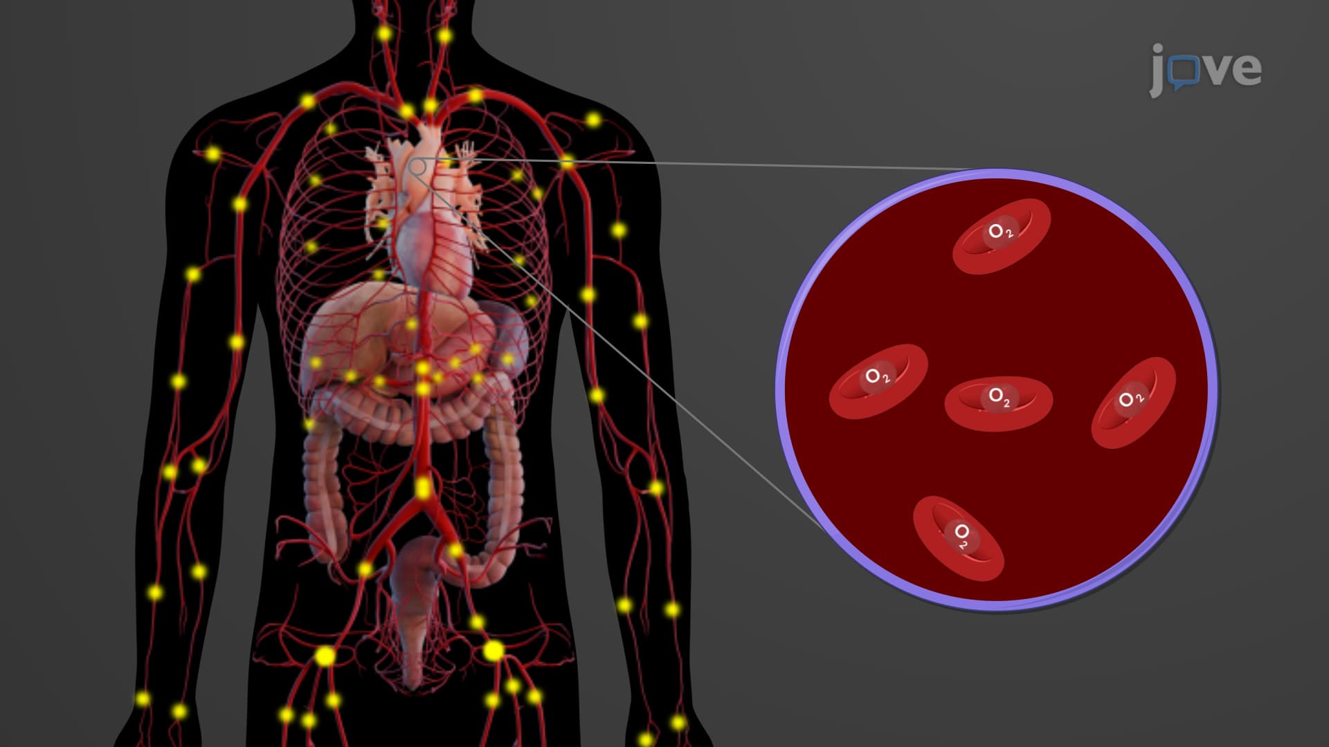
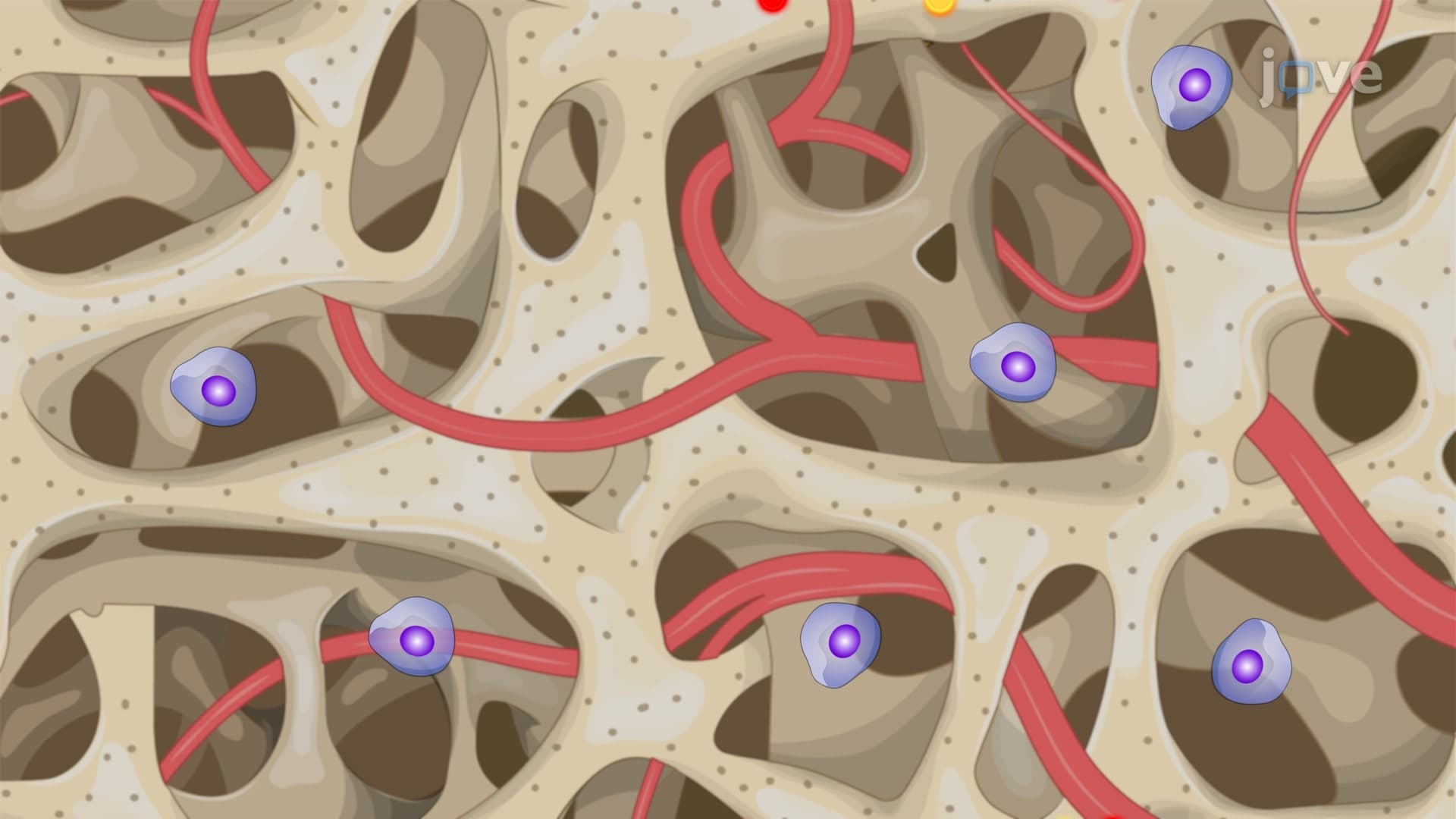
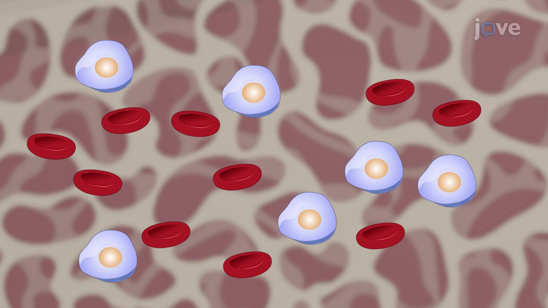
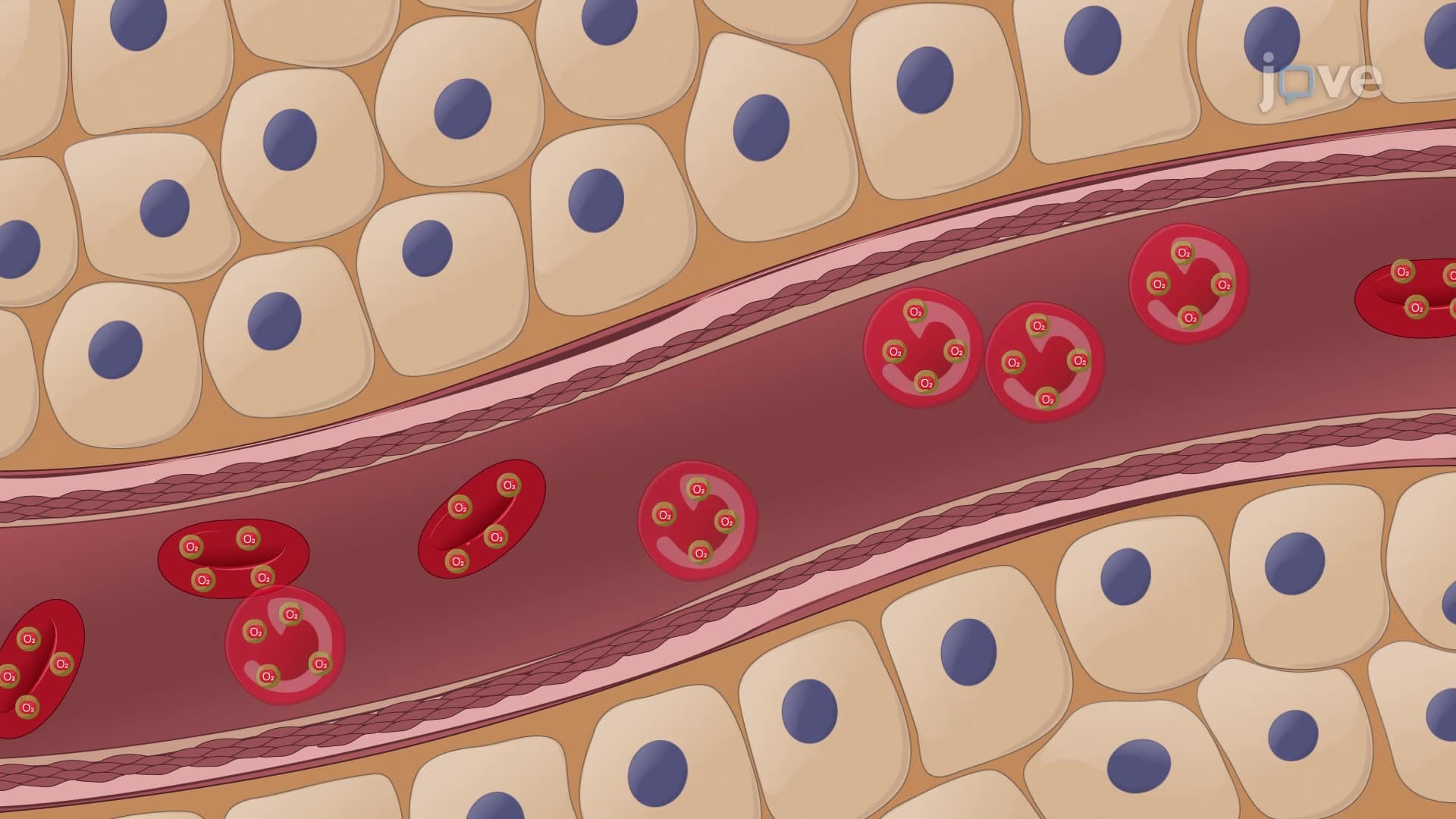
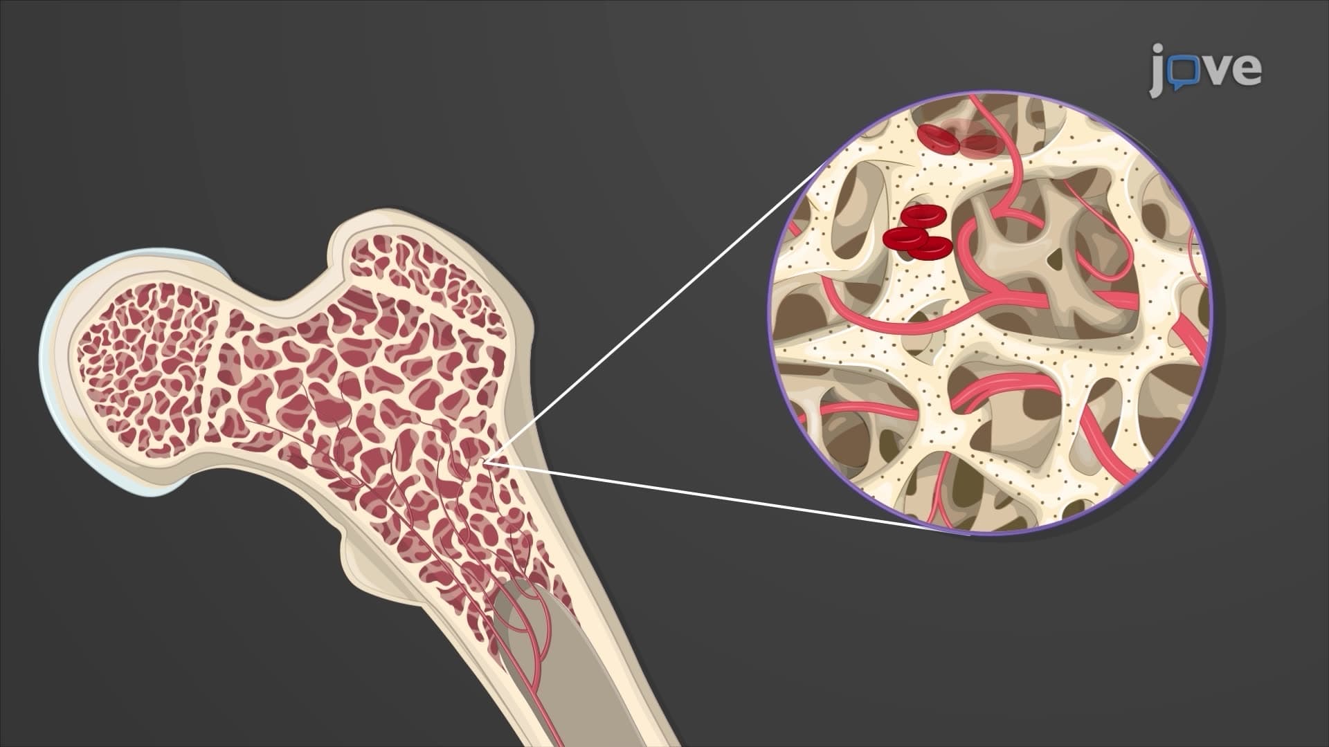
Reticulocyte to erythrocyte maturation occurs primarily in the bone marrow, where pluripotent stem cells differentiate through erythroblast stages. During this process, cells progressively lose their nuclei and organelles, transforming into the characteristic biconcave discs optimized for oxygen transport. This RBC 120 day lifespan begins when mature erythrocytes enter circulation, equipped with flexible membranes essential for navigating capillaries as narrow as 2-3 micrometers.
Students preparing for the MCAT should understand that this maturation process takes approximately 7 days, with reticulocytes spending their final 1-2 days in circulation before becoming fully mature erythrocytes. The biconcave shape maximizes surface area for gas exchange while maintaining flexibility-a critical concept tested on AP Biology exams.
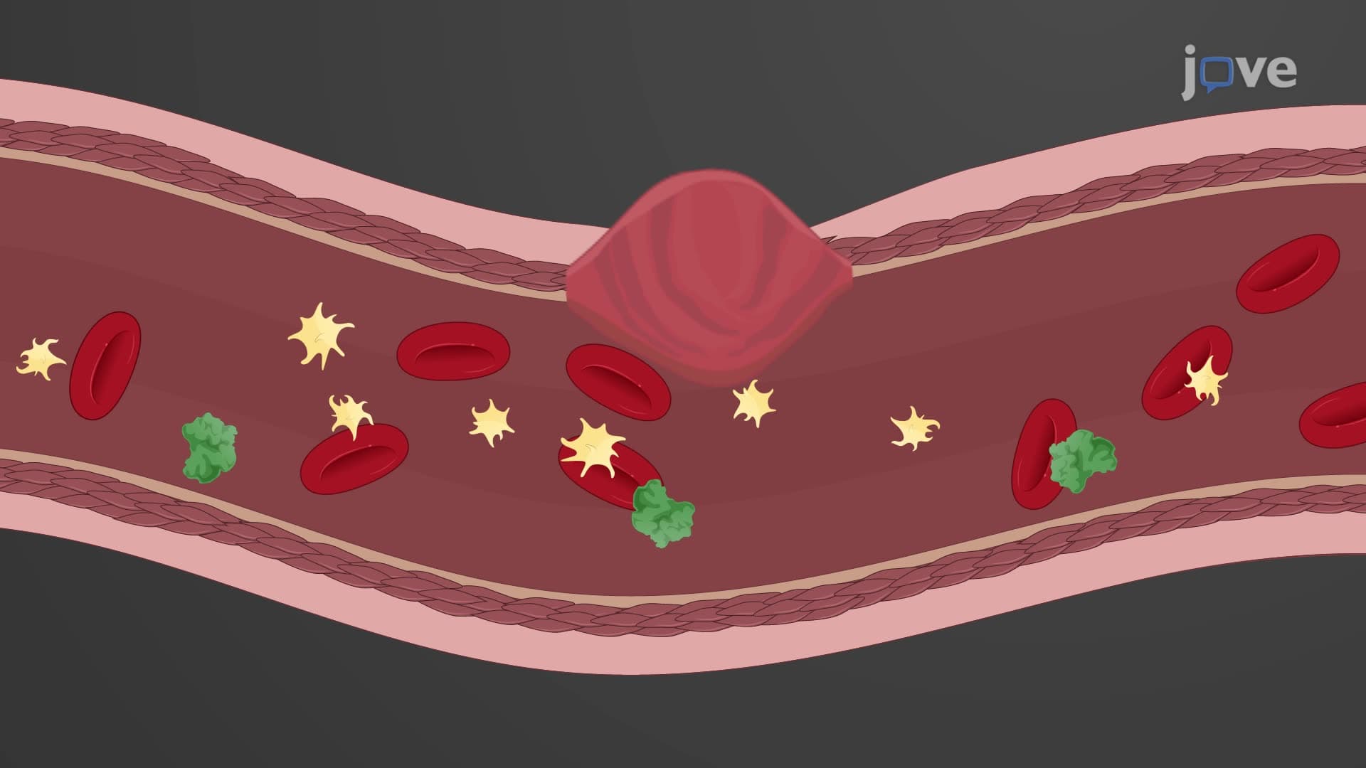
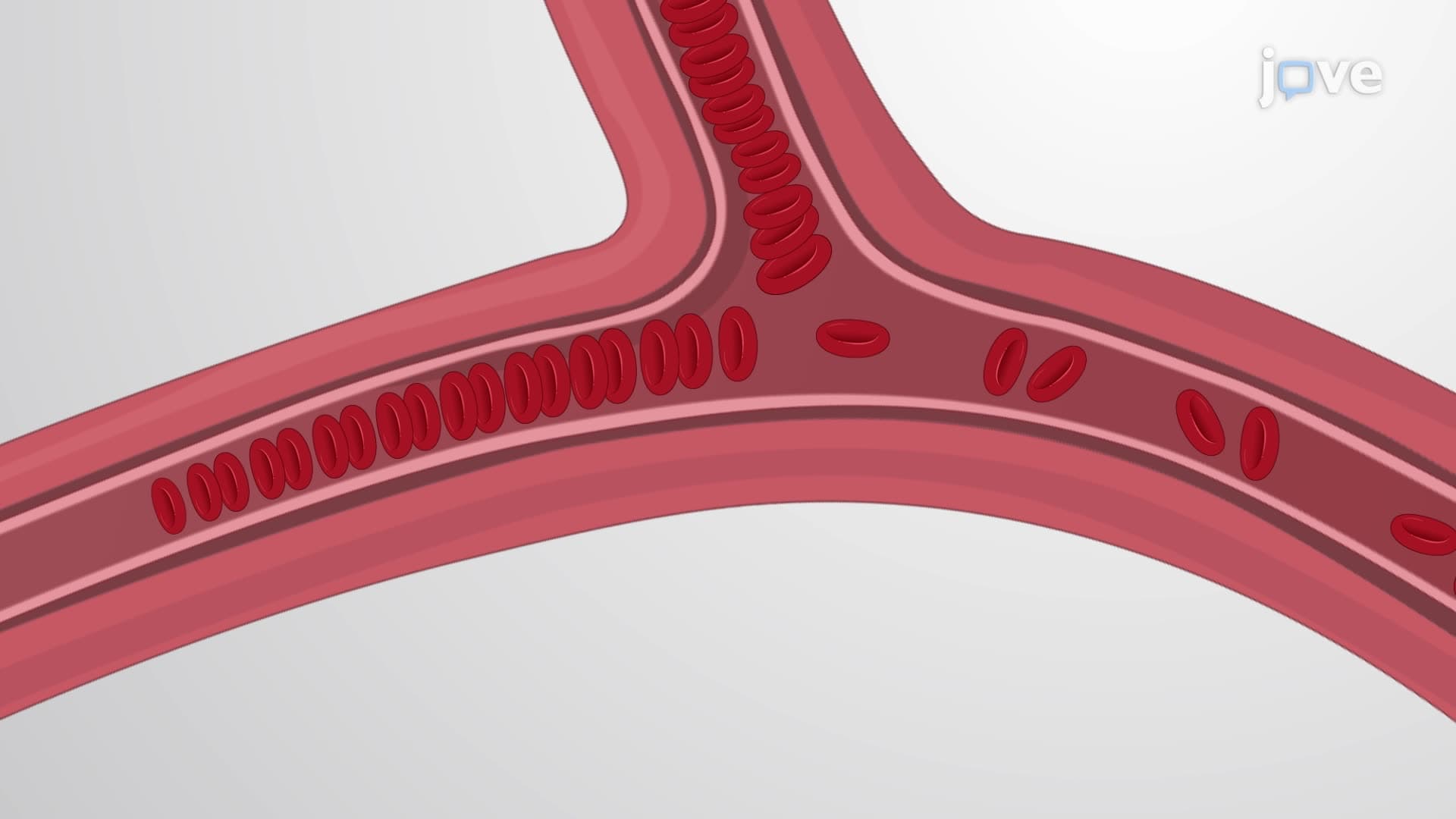
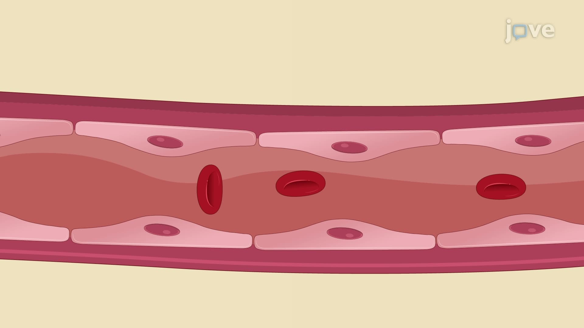
During circulation, erythrocytes endure tremendous mechanical stress, squeezing through approximately 10,000 capillaries daily. This constant deformation gradually damages plasma membranes, reducing cellular flexibility and metabolic efficiency. The red blood cell lifespan explained by this mechanical wear demonstrates why cells with compromised membranes, such as those in hereditary spherocytosis, have significantly shortened lifespans.
Aged RBC spleen removal occurs when erythrocytes become rigid and fragile after 100-120 days. Splenic macrophages possess sophisticated recognition mechanisms that identify senescent cells through membrane changes and reduced deformability. This quality control system prevents damaged cells from impeding circulation-a concept frequently tested in college physiology courses.
The final phase involves erythrocyte recycling hemoglobin through systematic breakdown by tissue macrophages. Iron recovery through ferritin storage and transferrin transport ensures 95% of cellular iron returns to bone marrow for new hemoglobin synthesis. Meanwhile, bilirubin RBC breakdown produces the yellow pigment responsible for jaundice when accumulation exceeds hepatic processing capacity.
Understanding this recycling efficiency helps students grasp why iron deficiency anemia develops slowly despite continuous RBC turnover, and why patients with hemolytic disorders like sickle cell disease require enhanced iron monitoring at major medical centers like Cleveland Clinic.
Related Micro-courses