Loading subject page…


68,318 views

Video Summary: Muscles Pelvic Floor and Perineum Explained
Ever wonder why childbirth classes at Johns Hopkins emphasize pelvic floor perineum muscles so heavily? These intricate muscle groups form your body's foundation, literally supporting vital organs while controlling essential bodily functions. The diamond-shaped perineum sits beneath the pelvic floor's muscular diaphragm, creating a complex anatomical region that medical students at Harvard and UCLA spend weeks mastering. Watch the full video on JoVE Coach to master this concept with expert-led visuals and step-by-step explanations.
The pelvic floor muscle anatomy represents one of medicine's most intricate architectural designs, forming the inferior boundary of your abdominopelvic cavity. Unlike the rigid bones above, this muscular floor must balance strength with flexibility, supporting organs while allowing passage for essential functions. At prestigious medical schools like Stanford and Duke, anatomy students dedicate entire laboratory sessions to understanding these relationships.
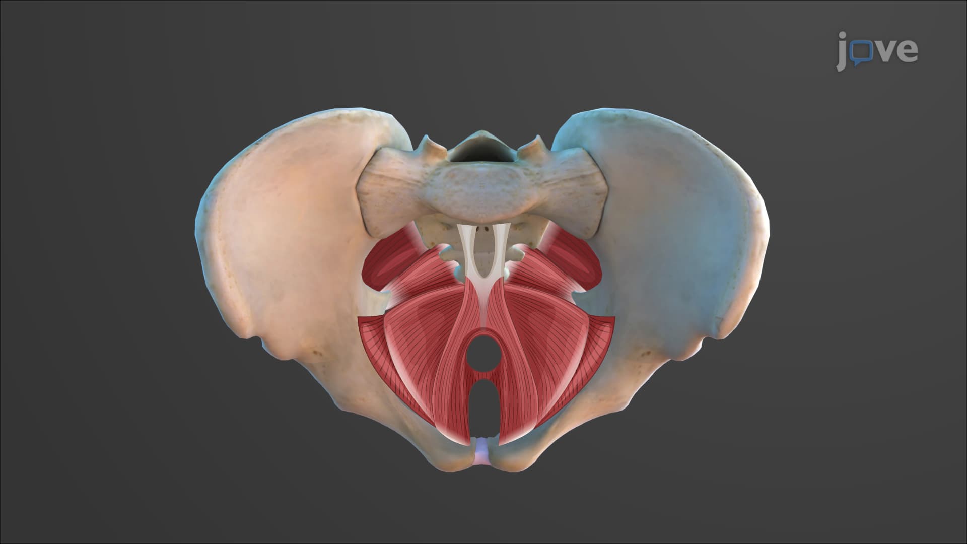
The levator ani pelvic floor serves as the primary structural component, comprising three distinct muscles that work in harmony. The pubococcygeus extends from the pubic bone to the coccyx, providing anterior support. The puborectalis forms a muscular sling around the rectum, creating the critical anorectal angle that maintains fecal continence. The iliococcygeus completes the posterior aspect, attaching to the iliac spine and coccyx.
Physical therapy programs at Northwestern and Emory Universities emphasize how the coccygeus pelvic muscle partners with the levator ani. This smaller but equally important muscle provides posterior stability and works with surrounding fascia to create a comprehensive support system.
The urogenital diaphragm perineum occupies the anterior triangle, housing critical muscles for sexual and urinary function. The bulbospongiosus muscle surrounds erectile tissues, while the ischiocavernosus muscles compress the crura of the corpora cavernosa. These muscles become especially relevant in urology residency programs at Mayo Clinic and Cleveland Clinic.
The posterior anal triangle contains the external anal sphincter, which medical boards like the MCAT frequently test. This voluntary muscle allows conscious control over defecation, working with the involuntary internal sphincter formed by smooth muscle.
For AP Biology students and pre-med undergraduates, understanding what are the muscles of the pelvic floor and perineum proves essential for MCAT success. The association between childbirth trauma and pelvic floor dysfunction appears regularly on USMLE Step 1 examinations. Physical therapy students preparing for NPTE boards must master how perineal body muscle damage affects overall pelvic stability.
Modern obstetrics at institutions like Johns Hopkins increasingly focuses on pelvic floor support muscle preservation during delivery, making this anatomy crucial for future healthcare providers.
Related Micro-courses