Loading video…


 20 Concepts
 68.3K views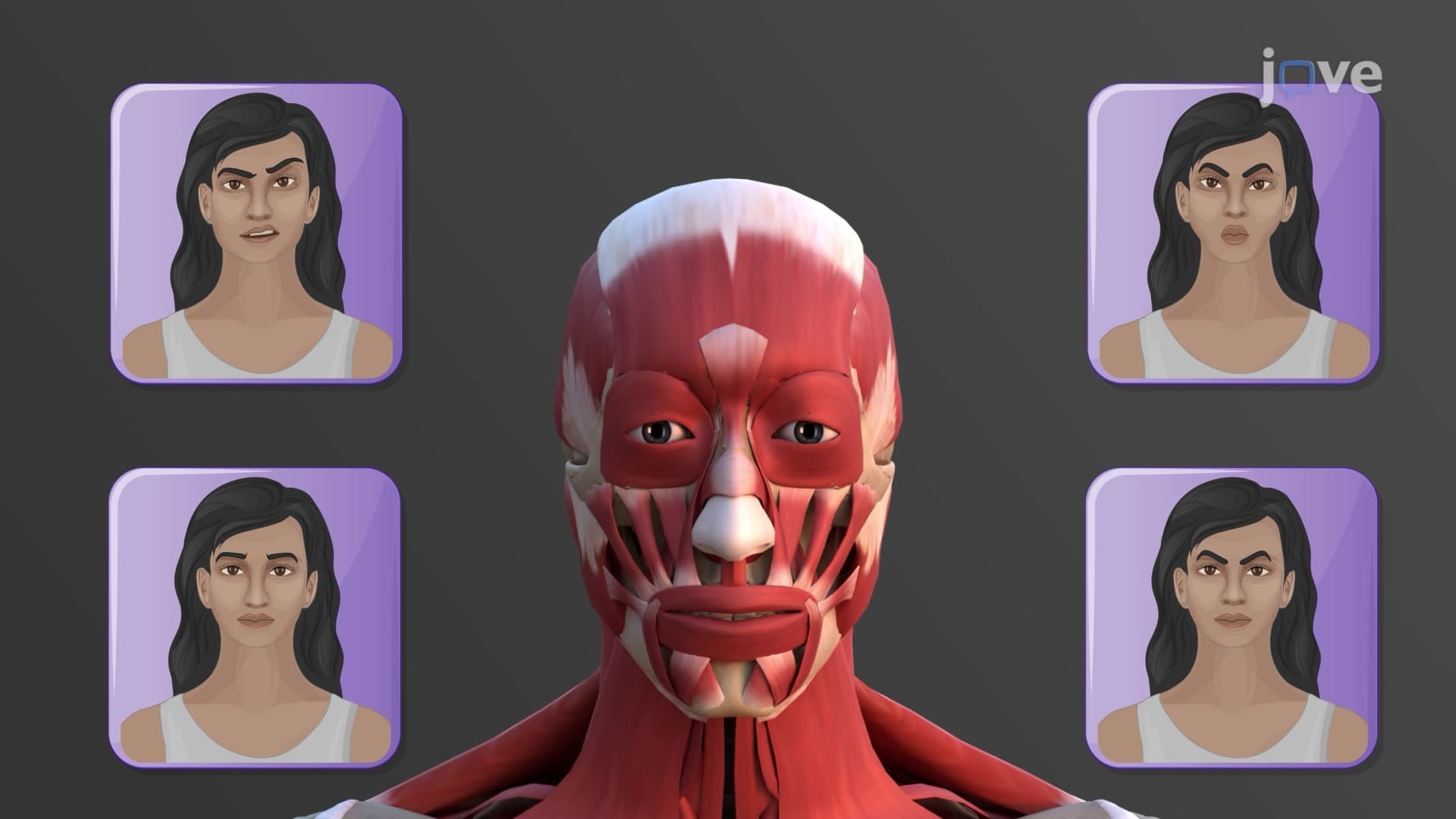

Video Summary: What are Muscles for Facial Expressions
Ever wonder why a genuine smile involves your whole face while a fake one looks forced? Facial expression muscles anatomy reveals that unlike other body muscles, these specialized fibers attach directly to skin rather than bone, creating the 43 distinct muscle contractions that form human expressions. Consider how actors at Juilliard School master micro-expressions by understanding what are the muscles involved in facial expressions - from the frontalis lifting eyebrows in surprise to the orbicularis oris forming perfect vowel sounds. Watch the full video on JoVE Coach to master this concept with expert-led visuals and step-by-step explanations.
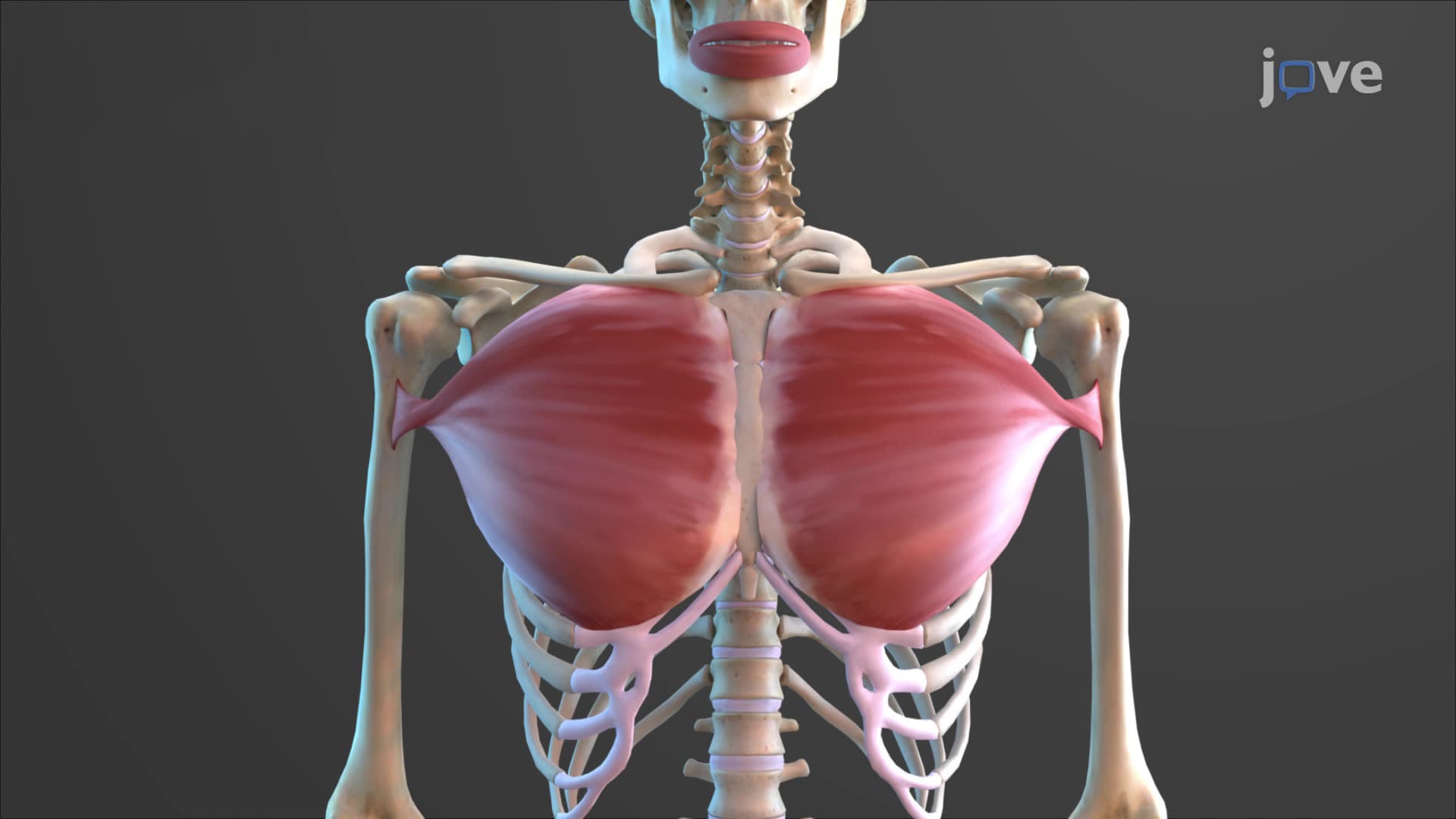
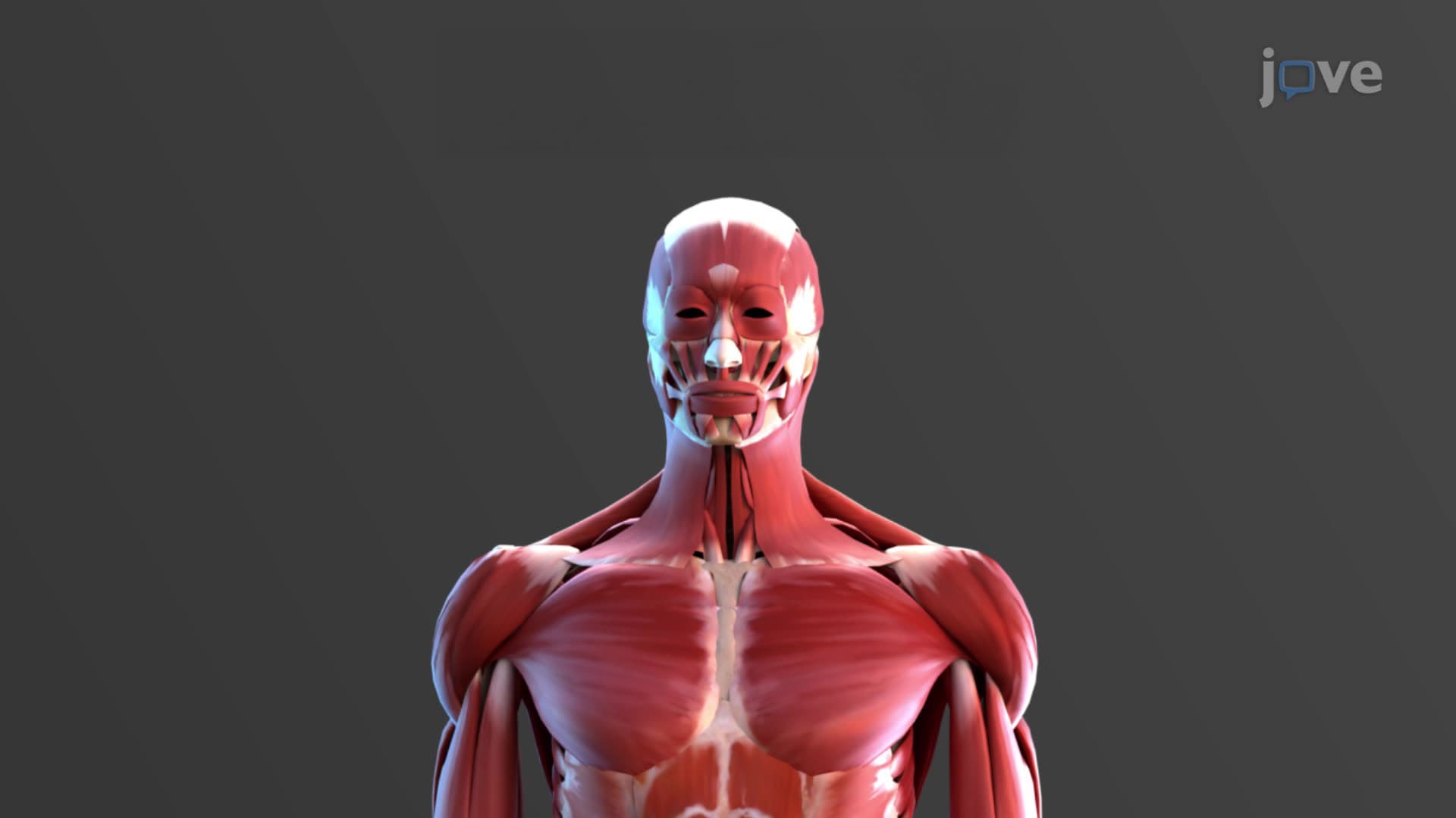
Facial expression muscles anatomy represents one of the most sophisticated muscular systems in human physiology, enabling the nuanced communication that distinguishes human social interaction. Unlike appendicular or axial skeletal muscles that primarily move bones, facial muscles create expression through direct skin manipulation. This unique architecture allows humans to produce over 10,000 distinct facial expressions, from the subtle eyebrow raise indicating skepticism to the complex muscle coordination required for genuine laughter.
The muscles controlling facial expression exhibit distinctive anatomical features that optimize them for their communicative function. Most originate from fascial planes of deeper muscles or directly from cranial bones, then insert into dermal layers rather than bony landmarks. This skin-to-skin attachment pattern enables the fine motor control necessary for emotional expression and non-verbal communication.
The occipitofrontalis exemplifies this specialized anatomy perfectly. This composite muscle consists of separate frontal and occipital bellies connected by the epicranial aponeurosis, a broad fibrous sheet spanning the scalp. When medical students at Stanford University study neuroanatomy, they learn how coordinated contraction of these bellies creates forehead wrinkles and eyebrow elevation - expressions crucial for conveying surprise, concern, or concentration during clinical interviews.
What are the muscles involved in facial expressions encompasses several functionally distinct groups. The orbicularis oris forms the muscular foundation of lip movement, originating from multiple facial muscles attached to the maxilla and mandible before inserting circumferentially around the mouth opening. This circular arrangement enables the precise lip positioning required for speech articulation - a concept emphasized in communication disorders programs at universities like Northwestern.
The buccinator muscle works synergistically with the orbicularis oris, originating from both the maxilla and mandible to insert into the corner of the mouth. Together, these muscles control essential functions from eating to speaking. Physical therapy students studying orofacial myology learn how buccinator weakness can affect swallowing patterns and speech clarity, making this knowledge clinically relevant for treating patients with facial nerve damage.
Understanding facial expression muscles anatomy proves essential for multiple healthcare disciplines. Pre-med students preparing for the MCAT encounter questions about facial muscle innervation, particularly the role of cranial nerve VII (facial nerve) in controlling expression muscles. Similarly, nursing students taking the NCLEX must understand how facial muscle assessment reveals neurological function.
This knowledge also supports performance arts education. Drama students at Yale School of Drama study facial muscle control to enhance character portrayal, while speech-language pathology programs emphasize how facial muscle coordination affects articulation and social communication skills.
Related Micro-courses