Loading video…


 20 Concepts
 68.3K views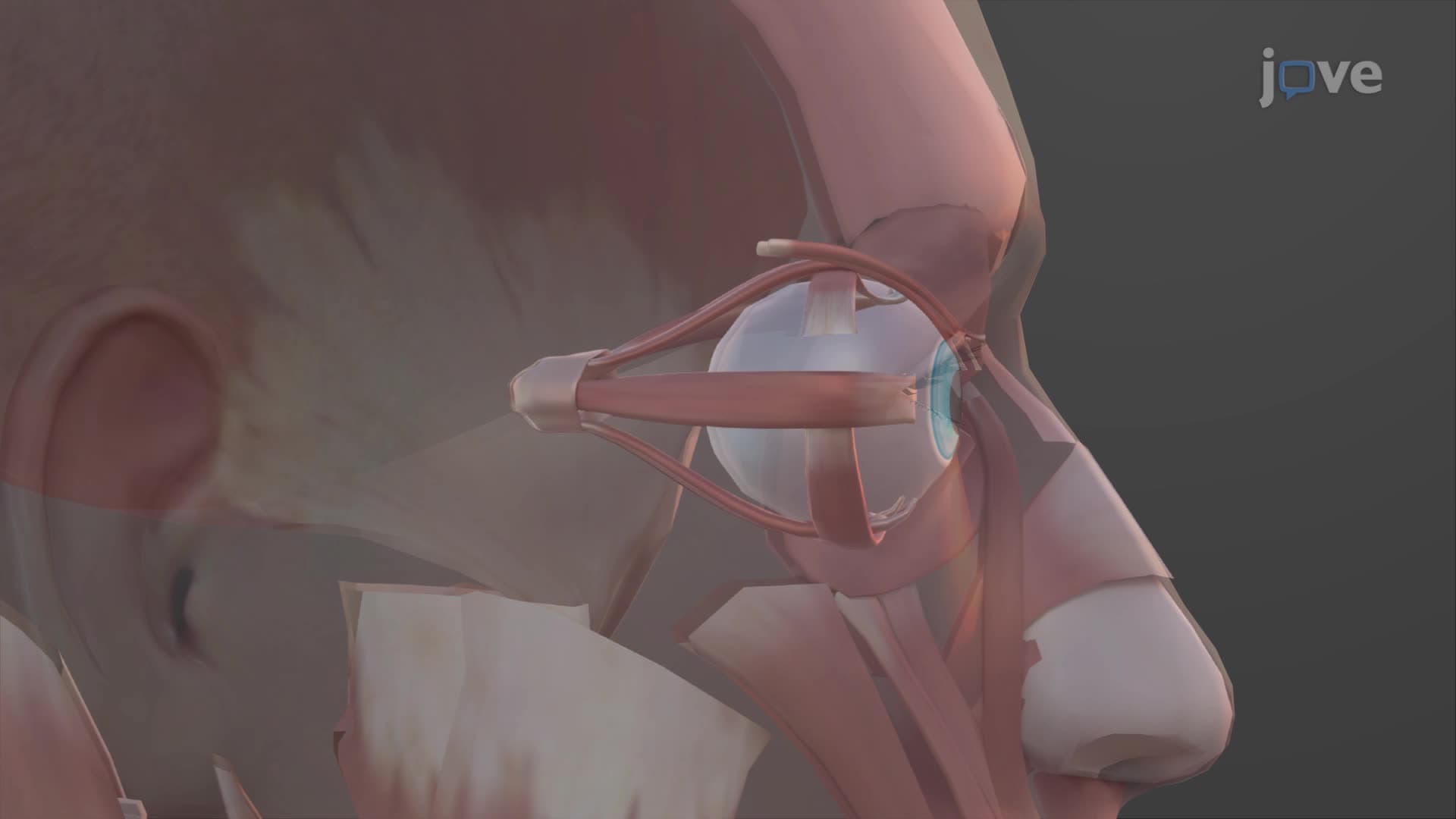

Video Summary: What Is Muscles of the Eye
Ever wonder how your eyes can track a baseball flying through the air during a Yankees game? Eye muscles anatomy physiology involves a complex system of ten specialized muscles per eye working in perfect coordination. Each eye contains six extraocular muscles that control movement, three smooth intraocular muscles managing focus and light intake, plus the levator palpebrae superioris for eyelid opening. Understanding what is muscles of the eye reveals how these tiny powerhouses enable everything from reading textbooks to driving safely. Watch the full video on JoVE Coach to master this concept with expert-led visuals and step-by-step explanations.
Eye muscles anatomy physiology encompasses a sophisticated network of ten muscles per eye, each serving distinct but interconnected functions. This muscular system enables the precise control necessary for activities ranging from reading SAT questions to tracking moving objects during sports.
The six extraocular eye muscles form the primary system for eye movement control. Four rectus muscles-medial lateral rectus eye muscles along with superior inferior rectus muscles-originate from the common tendinous ring surrounding the optic foramen. These muscles insert directly onto the sclera, creating the primary axes of movement.
The medial lateral rectus eye pair controls horizontal movements essential for reading and tracking. The medial rectus enables inward rotation (adduction), while the lateral rectus pulls the eye outward (abduction). Students preparing for the MCAT should note that damage to cranial nerve VI affects lateral rectus function, causing inward eye deviation.
The superior inferior rectus muscles primarily control vertical movements but also contribute to rotational movements due to their anatomical positioning. When studying for AP Biology exams, remember that these muscles work in conjunction with oblique muscles to achieve complex diagonal movements.
Superior inferior oblique muscles provide fine-tuned rotational control and assist in vertical movements. The superior oblique, innervated by cranial nerve IV (trochlear), passes through a pulley-like structure called the trochlea, giving it unique biomechanical properties. The inferior oblique originates from the orbital floor, making it anatomically distinct from other extraocular muscles.
The three smooth intraocular muscles-ciliary muscle, dilator pupillae, and sphincter pupillae-control accommodation and pupillary responses. These muscles are crucial for USMLE Step 1 questions about autonomic nervous system control of vision.
Understanding eye movement muscle control proves essential for diagnosing conditions like strabismus, which affects approximately 4% of US children. Ophthalmologists use this knowledge when treating diplopia (double vision) or planning corrective surgeries at institutions like Mayo Clinic and Cleveland Clinic.
Related Micro-courses