Loading video…


 20 Concepts
 68.3K views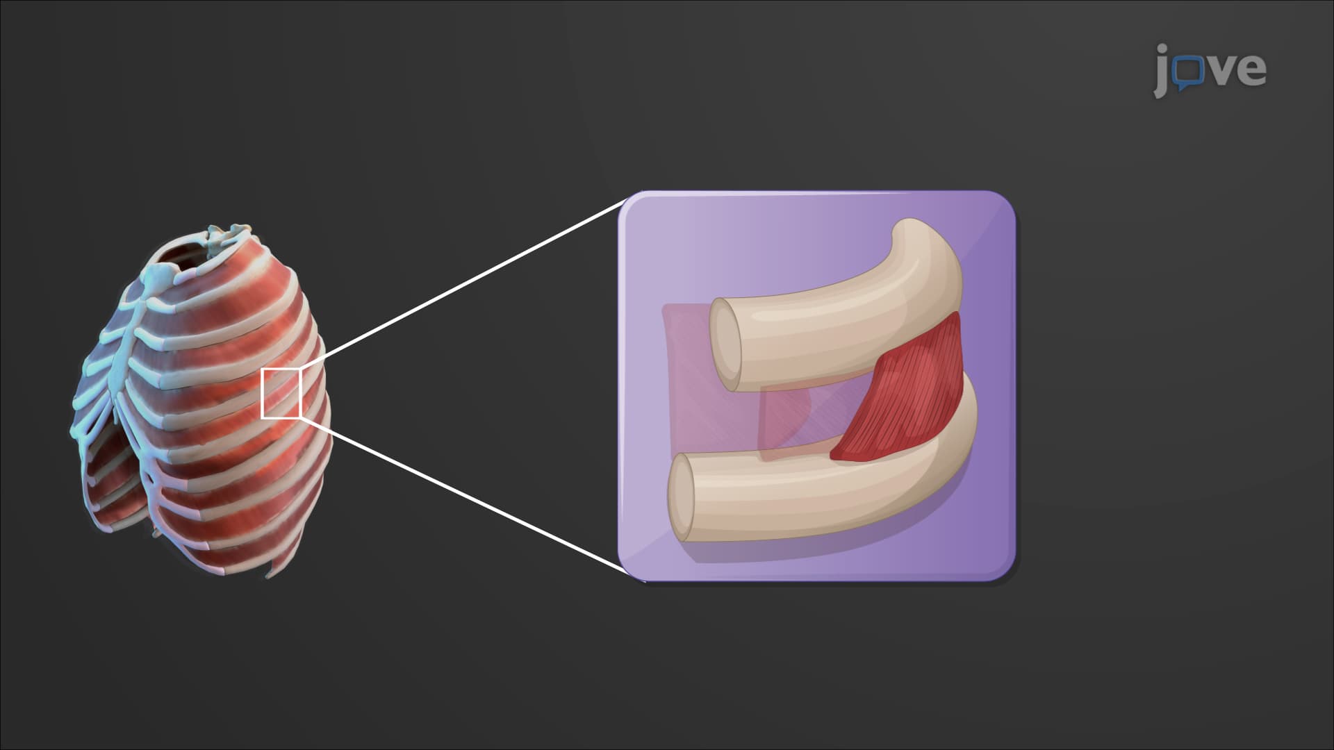

Video Summary: What Is Muscles of the Thorax
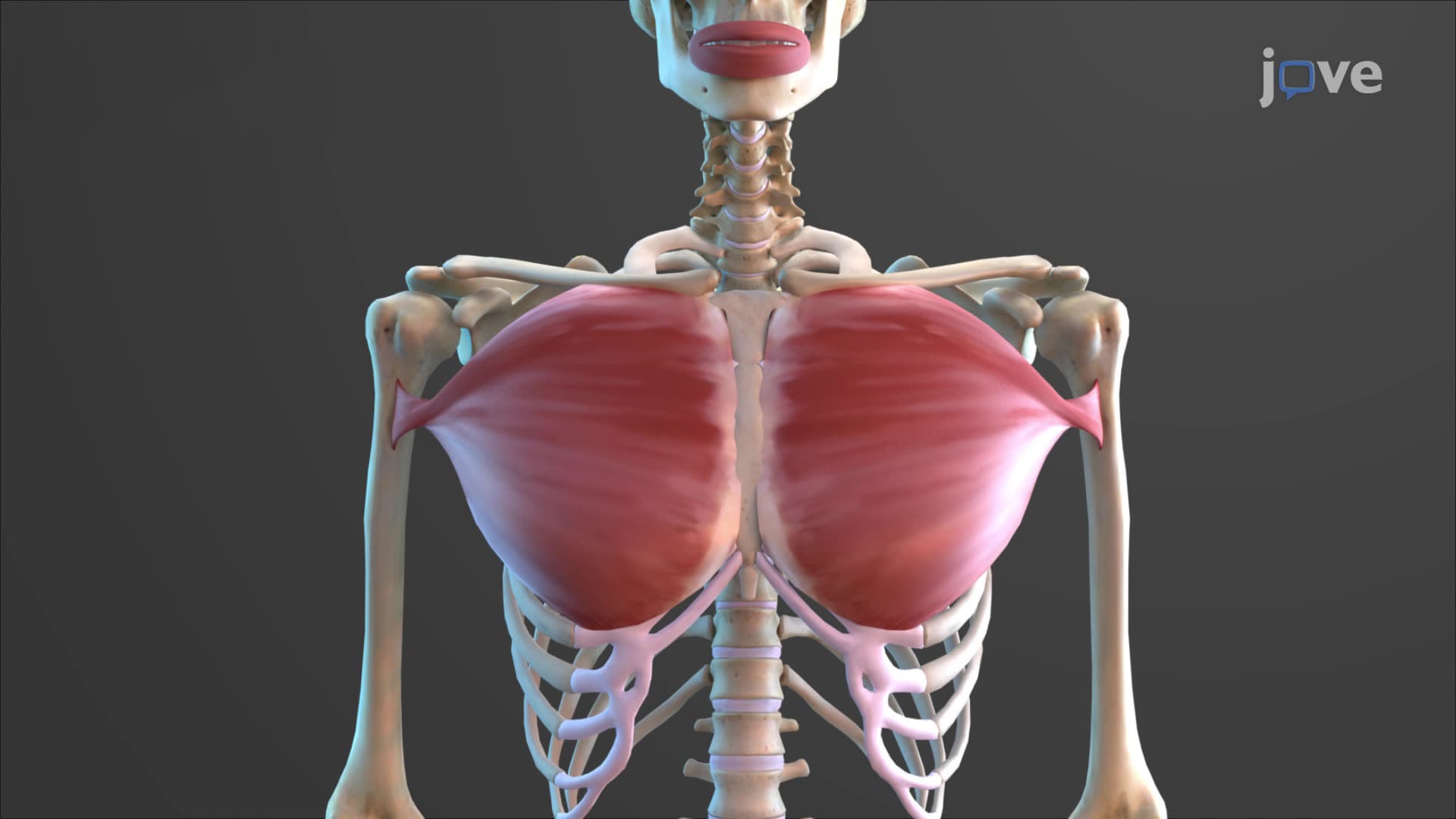
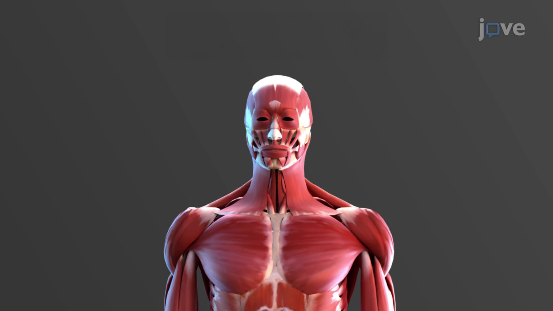
Every time you take a breath during a basketball game at Madison Square Garden, your thorax muscles anatomy physiology coordinates a complex mechanical dance involving over 22 different muscles. The muscles of the thorax include the dome-shaped diaphragm and three layers of intercostal muscles that work together to expand and compress your chest cavity during breathing. Watch the full video on JoVE Coach to master this concept with expert-led visuals and step-by-step explanations.
The thorax muscles anatomy physiology represents one of the most precisely coordinated muscular systems in the human body. These muscles form the mechanical foundation for breathing, a process so automatic that most people never consider the complex muscular choreography occurring 15-20 times per minute.
The diaphragm stands as the most important muscle in the chest wall muscles explained system. This dome-shaped muscular partition separates your thoracic cavity from your abdominal cavity, functioning like a biological piston. Originating from the lower ribs, sternum, and lumbar vertebrae, the diaphragm inserts into its own central tendon - a unique arrangement that allows it to flatten dramatically during contraction.
When the diaphragm contracts during inspiration, it can descend up to 10 centimeters in healthy adults. This downward movement increases thoracic cavity volume by approximately 500 milliliters during quiet breathing. For students preparing for the MCAT or AP Biology exams, understanding that diaphragmatic paralysis can reduce lung capacity by 25-30% demonstrates the muscle's critical importance.
What are the muscles of the thorax in anatomy extends beyond just the diaphragm to include 11 pairs of intercostal muscles arranged in three distinct layers. The external internal intercostal muscles work as functional antagonists - external intercostals lift the ribs during inspiration while internal intercostals pull ribs together during forced expiration.
The external intercostals run obliquely downward and forward between adjacent ribs, following the same direction as your fingers when you place your hands in your pockets. During inspiration, these muscles contract to elevate the ribs, increasing both the anteroposterior and lateral dimensions of the thoracic cavity - a movement respiratory therapists call the "bucket handle" motion.
Internal intercostals run perpendicular to the externals, contracting during active expiration to compress the thoracic cavity. The innermost intercostals, the deepest layer, assist the internal intercostals and contain important neurovascular bundles that physicians must consider during procedures like chest tube insertion.
Understanding diaphragm breathing muscle function proves essential for healthcare professionals. Patients with spinal cord injuries above C3-C5 lose diaphragmatic function, requiring mechanical ventilation. This knowledge frequently appears on USMLE Step 1 examinations and NCLEX questions related to respiratory assessment.
The transversus thoracis muscle, though smaller, assists in forced expiration and plays a role in coughing - a protective reflex that can generate intrathoracic pressures exceeding 100 mmHg. For pre-med students, recognizing how thoracic muscle dysfunction contributes to conditions like restrictive lung disease provides crucial clinical context for understanding thoracic muscle breathing mechanics.
Related Micro-courses