Loading subject page…


68,318 views

Video Summary: What Is Muscles Vertebral Column
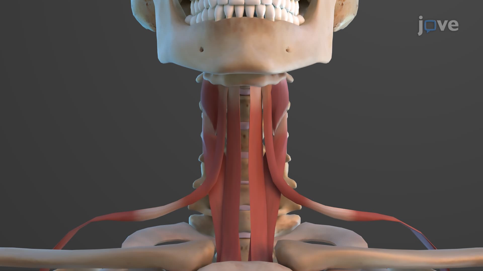
Ever wondered why professional football players like those in the NFL can absorb massive tackles without spinal injury? The vertebral column muscles anatomy forms a sophisticated three-layered system that protects and moves your spine with remarkable precision. From the superficial splenius muscles controlling head movement to the deep multifidus stabilizing individual vertebrae, these back muscles spine explained work together like a complex engineering framework. Watch the full video on JoVE Coach to master this concept with expert-led visuals and step-by-step explanations.
The vertebral column muscles anatomy represents one of the most sophisticated muscular systems in the human body, organized into three distinct layers that work in perfect harmony. This architectural marvel enables everything from a baseball pitcher's wind-up to a dancer's graceful spinal extension. For students preparing for the MCAT or AP Biology exams, understanding this layered organization is crucial for mastering musculoskeletal physiology.
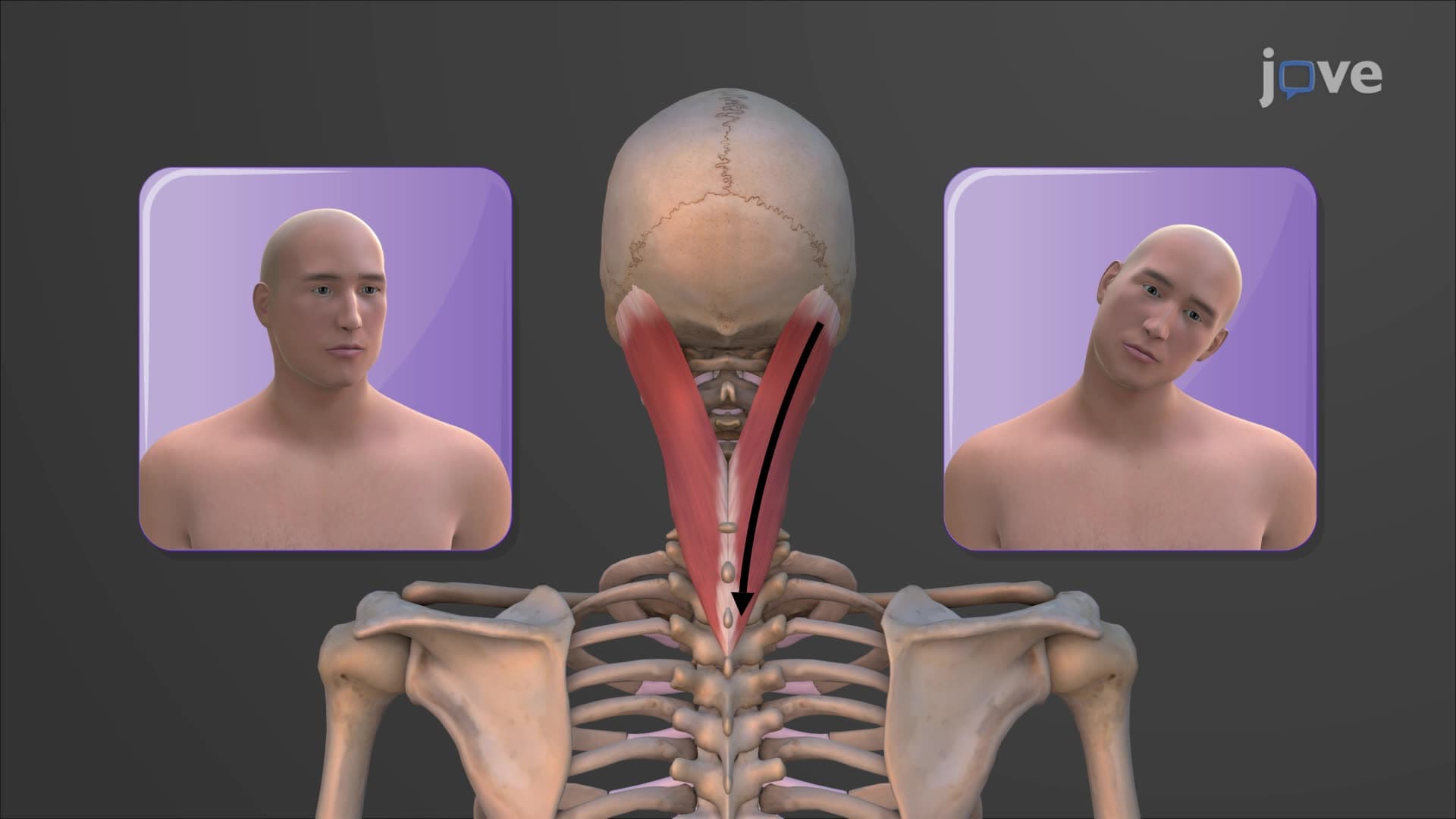
The superficial layer houses the splenius muscles, which serve as the primary controllers of head and neck movement. These muscles are particularly important for students studying for the USMLE Step 1, as they frequently appear in questions about cervical spine function. The splenius capitis and splenius cervicis work together to rotate and extend the head, movements essential for activities ranging from driving (checking blind spots) to sports performance.
The erector spinae group forms the intermediate layer and represents the powerhouse of spinal movement. This back muscles spine explained system includes three major columns: iliocostalis, longissimus, and spinalis. Each column subdivides based on anatomical regions-for example, the iliocostalis longissimus spinalis system includes the iliocostalis cervicis, thoracis, and lumborum. These muscles are responsible for the bilateral extension that allows you to stand upright against gravity, a concept frequently tested in college anatomy courses and the HESI A2 exam.
Understanding bilateral versus unilateral contraction is critical for exam success. When both sides contract simultaneously, the spine extends backward. When only one side contracts, lateral flexion occurs toward that side-imagine a tennis player's powerful serve motion.
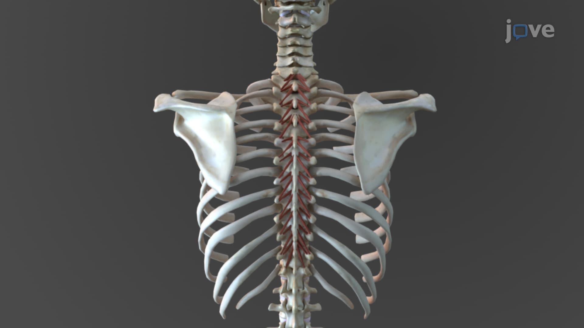
The deep layer contains the most clinically significant muscles: semispinalis, multifidus rotatores back, and rotatores. These deep back muscle vertebral stabilizers are essential for maintaining proper vertebral alignment during movement. The multifidus, in particular, is crucial for preventing lower back injuries-a topic increasingly emphasized in nursing programs and physical therapy curricula.
For NCLEX preparation, understanding how these muscles provide segmental stability is vital. Each vertebra requires individual stabilization during complex movements, and dysfunction in these deep muscles often leads to the chronic back pain affecting over 80% of Americans at some point in their lives.
Related Micro-courses