Loading subject page…

- Cell Biology
- Visualizing Cells, Tissues, and Molecules
- Electron Microscope Tomography and Single-particle Reconstruction
Micro-course: Visualizing Cells, Tissues, and Molecules
138,512 views
Learn key concepts and earn your certificate.


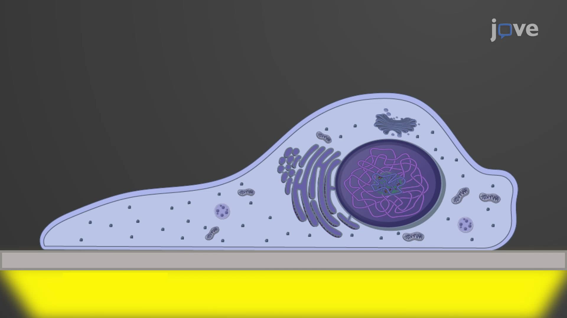
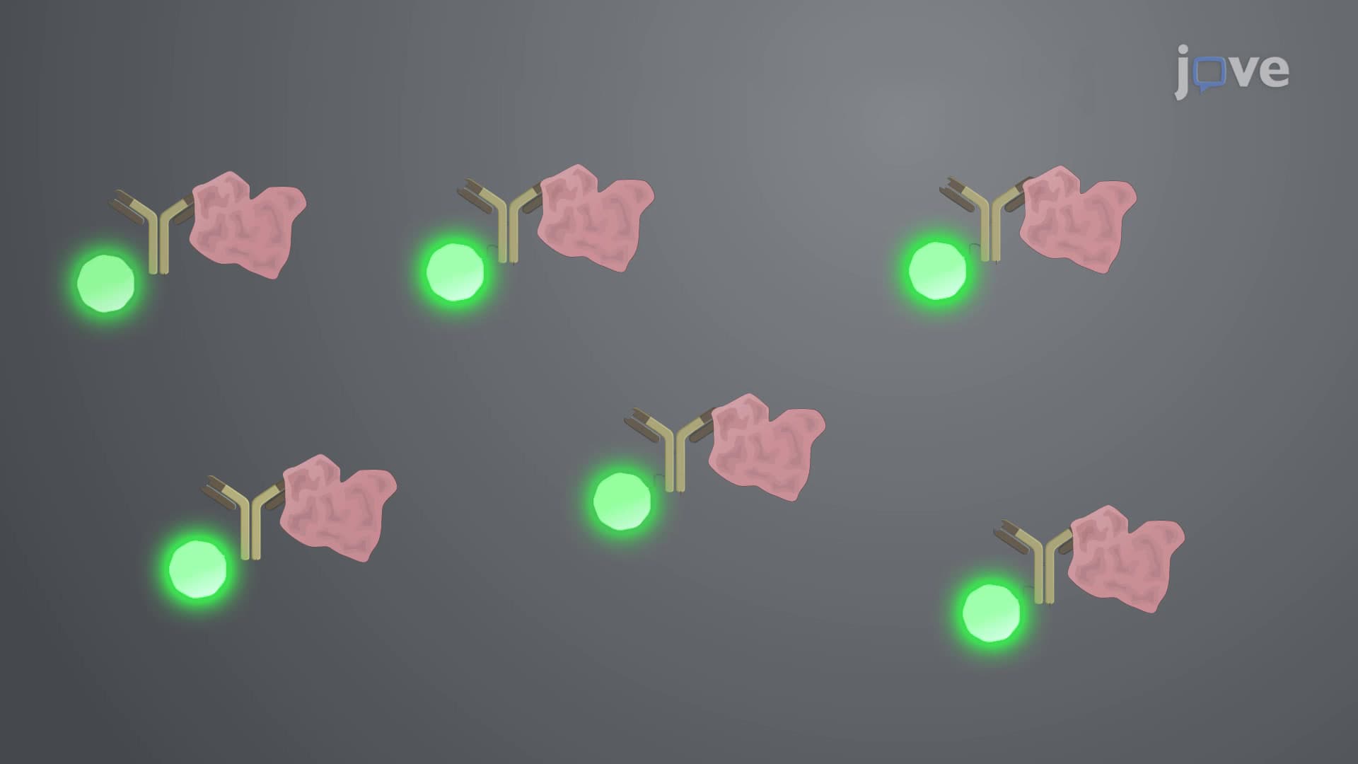


Electron Microscope Tomography and Single-particle Reconstruction

00:00
01:07
Video Summary: Electron Microscope Tomography and Single Guide
Did you know that scientists at Johns Hopkins University used electron microscope tomography and single particle reconstruction to reveal how SARS-CoV-2 spike proteins change shape when infecting cells? Electron microscope tomography and single particle reconstruction are revolutionary 3D imaging techniques that allow researchers to visualize biological structures at the molecular level. These cryo-electron microscopy methods help decode everything from virus mechanisms to protein folding diseases like Alzheimer's. Watch the full video on JoVE Coach to master this concept with expert-led visuals and step-by-step explanations.
- Understand the fundamental principles of electron microscope tomography and single particle reconstruction as complementary 3D imaging techniques
- Identify the key differences between electron tomography's tilting methodology and single particle reconstruction's averaging approach
- Analyze how electron tomography preserves native cellular environments while maintaining structural integrity
- Compare the applications of in vivo electron tomography versus in vitro single particle reconstruction
- Evaluate the advantages and limitations of each technique for different biological research questions
- Apply knowledge of sample preparation requirements for both tomography and single particle methods
- Recognize the computational processes involved in 3D reconstruction from 2D electron microscopy images
- Explore real-world applications of these techniques in drug discovery, structural biology, and disease research
Understanding Electron Microscope Tomography and Single Particle Reconstruction
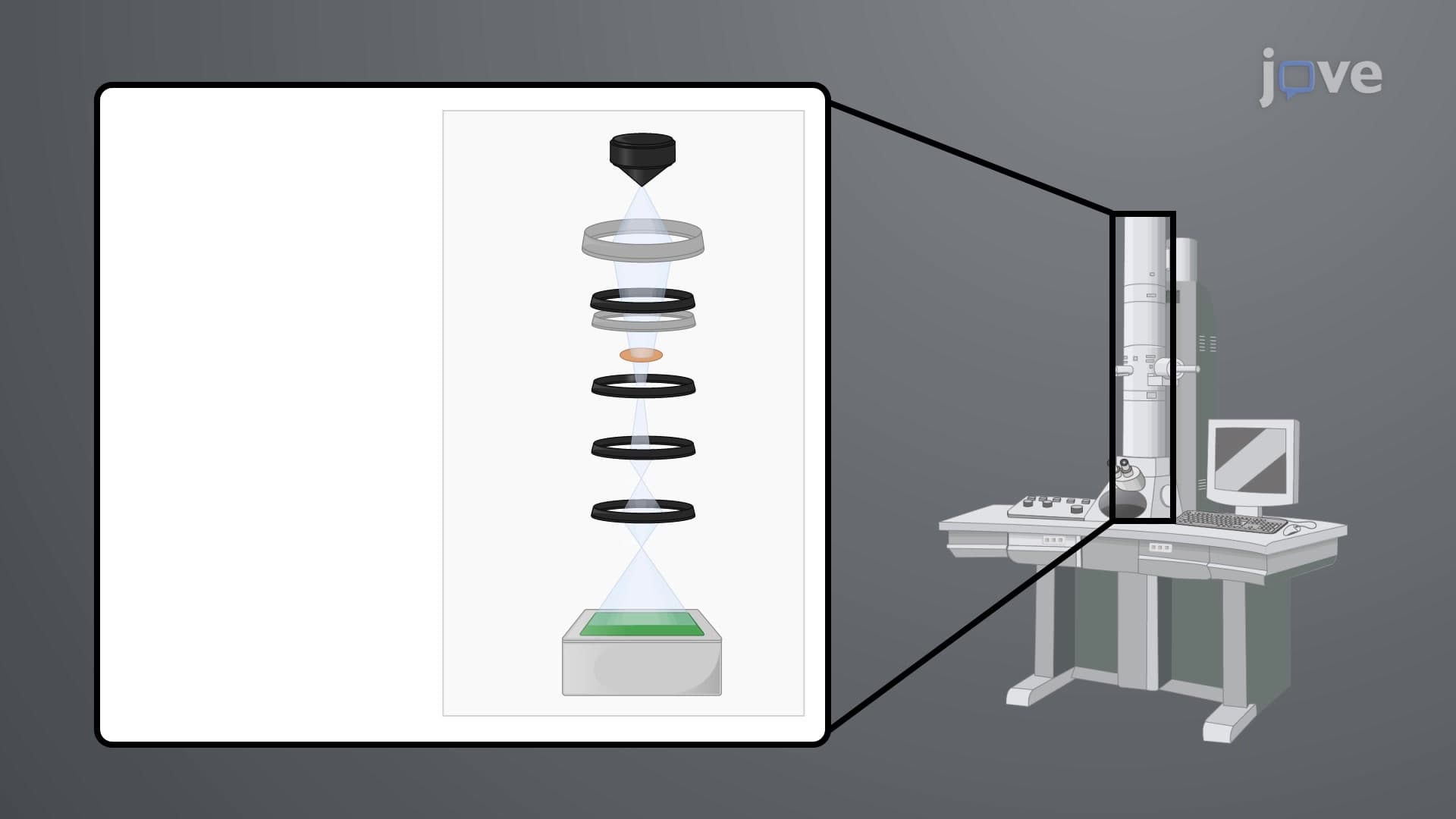
Electron microscope tomography and single particle reconstruction represent two groundbreaking approaches to three-dimensional biological imaging that have revolutionized our understanding of cellular architecture. These techniques overcome the fundamental limitation of traditional electron microscopy-its inherently two-dimensional nature-by employing sophisticated computational methods to reconstruct three-dimensional structures from multiple 2D images.
Electron Tomography: Preserving Native Cellular Architecture
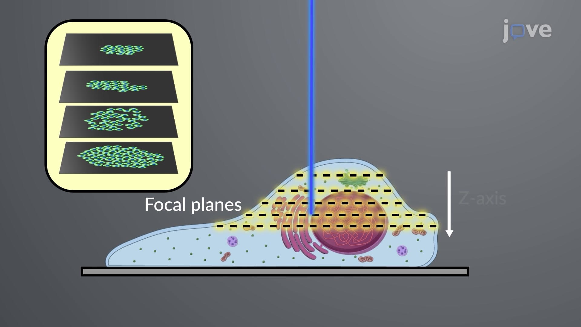
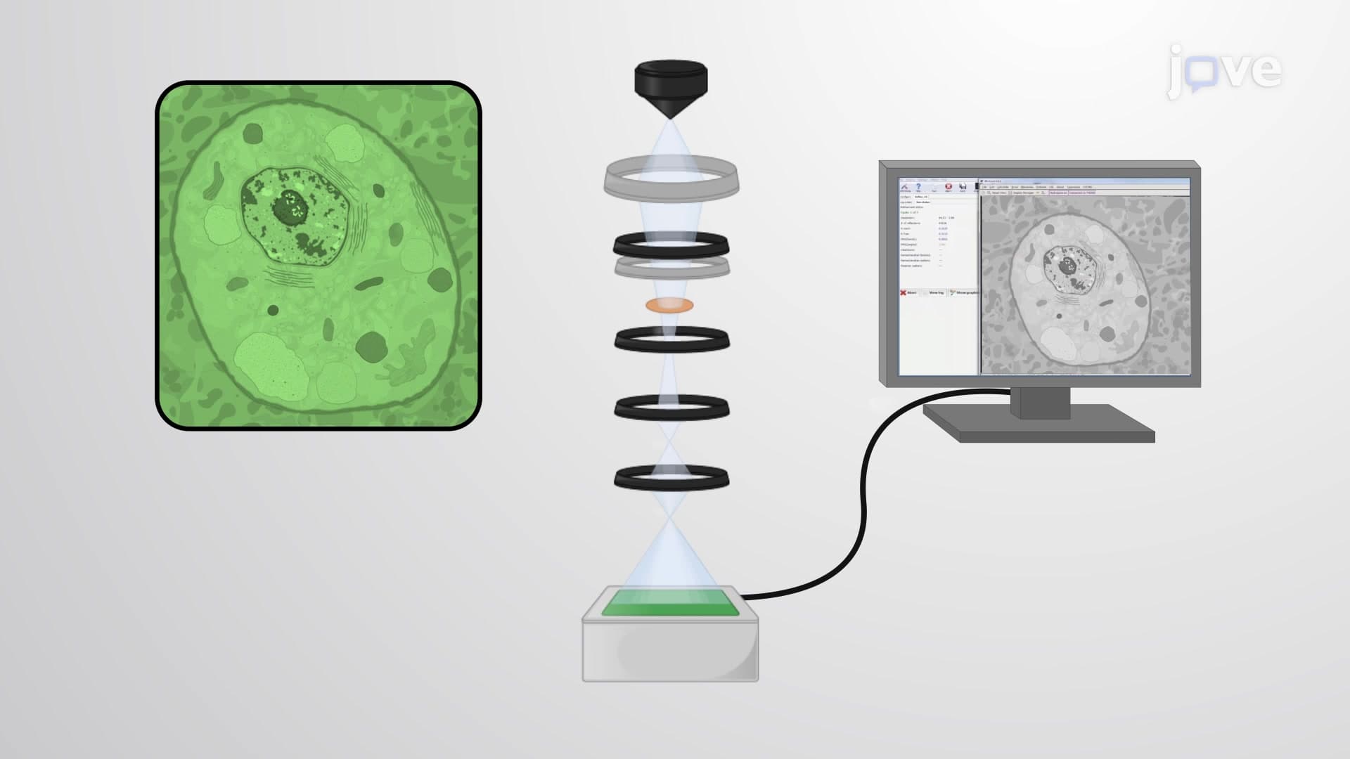
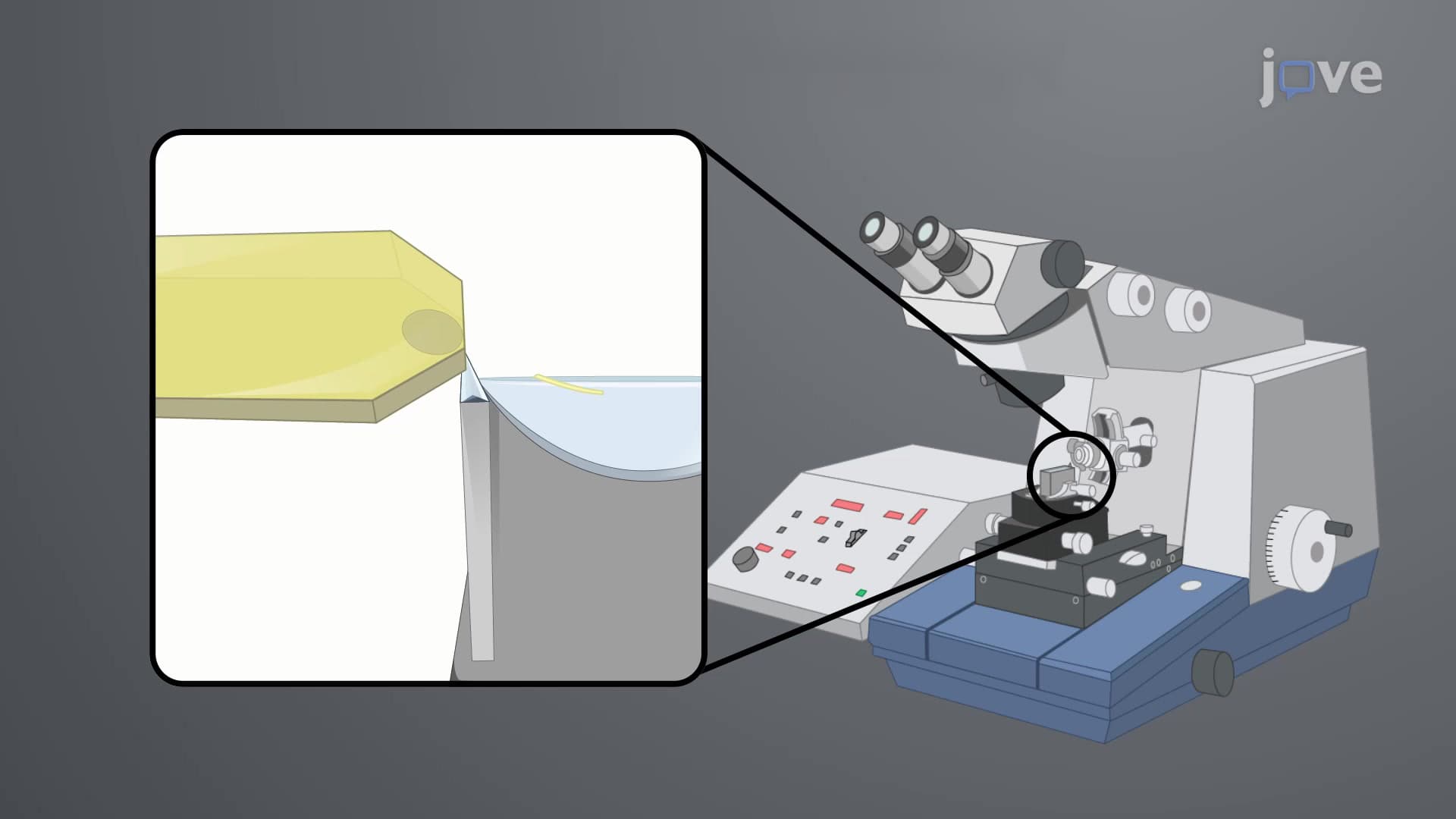
Electron tomography operates on the principle of progressive sample tilting, much like a medical CT scan but at the nanometer scale. The biological sample remains frozen in a near-native state while being incrementally rotated relative to the electron beam, typically through angles ranging from -60° to +60°. This process generates a tilt series of 2D projection images that computational algorithms then reconstruct into a three-dimensional tomogram.
The technique's greatest strength lies in its ability to maintain the in vivo structural integrity of biological samples. Researchers at institutions like Harvard Medical School and Stanford University regularly use electron tomography to study organelle interactions, viral infection mechanisms, and protein complex arrangements within their natural cellular environments. For students preparing for AP Biology or college-level cell biology courses, understanding electron tomography is crucial for grasping modern approaches to cellular structure determination.
Single Particle Reconstruction: Molecular-Level Precision
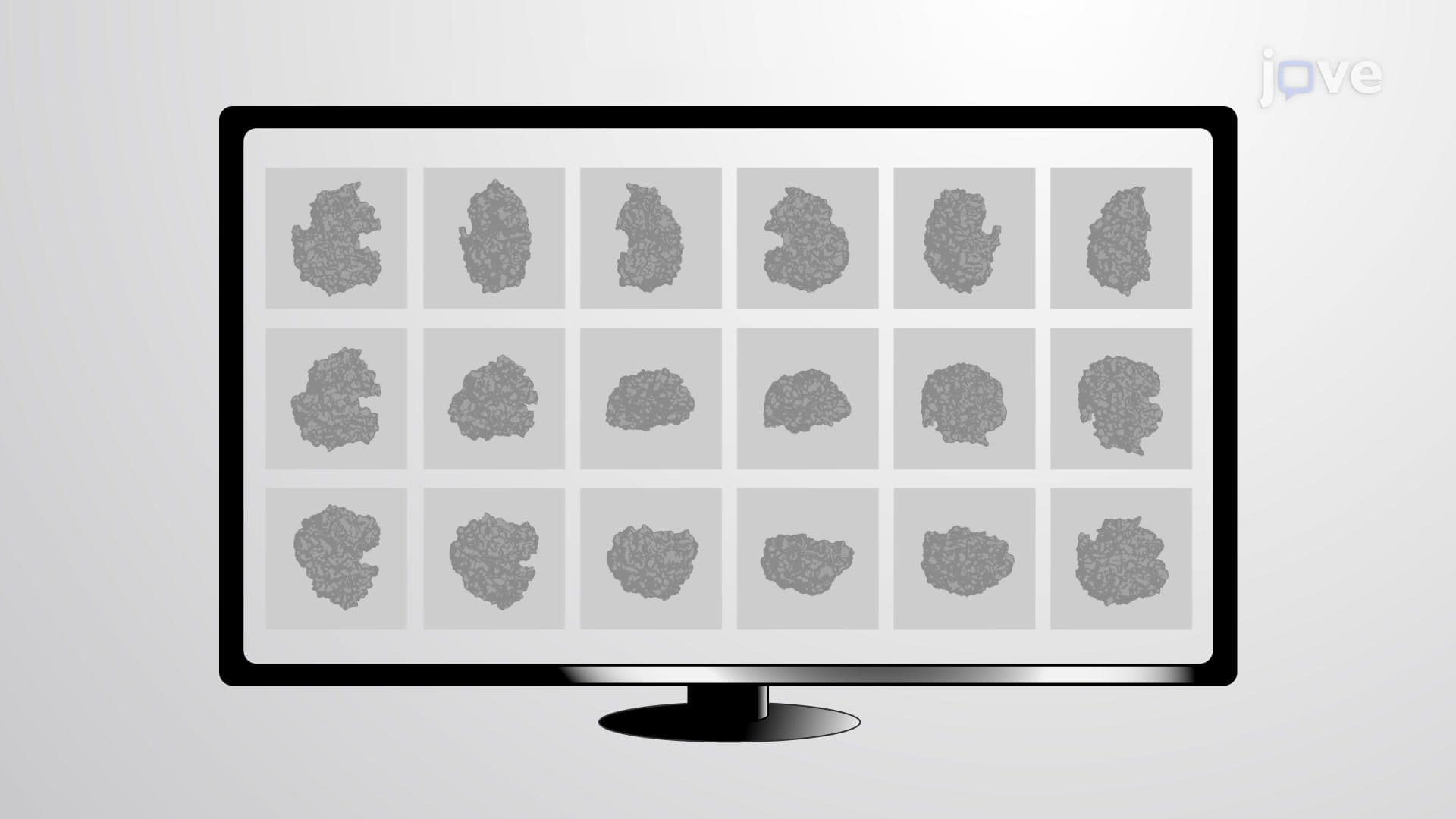
Single particle reconstruction takes a fundamentally different approach, focusing on isolated, purified biological molecules distributed randomly across electron microscopy grids. This technique captures thousands of 2D images of individual particles in various orientations, then uses sophisticated algorithms to classify, align, and average these images into high-resolution 3D structures.
The power of single particle reconstruction lies in its ability to achieve near-atomic resolution-often better than 2-3 angstroms-making it invaluable for drug design and structural biology. The 2017 Nobel Prize in Chemistry recognized the developers of cryo-electron microscopy, highlighting single particle reconstruction's importance in modern biochemistry. Students encountering this concept in MCAT preparation should focus on understanding how computational averaging eliminates noise and enhances structural details.
Practical Applications and Complementary Strengths
These techniques serve different but complementary roles in biological research. Electron tomography excels at revealing cellular organization, organelle interactions, and macromolecular complexes in their native states, making it ideal for studying processes like autophagy, mitochondrial dynamics, and viral replication cycles. Single particle reconstruction, conversely, provides the atomic-level detail necessary for understanding enzyme mechanisms, protein-drug interactions, and conformational changes.
Both techniques require sophisticated computational resources and specialized software packages, reflecting the integration of biology, physics, and computer science that characterizes modern structural biology. For students pursuing STEM careers, these methods exemplify how interdisciplinary approaches drive scientific breakthroughs.
Frequently Asked Questions
These are advanced 3D imaging techniques using electron microscopy to visualize biological structures at the molecular level. Electron tomography tilts samples to capture multiple angles, creating 3D images of cells and organelles in their natural state. Single particle reconstruction averages thousands of images of isolated molecules to achieve near-atomic resolution structures.
MCAT questions often focus on comparing these methods' resolution capabilities, sample preparation requirements, and applications in drug discovery. AP Biology may test understanding of how these techniques advance cell biology research. Both exams emphasize connecting structural information to biological function and experimental design principles.
Most college exams emphasize single particle reconstruction because of its relevance to protein structure determination and drug design. However, understanding both techniques' complementary roles demonstrates mastery of modern structural biology. Focus on their different applications, resolution limits, and how each contributes to our understanding of biological processes.
The NIH and pharmaceutical companies like Pfizer use these techniques extensively for drug development. During COVID-19, researchers used single particle reconstruction to determine spike protein structures for vaccine design. Cancer research centers use electron tomography to study how drugs affect cellular organelles and membrane structures.
The basic principles require only high school-level geometry and physics understanding. While the computational algorithms involve complex mathematics, introductory courses focus on conceptual understanding rather than mathematical derivations. Students should concentrate on applications, advantages, and limitations rather than computational details.
Create comparison charts highlighting each method's sample requirements, resolution capabilities, and typical applications. Practice identifying which technique would best answer specific research questions. Use visual aids and diagrams to understand the geometric principles underlying tomographic reconstruction and particle averaging processes.
These methods complement X-ray crystallography and NMR spectroscopy in structural biology. Understanding their relative strengths helps explain why researchers choose specific techniques for different questions. This knowledge connects to broader themes in biochemistry about structure-function relationships and experimental methodology.
Consider studying cryo-electron microscopy sample preparation, computational image processing algorithms, and hybrid structural approaches combining multiple techniques. Advanced topics include subtomogram averaging, focused ion beam milling for cellular tomography, and machine learning applications in structure determination.
Related Micro-courses