Loading subject page…
- Microbiology
- Microbial Growth
- Microbial Growth Measurement: Direct Methods
Micro-course: Microbial Growth
10,971 views
Learn key concepts and earn your certificate.


Microbial Growth Measurement: Direct Methods

00:00
01:23
Video Summary: Microbial Growth Measurement Direct Methods Guide
Did you know that a single drop of pond water can contain over 50,000 bacterial cells invisible to the naked eye? Microbial growth measurement direct techniques allow scientists to count and quantify these microscopic organisms with remarkable precision. From testing water quality at the EPA to quality control in pharmaceutical manufacturing, the Microbial Growth Measurement Direct Methods Guide provides essential skills for laboratory professionals nationwide. Watch the full video on JoVE Coach to master this concept with expert-led visuals and step-by-step explanations.
- Understand the fundamental principles of Microbial Growth Measurement Direct Methods Guide and distinguish between direct and indirect measurement approaches
- Identify the two primary direct counting techniques: microscopic cell counting and colony-forming unit (CFU) enumeration
- Analyze the advantages and limitations of Petroff-Hausser chamber methodology for immediate cell density determination
- Compare serial dilution protocols and calculate dilution factors for accurate microbial quantification
- Evaluate sources of error in direct counting methods, including dead cell interference and cell clumping artifacts
- Apply plate counting techniques to determine viable cell concentrations in laboratory samples
- Recognize real-world applications of direct measurement in clinical diagnostics, food safety, and environmental monitoring
- Explore quality control procedures essential for reproducible and accurate microbial enumeration results
Understanding Microbial Growth Measurement Direct Methods
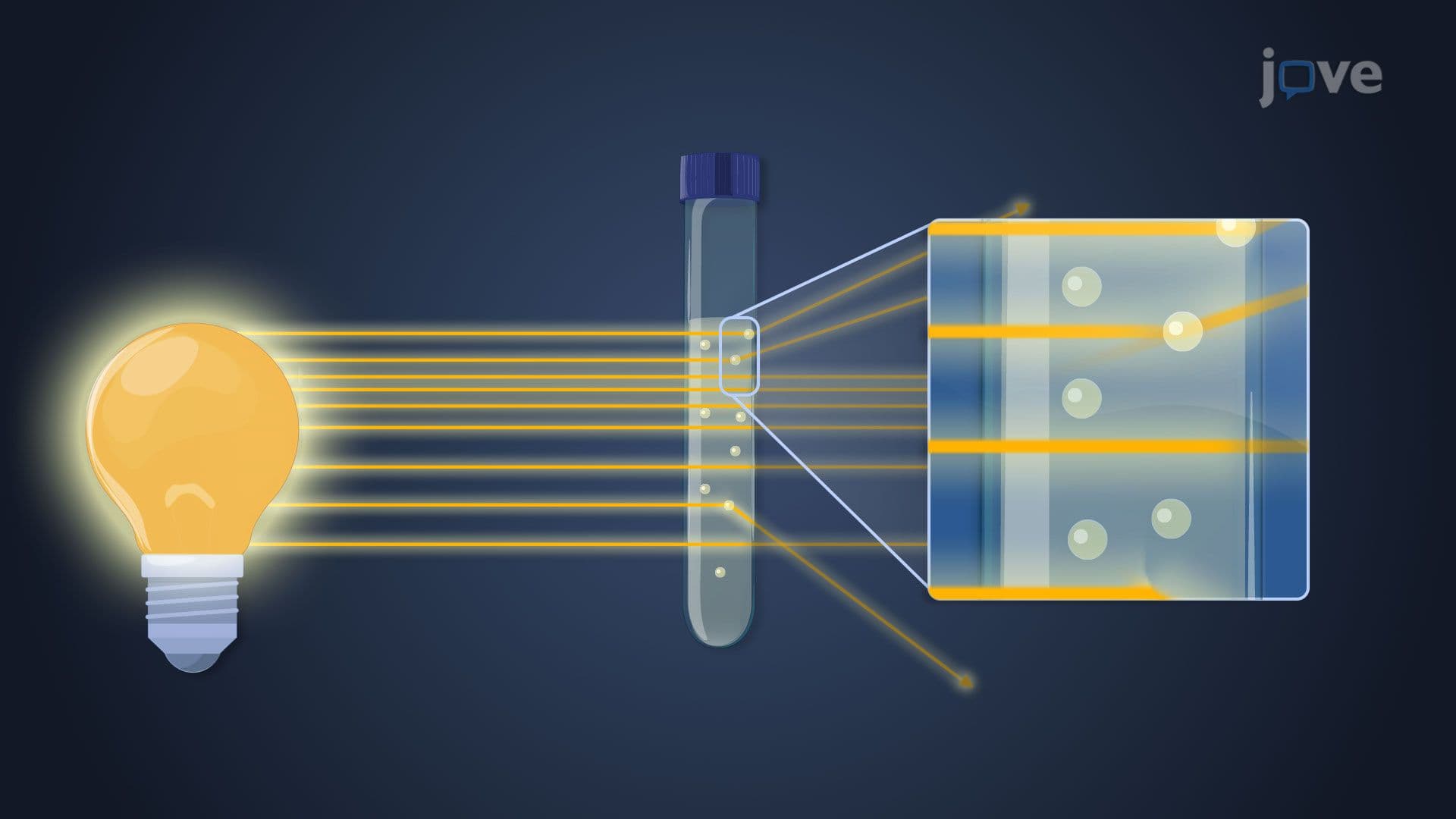
Microbial growth measurement direct approaches form the cornerstone of quantitative microbiology, enabling precise enumeration of microorganisms in diverse samples. Unlike indirect methods that measure metabolic byproducts or biomass, direct techniques physically count individual cells or colonies, providing definitive quantitative data essential for clinical diagnostics, pharmaceutical quality control, and environmental monitoring.
Microscopic Direct Counting with Petroff-Hausser Chambers
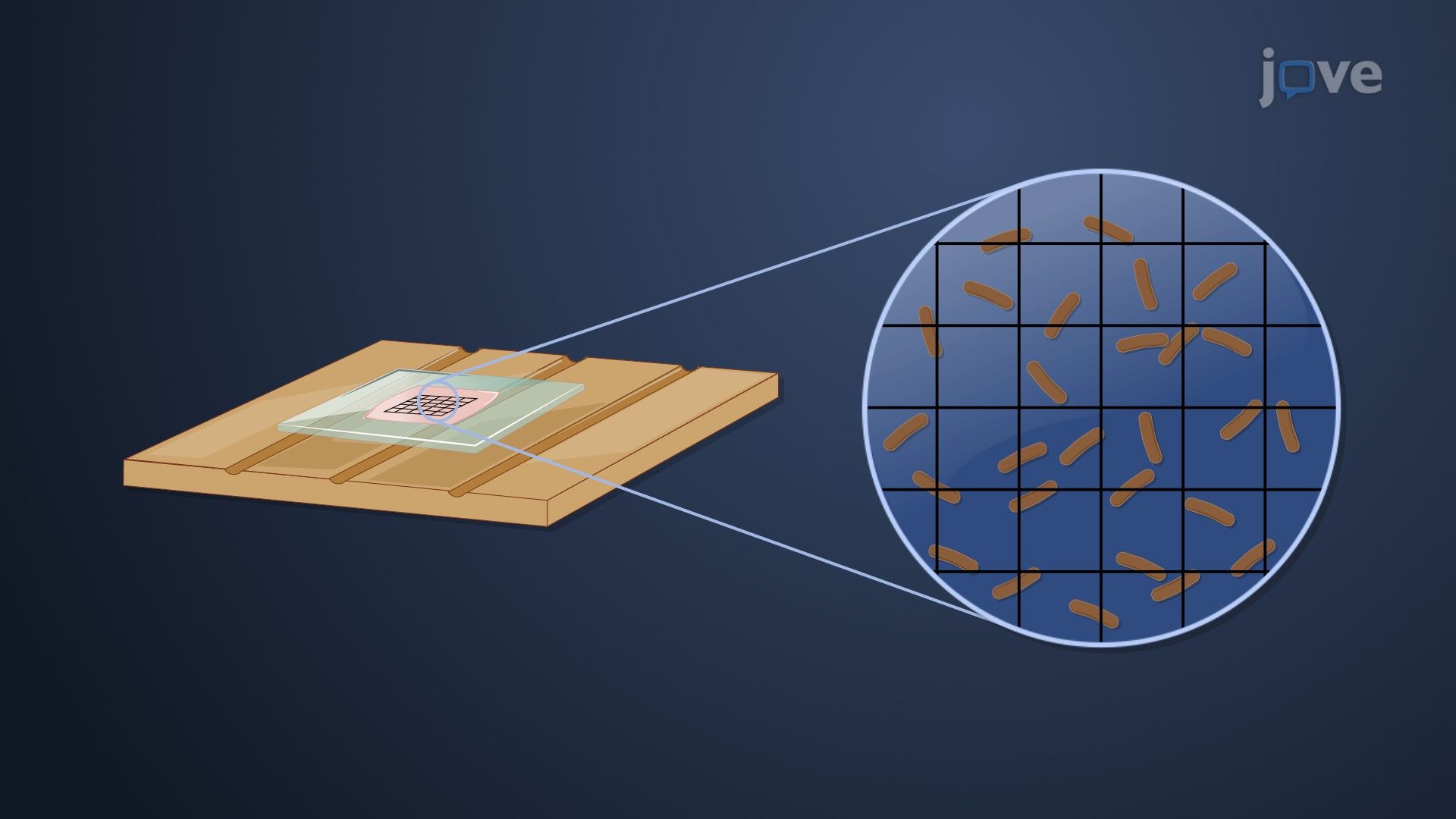
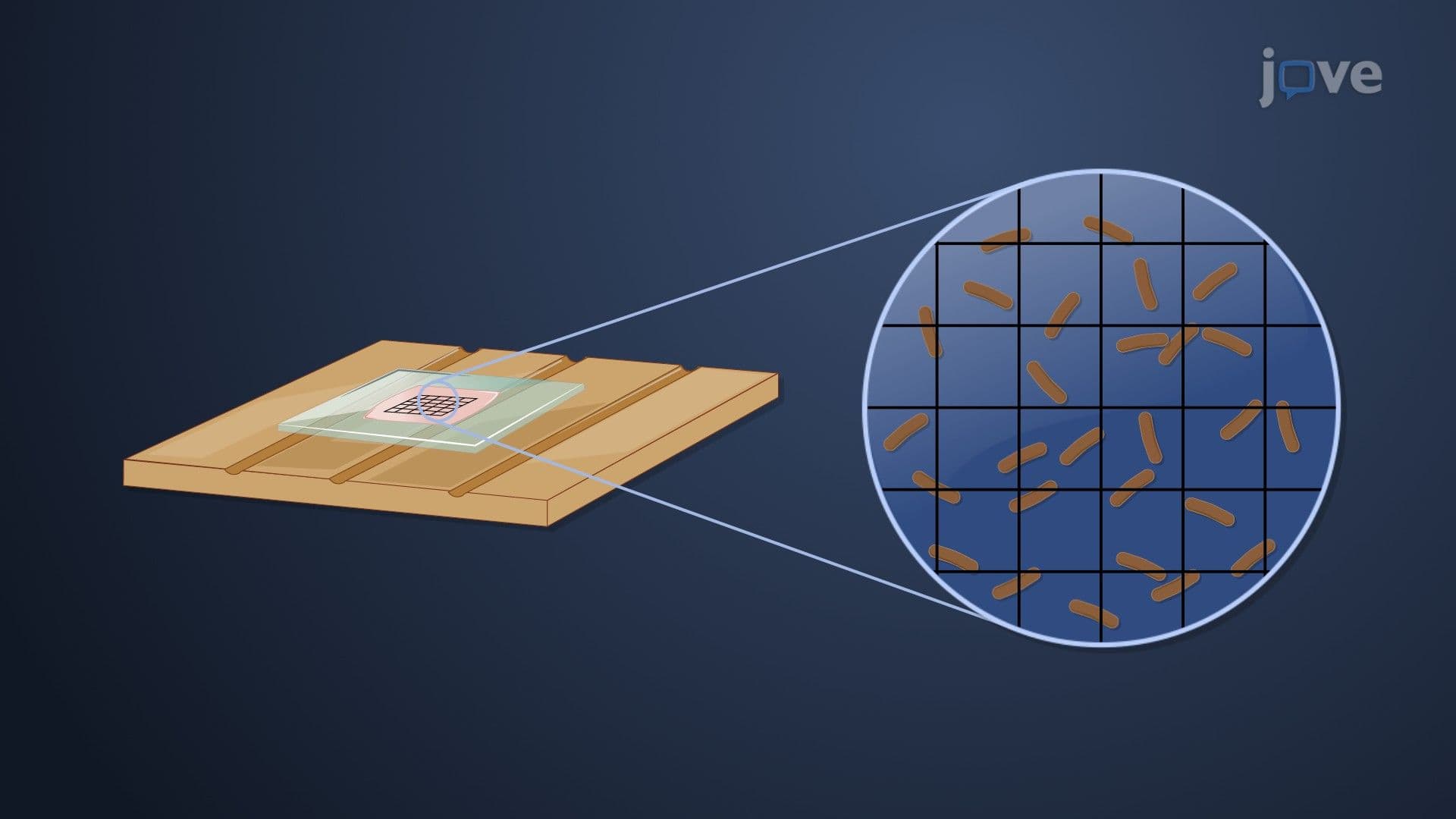
The Petroff-Hausser counting chamber represents a fundamental tool in microbiology laboratories across American universities and research institutions. This specialized microscope slide contains precisely etched grids creating defined volumes, typically 0.02 mm³ per large square. Laboratory technicians at facilities like the CDC utilize this method for rapid bacterial enumeration in clinical specimens.
The technique involves loading a diluted microbial suspension onto the chamber and systematically counting cells within predetermined grid squares. Cell density calculations follow the formula: Cells/mL = (Average count per square × Dilution factor) / (Volume per square in mL). This immediate counting method proves invaluable when time-sensitive results are required, such as monitoring bacterial contamination in pharmaceutical manufacturing.
However, significant limitations affect accuracy. Dead cells appear morphologically identical to viable organisms under standard light microscopy, potentially overestimating living populations. Debris particles may be miscounted as cells, while motile bacteria can move out of the focal plane during counting, introducing variability.
Colony-Forming Unit (CFU) Enumeration Through Plate Counting
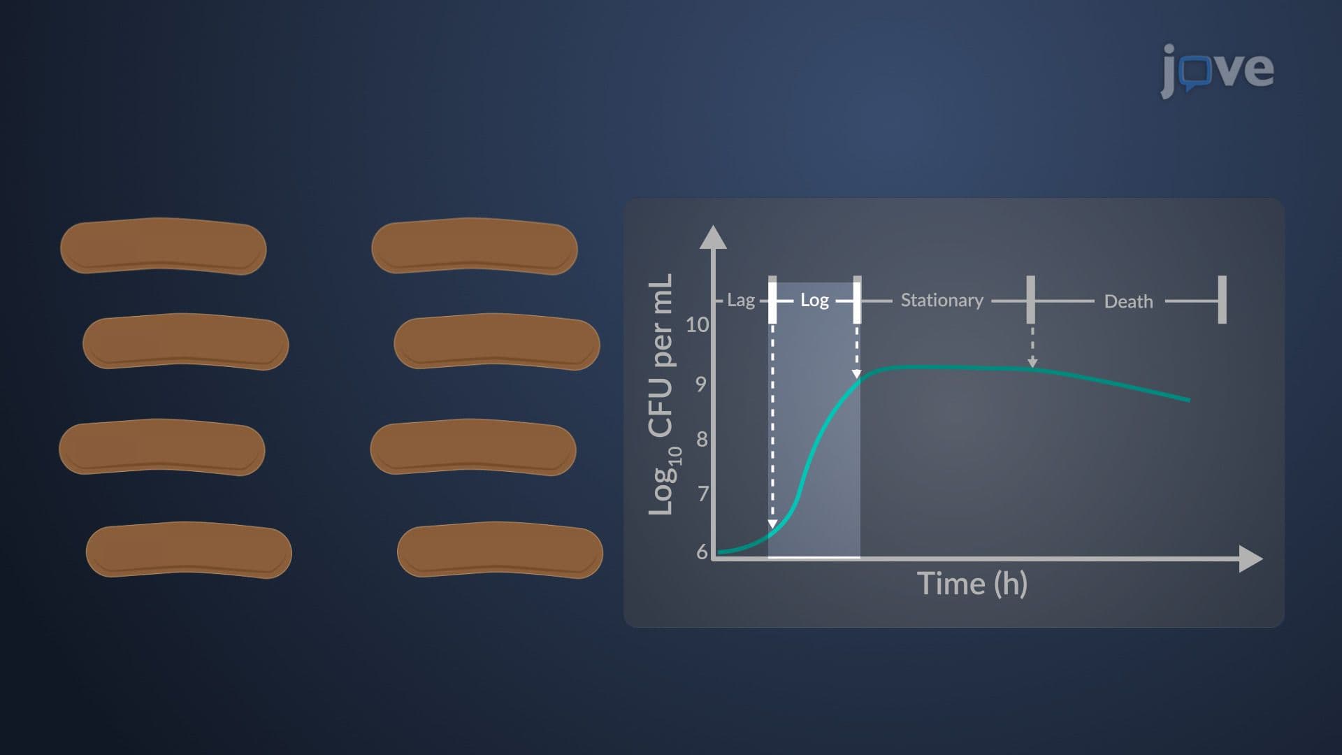
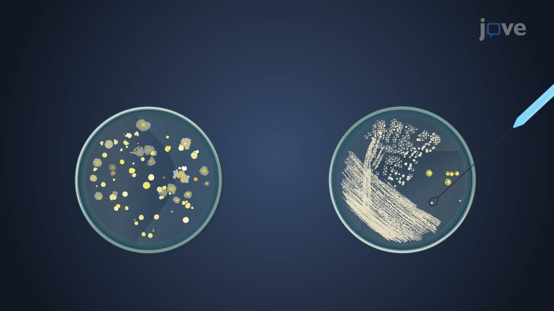
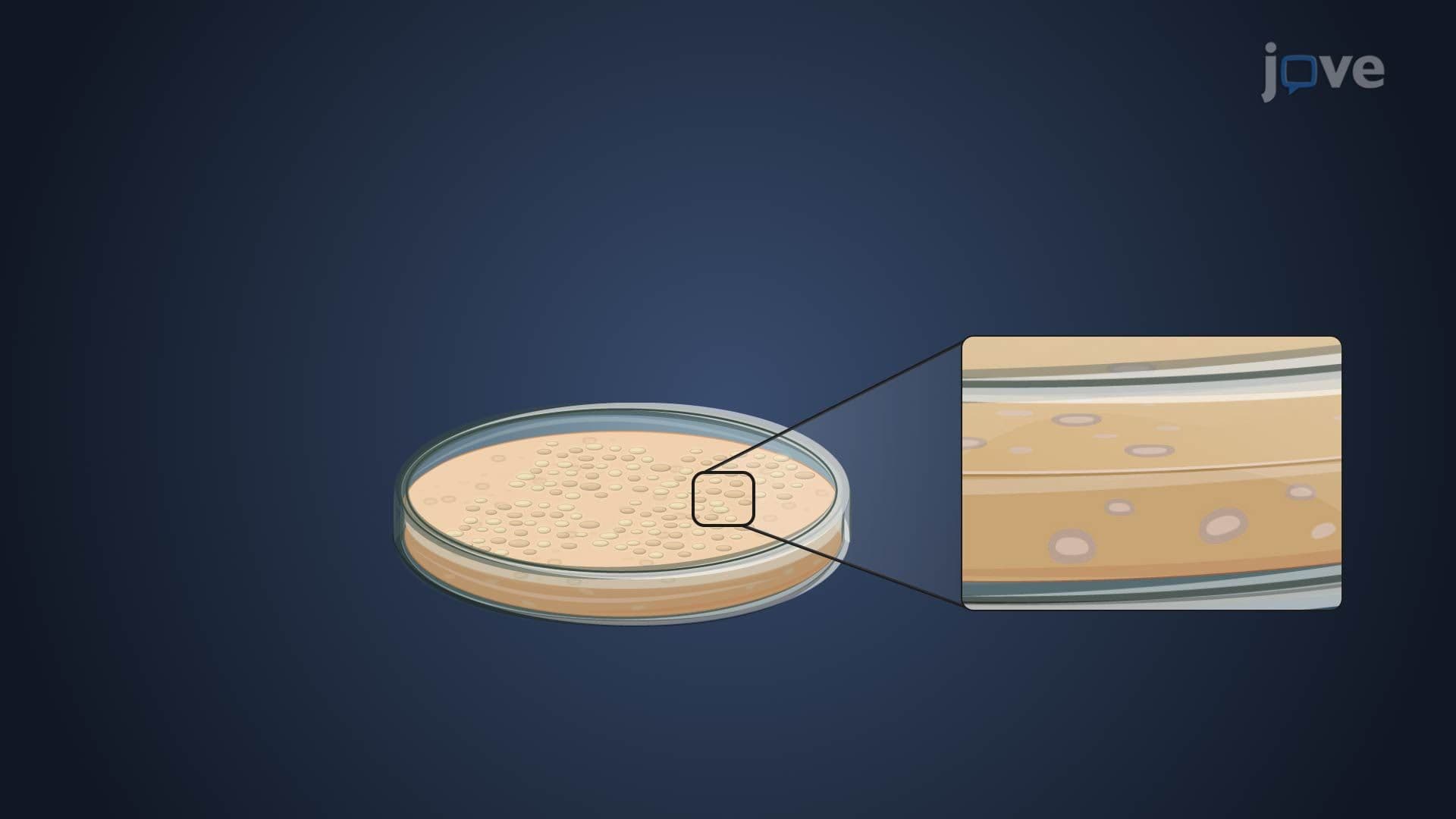
Plate counting methodology addresses viability concerns by measuring only cells capable of reproduction. This technique assumes each viable cell generates one visible colony after incubation on nutrient agar. Serial dilution protocols, typically using 10-fold reductions (1:10, 1:100, 1:1,000), reduce cell concentrations to countable ranges of 30-300 colonies per plate.
American clinical laboratories routinely employ this method for diagnosing urinary tract infections, where bacterial counts exceeding 100,000 CFU/mL indicate significant bacteriuria. Food safety inspectors use similar techniques to assess contamination levels in products regulated by the FDA.
The total dilution factor equals the product of all successive dilutions. For example, three consecutive 1:10 dilutions yield a 1:1,000 total dilution. Original cell concentration calculations use: CFU/mL = (Colony count × Total dilution factor) / Volume plated.
Applications in Academic and Professional Settings
These direct measurement techniques appear frequently on standardized examinations, including AP Biology, MCAT microbiology sections, and undergraduate laboratory practicals. Students must understand both theoretical principles and practical applications, as questions often involve calculating cell concentrations from experimental data or identifying appropriate methods for specific scenarios.
Frequently Asked Questions
Microbial growth measurement direct methods involve physically counting individual cells or colonies to determine exact population sizes in samples. These techniques provide precise quantitative data essential for clinical diagnostics, pharmaceutical quality control, and research applications. Unlike indirect methods measuring metabolic byproducts, direct counting gives definitive cell numbers, making it the gold standard for microbial enumeration in American laboratories.
These concepts frequently appear in MCAT biological sciences sections and AP Biology laboratory questions, typically involving calculation problems with serial dilutions and CFU determinations. Students must interpret experimental data, calculate original cell concentrations, and identify appropriate counting methods for different scenarios. Practice problems often mirror real laboratory situations encountered in clinical or research settings.
Master these key formulas: Cell density = (Average count × Dilution factor) / Volume counted, and CFU/mL = (Colony count × Total dilution factor) / Volume plated. Understanding dilution factor calculations is crucial-multiply all successive dilution ratios together. Most professors emphasize problem-solving over memorization, so focus on understanding when and how to apply each formula in different experimental contexts.
Clinical labs use plate counting to diagnose infections like urinary tract infections, where bacterial counts above 100,000 CFU/mL indicate significant disease. Blood culture analysis employs similar techniques to detect sepsis-causing organisms. Food safety laboratories use direct counting to ensure products meet FDA safety standards, while pharmaceutical companies apply these methods for sterility testing of injectable medications.
The mathematical concepts involve only basic multiplication and division suitable for high school algebra students. Start with simple 1:10 dilutions and practice calculating total dilution factors step-by-step. Most students master these calculations quickly with consistent practice. Focus on understanding the logic behind dilutions rather than memorizing formulas, and the mathematical relationships become intuitive.
Create practice problems using real laboratory scenarios, like calculating bacterial concentrations from plate count data. Draw diagrams showing serial dilution schemes and practice both Petroff-Hausser chamber and plate counting calculations. Form study groups to work through complex problems together, and review common error sources like cell clumping and dead cell interference that affect accuracy.
Begin with plate counting (CFU method) as it's more commonly used in clinical applications and appears frequently on standardized exams. The calculations are straightforward, and the concept of viable cell enumeration is fundamental to microbiology. Once comfortable with plate counting principles and serial dilutions, microscopic counting methods become easier to understand and apply.
Progress to indirect growth measurement techniques like turbidity and metabolic assays, which complement direct methods in comprehensive microbial analysis. Study microbial growth curves and generation time calculations, followed by antimicrobial susceptibility testing methods. These topics build naturally on direct counting foundations and frequently appear together in advanced microbiology coursework and professional laboratory settings.
Related Micro-courses

6 Concepts

31 Concepts

37 Concepts