Loading subject page…
- Microbiology
- Introduction to Microbiology
- Special Staining Techniques
Micro-course: Introduction to Microbiology
15,421 views
Learn key concepts and earn your certificate.

Special Staining Techniques

00:00
01:13
Video Summary: What are Special Staining Techniques
Ever wondered how scientists at the CDC identify dangerous bacteria like *Clostridium difficile* in hospital outbreaks? Special staining techniques reveal hidden bacterial structures invisible to standard microscopy methods. These advanced visualization methods, including capsule staining, endospore staining, and flagella staining, help microbiologists identify specific bacterial characteristics crucial for diagnosis and treatment. Watch the full video on JoVE Coach to master this concept with expert-led visuals and step-by-step explanations.
- Understand what are special staining techniques and their importance in bacterial identification beyond basic staining methods
- Identify the three major categories of special staining: capsule staining, endospore staining, and flagella staining
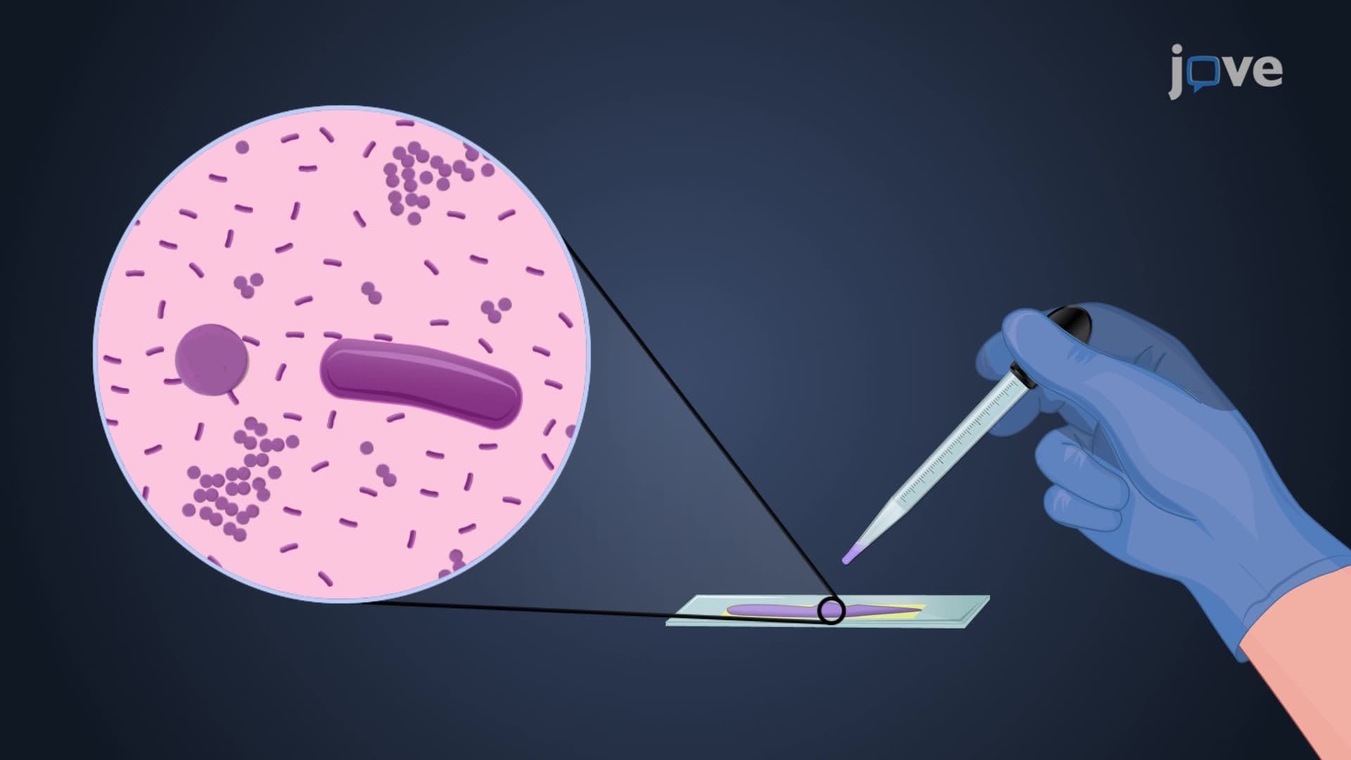
- Analyze how negative staining in capsule detection creates clear halos around bacterial cells using India ink or nigrosin
- Compare the Schaeffer-Fulton endospore staining method with standard staining techniques for spore-forming bacteria
- Evaluate the clinical significance of capsule detection in identifying virulent bacterial strains
- Apply knowledge of flagella staining to determine bacterial motility and classification characteristics
- Recognize the specific reagents and procedures used in each special staining technique
- Explore the relationship between bacterial structures and pathogenicity in medical microbiology
Understanding Special Staining Techniques in Microbiology
Special staining techniques represent advanced microscopic methods that reveal bacterial structures invisible through conventional staining. While basic stains like methylene blue or crystal violet color entire bacterial cells uniformly, special stains target specific cellular components, providing critical diagnostic information for medical professionals and researchers.
Capsule Staining: Visualizing Bacterial Defense Systems
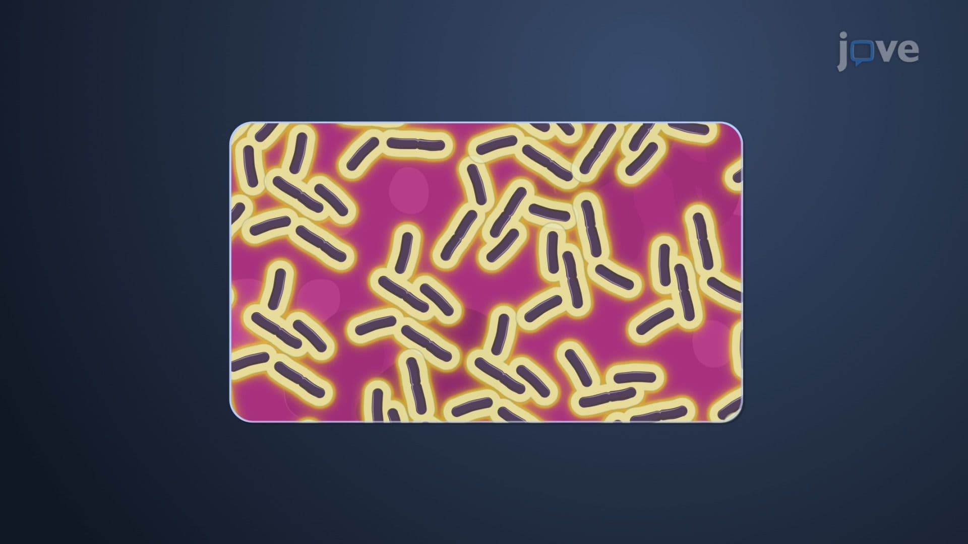
Capsule staining employs negative staining principles to detect polysaccharide capsules surrounding bacterial cells. Unlike positive stains that color the bacteria directly, negative staining with India ink or nigrosin darkens the background while leaving capsules unstained, creating distinctive clear halos. This technique proves invaluable in clinical settings-for instance, identifying *Streptococcus pneumoniae* in pneumonia cases, where capsulated strains show increased virulence and vaccine resistance. The capsule's composition, primarily consisting of polysaccharides, helps bacteria evade immune system recognition, making capsule detection crucial for appropriate antibiotic selection.
Endospore Staining: Detecting Bacterial Survival Mechanisms
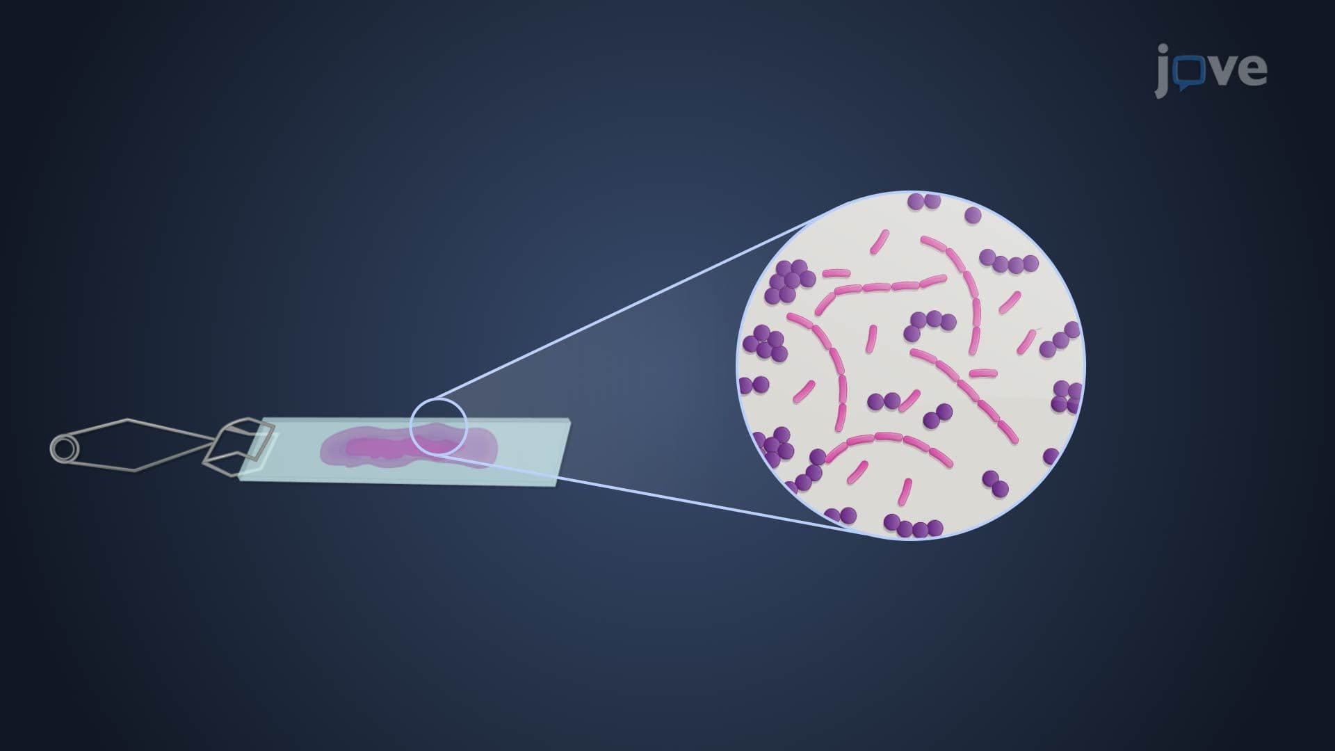
The Schaeffer-Fulton endospore staining method specifically targets bacterial endospores-highly resistant structures formed by genera like *Bacillus* and *Clostridium*. This technique requires heat application to drive malachite green through the tough endospore coat, followed by safranin counterstaining to color vegetative cells pink while endospores remain green. Medical laboratories frequently use this method when investigating food poisoning cases potentially caused by *Clostridium botulinum* or identifying *Bacillus anthracis* in bioterrorism investigations. Understanding endospore staining becomes particularly relevant for pre-med students preparing for the MCAT, where bacterial pathogenesis questions often focus on spore-forming pathogens.
Flagella Staining: Revealing Bacterial Motility Patterns
Flagella staining overcomes the challenge of visualizing bacterial flagella, which are too thin for standard light microscopy. The technique employs mordants like tannic acid to thicken flagella before applying basic fuchsin stain, making these structures visible. The number and arrangement of flagella-monotrichous, lophotrichous, amphitrichous, or peritrichous-serve as taxonomic characteristics essential for bacterial classification. This proves particularly important in clinical diagnostics, such as distinguishing motile *Salmonella* species from non-motile *Shigella* in gastroenteritis cases.
Frequently Asked Questions
Special staining techniques are advanced microscopic methods that target specific bacterial structures like capsules, endospores, and flagella. Unlike basic stains that color entire bacterial cells uniformly, special stains reveal hidden structures crucial for bacterial identification and pathogenicity assessment. They provide diagnostic information essential for medical treatment decisions.
The MCAT commonly tests capsule staining and endospore staining, particularly in microbiology and pathophysiology sections. AP Biology focuses on understanding staining principles rather than specific techniques. College microbiology courses typically require mastery of all three major special staining methods for laboratory practicals and written exams.
Hospital labs use capsule staining to identify virulent pneumonia-causing bacteria like Streptococcus pneumoniae. Endospore staining helps detect dangerous spore-forming bacteria such as Clostridium difficile in intestinal infections. Emergency departments may use flagella staining to distinguish between similar-appearing bacteria causing food poisoning outbreaks.
Special staining techniques build logically on basic staining knowledge taught in introductory biology. The concepts require understanding of bacterial cell structure and chemical principles, but the procedures themselves are straightforward. Most students master these techniques through hands-on laboratory practice and visual learning.
Create memory associations between staining methods and their purposes-capsule staining shows "halos" around virulent bacteria, endospore staining uses "heat-resistant green" for heat-resistant spores. Practice drawing the staining steps and results. Focus on understanding why each reagent is used rather than just memorizing the sequence.
Special staining enables rapid bacterial identification before culture results are available, allowing physicians to start appropriate antibiotic treatment sooner. Capsule detection indicates increased virulence, endospore detection suggests environmental persistence, and flagella patterns aid in species identification. This knowledge accelerates patient treatment and improves outcomes.
Advance to bacterial biochemical testing, antimicrobial susceptibility testing, and molecular diagnostic methods like PCR. Understanding special staining provides the foundation for studying bacterial pathogenesis mechanisms and host-pathogen interactions. These concepts connect directly to immunology and infectious disease coursework.
Related Micro-courses

13 Concepts
26 Concepts
13 Concepts