Loading subject page…
- Microbiology
- Introduction to Microbiology
- Differential Staining Technique
Micro-course: Introduction to Microbiology
15,421 views
Learn key concepts and earn your certificate.




Differential Staining Technique

00:00
01:26
Video Summary: What Is Differential Staining Technique
Every time a doctor diagnoses strep throat, they're relying on differential staining technique to identify the specific bacteria causing infection. This powerful laboratory method allows scientists to distinguish between different types of microorganisms based on their unique cell wall properties, making bacterial identification possible in clinical settings across the United States. Watch the full video on JoVE Coach to master this concept with expert-led visuals and step-by-step explanations.
- Understand what is differential staining technique and its fundamental role in microbiology
- Identify the key differences between Gram-positive and Gram-negative bacteria through staining results
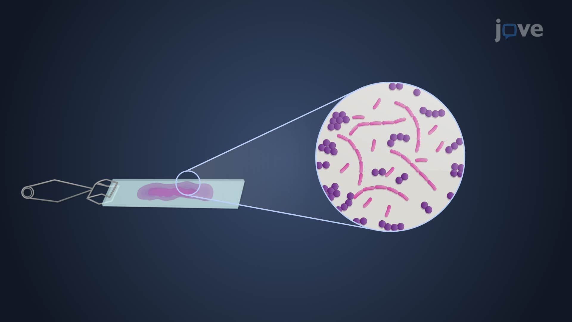
- Analyze the step-by-step process of Gram staining, from crystal violet application to safranin counterstaining
- Compare the cell wall structures that determine staining outcomes in different bacterial species
- Evaluate how acid-fast staining specifically detects Mycobacterium species in clinical diagnostics
- Apply differential staining principles to interpret laboratory results and bacterial classifications
- Recognize the clinical significance of staining techniques in medical diagnosis and treatment decisions
- Explore the relationship between bacterial cell wall composition and antibiotic susceptibility patterns
Understanding Differential Staining Technique Fundamentals
Differential staining technique serves as one of microbiology's most essential tools, enabling scientists and medical professionals to classify bacteria into distinct groups based on their cellular characteristics. Unlike simple stains that color all bacteria uniformly, differential staining creates contrast between different bacterial types, revealing critical information about their cell wall structure and composition.
This technique proves invaluable in clinical laboratories across the United States, where rapid bacterial identification can mean the difference between effective treatment and prolonged illness. The method's reliability has made it a cornerstone of medical education, appearing regularly on MCAT exams and forming a fundamental component of AP Biology curricula.
Gram Staining: The Gold Standard Method
Gram staining, developed by Danish bacteriologist Hans Christian Gram, remains the most widely used differential staining procedure in American hospitals and research facilities. The technique divides bacteria into two major categories: Gram-positive and Gram-negative organisms.
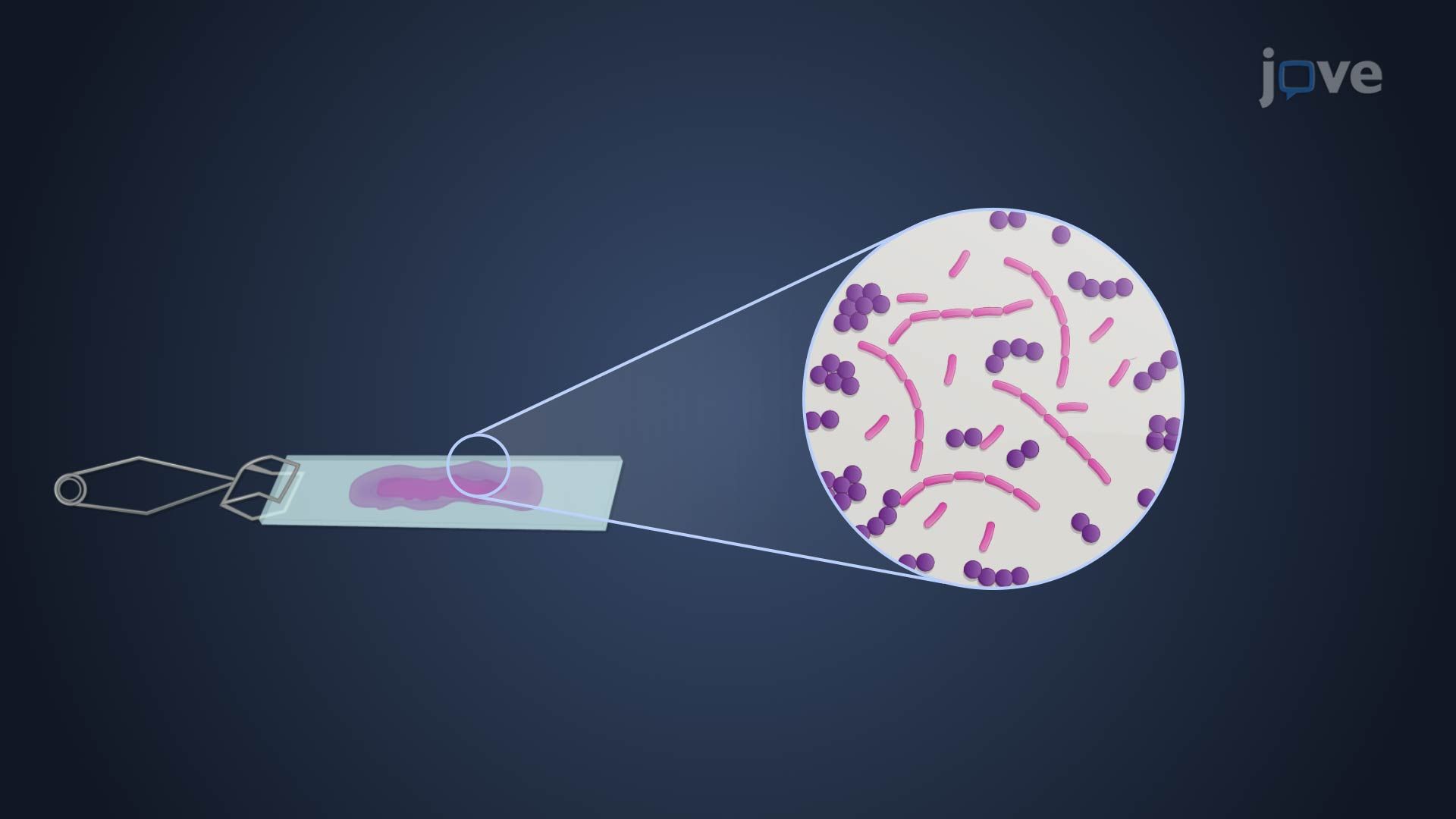
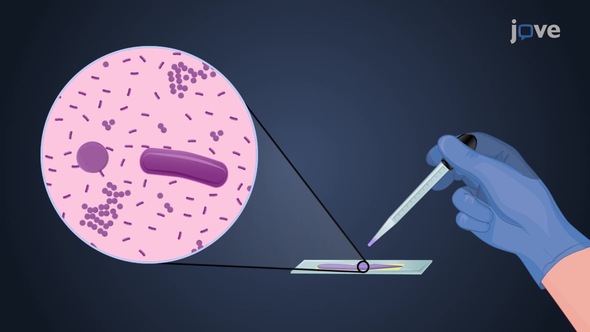
The process begins with creating a bacterial smear on a glass slide, followed by heat fixation to secure the cells. Crystal violet serves as the primary stain, coloring all bacteria purple initially. An iodine treatment then forms a complex with the crystal violet, creating a more stable color compound within the bacterial cells.
The critical decolorization step uses alcohol or acetone to remove the crystal violet-iodine complex from certain bacteria while leaving others unchanged. Gram-positive bacteria, possessing thick peptidoglycan cell walls, retain the purple coloration. Gram-negative bacteria lose their purple color as the alcohol disrupts their thinner peptidoglycan layer and outer membrane structure.
Clinical Applications and Acid-Fast Staining
Beyond Gram staining, acid-fast staining represents another crucial differential technique, particularly important for detecting tuberculosis-causing Mycobacterium tuberculosis. This method proves essential in American public health laboratories, where TB screening remains a critical diagnostic service.
Mycobacterium species possess unique waxy cell walls rich in mycolic acids, making them resistant to standard decolorization procedures. The acid-fast staining process uses carbol fuchsin, a red dye that penetrates these waxy barriers. Even after acid-alcohol treatment, acid-fast bacteria retain their red coloration, while other bacteria are counterstained blue with methylene blue.
Educational and Professional Applications
Students preparing for the MCAT will encounter differential staining concepts in both biology and biochemistry sections. College microbiology courses typically dedicate significant time to mastering these techniques, as they form the foundation for understanding bacterial pathogenesis and antibiotic mechanisms.
In professional healthcare settings, differential staining results guide treatment decisions daily. Emergency room physicians rely on Gram stain results to initiate appropriate antibiotic therapy before culture results become available, potentially saving lives through rapid bacterial classification.
Frequently Asked Questions
Differential staining technique is a laboratory method that uses specific dyes to distinguish between different types of bacteria based on their cell wall properties. It's crucial because it allows rapid bacterial identification in clinical settings, helping doctors choose appropriate antibiotics and treatments. This technique forms the backbone of bacterial classification systems used in hospitals and research laboratories worldwide. Students studying for AP Biology or college microbiology courses will find this concept fundamental to understanding bacterial diversity.
The MCAT frequently tests differential staining through questions about Gram staining mechanisms and bacterial classification. You'll likely encounter scenarios asking you to predict staining results based on bacterial cell wall composition or interpret laboratory findings. Focus on understanding the relationship between peptidoglycan thickness and staining outcomes. Practice questions often integrate this topic with antibiotic mechanisms and bacterial pathogenesis for comprehensive testing.
Gram-positive bacteria retain the purple crystal violet stain due to their thick peptidoglycan cell walls, while Gram-negative bacteria lose this stain during decolorization and appear pink after safranin counterstaining. This difference reflects fundamental structural variations that affect antibiotic susceptibility and treatment approaches. Understanding this distinction helps explain why certain antibiotics work better against specific bacterial types.
American hospitals use differential staining daily for rapid diagnosis of bacterial infections, particularly in emergency departments where quick treatment decisions are critical. For example, when a patient presents with pneumonia symptoms, a Gram stain of sputum can immediately indicate whether the likely cause is Streptococcus pneumoniae (Gram-positive) or Klebsiella pneumoniae (Gram-negative), guiding initial antibiotic choice. This technique also helps identify serious infections like meningitis within hours rather than days.
Differential staining technique is very accessible to high school students, requiring only basic chemistry knowledge about how molecules interact with cellular structures. The visual nature of the results makes it engaging and easy to understand. Most students successfully grasp the concept after seeing the dramatic color differences between bacterial types. The technique builds naturally on simple staining methods you may have already learned in biology class.
Create visual study aids showing the step-by-step staining process and resulting colors for different bacterial types. Practice drawing bacterial cell wall structures to understand why staining results differ. Use flashcards to memorize specific examples of Gram-positive and Gram-negative bacteria commonly tested. Focus on connecting staining results to clinical applications and antibiotic choices, as professors often test these connections on midterm and final exams.
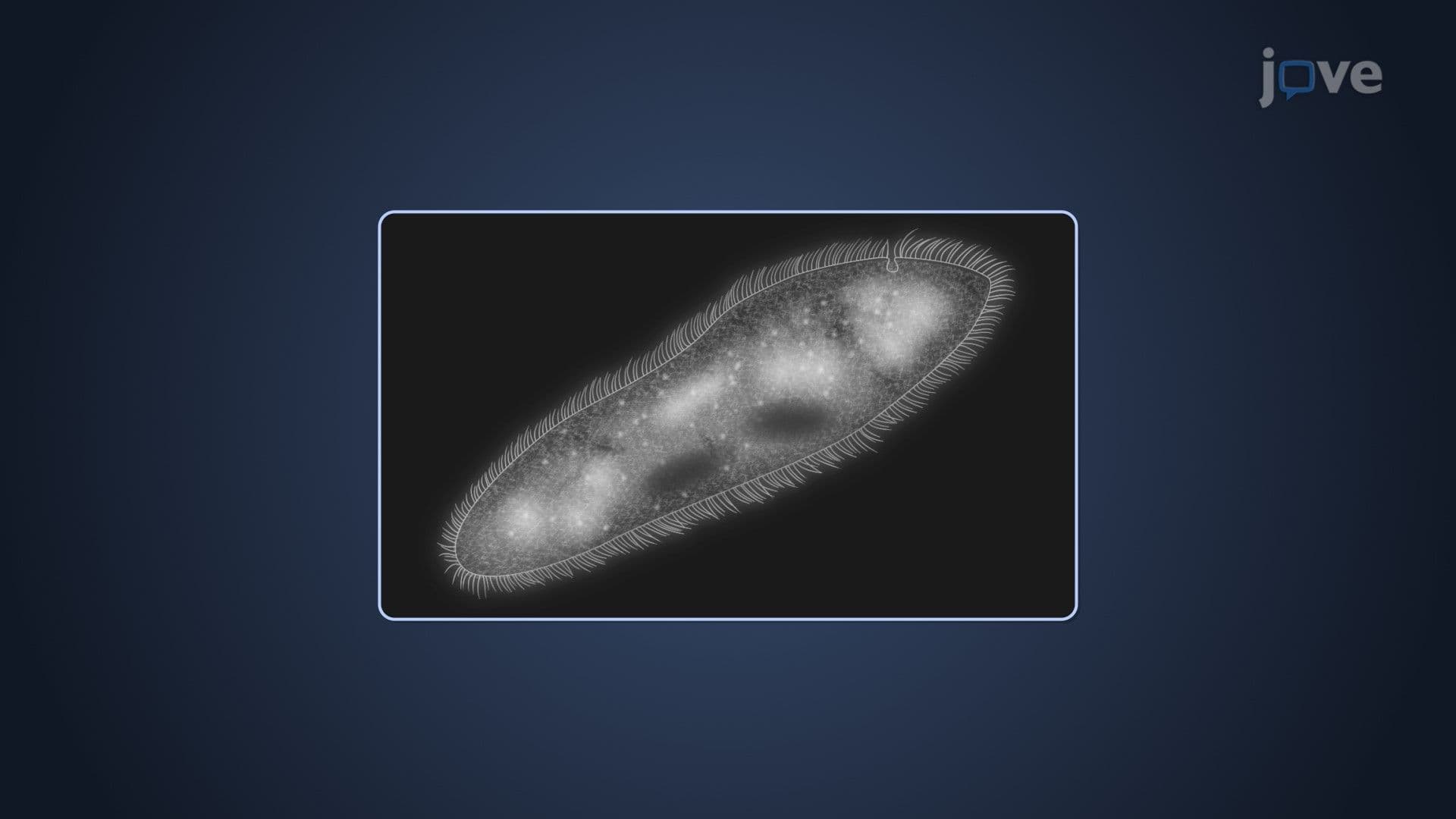
After mastering differential staining, explore fluorescent staining methods used in research laboratories and specialized clinical applications like immunofluorescence. Study how molecular techniques like PCR and DNA sequencing complement traditional staining for bacterial identification. Advanced microbiology courses often cover automated staining systems and digital microscopy techniques used in modern clinical laboratories. These topics become important for students pursuing careers in medical laboratory science or research.
Acid-fast staining specifically targets bacteria with waxy, mycolic acid-rich cell walls that resist standard decolorization procedures, particularly Mycobacterium species causing tuberculosis. Unlike Gram staining that works on most bacteria, acid-fast staining is selective for these unique organisms. The technique uses stronger chemicals and heat to penetrate waxy barriers, making it essential for TB diagnosis in American public health laboratories. This specialized approach demonstrates how different bacterial structures require tailored staining methods.
Related Micro-courses

13 Concepts
26 Concepts

13 Concepts