Loading subject page…
- Microbiology
- Introduction to Microbiology
- Simple Staining Technique
Micro-course: Introduction to Microbiology
15,421 views
Learn key concepts and earn your certificate.

Simple Staining Technique

00:00
01:24
Video Summary: What Is Simple Staining Technique
Ever wonder how researchers at the CDC can identify bacterial infections under a microscope? The simple staining technique transforms invisible microorganisms into clearly visible specimens using a single colored dye. This fundamental laboratory method allows scientists at institutions like Johns Hopkins to quickly assess bacterial size, shape, and arrangement in clinical samples. What is simple staining technique? It's the gateway to microbial identification that every biology student must master. Watch the full video on JoVE Coach to master this concept with expert-led visuals and step-by-step explanations.
- Understand what is simple staining technique and its role in basic microscopy for identifying microbial morphology
- Identify the essential materials needed including basic dyes like crystal violet, methylene blue, and safranin
- Analyze the electrostatic interactions between positively charged chromophores and negatively charged cellular components
- Apply proper heat fixation procedures to preserve bacterial cell structure before staining
- Recognize the step-by-step protocol from smear preparation through final observation
- Evaluate the advantages and limitations of simple staining compared to differential staining methods
- Compare different basic dyes and their effectiveness in highlighting various microbial features
- Explore real-world applications in clinical diagnostics and research laboratory settings
Understanding Simple Staining Technique Fundamentals
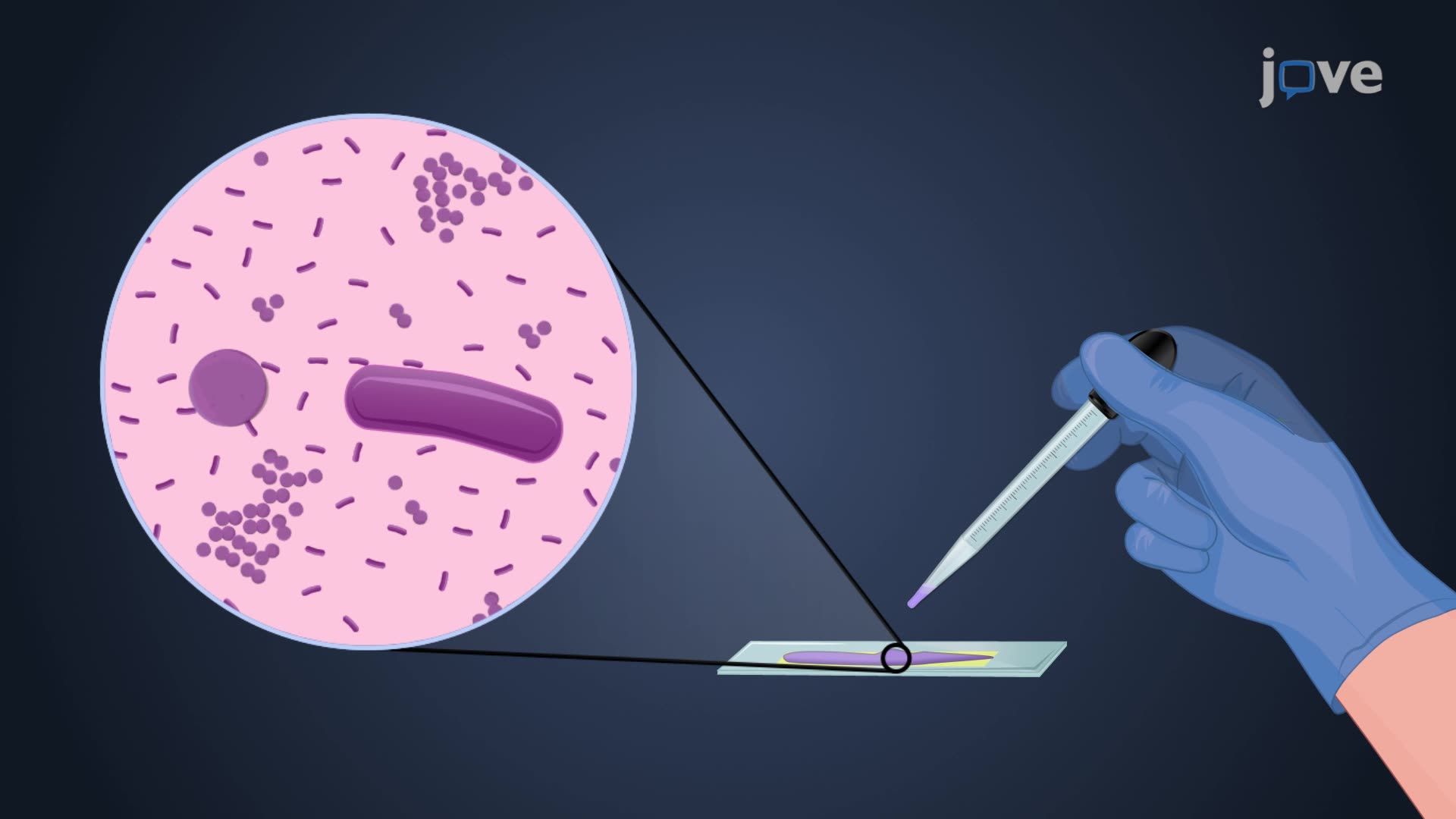
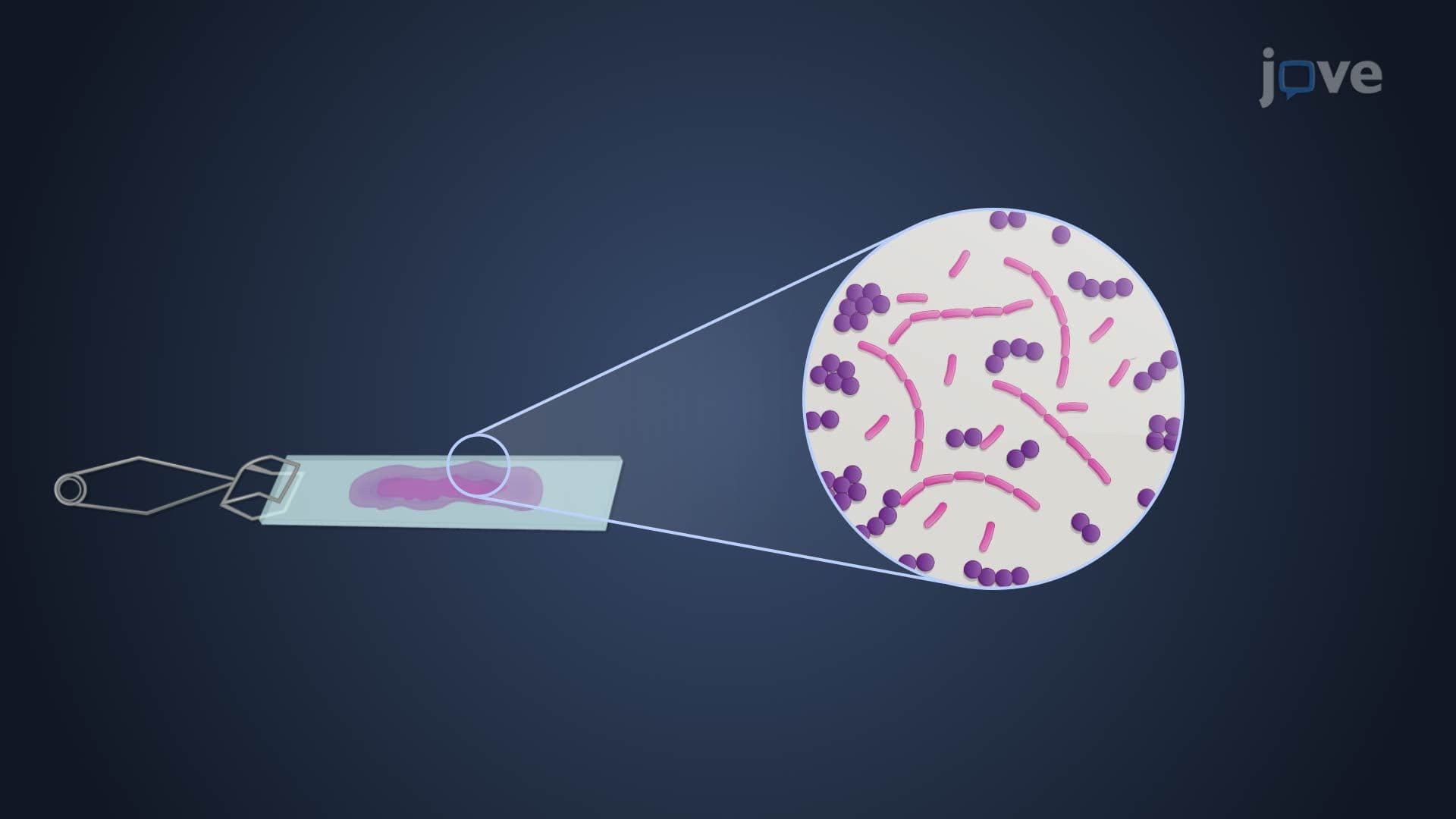
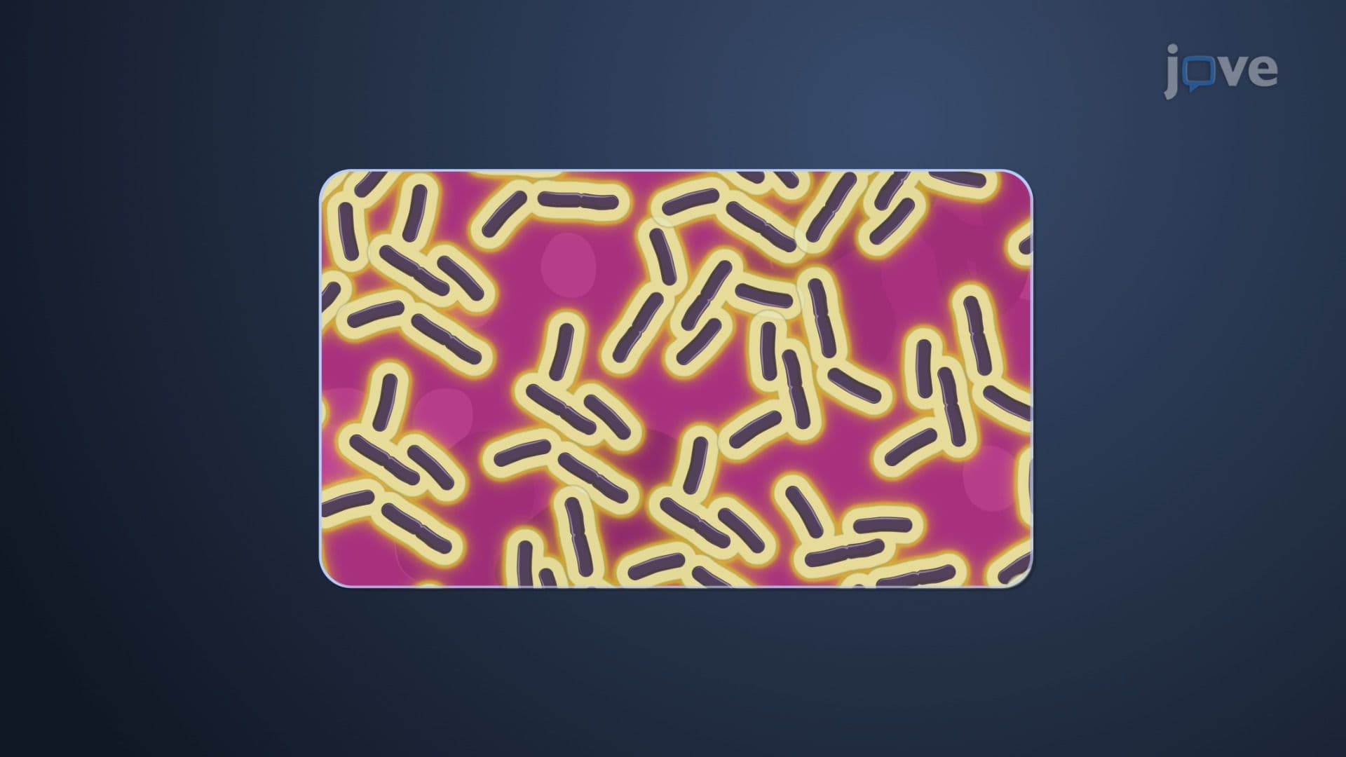
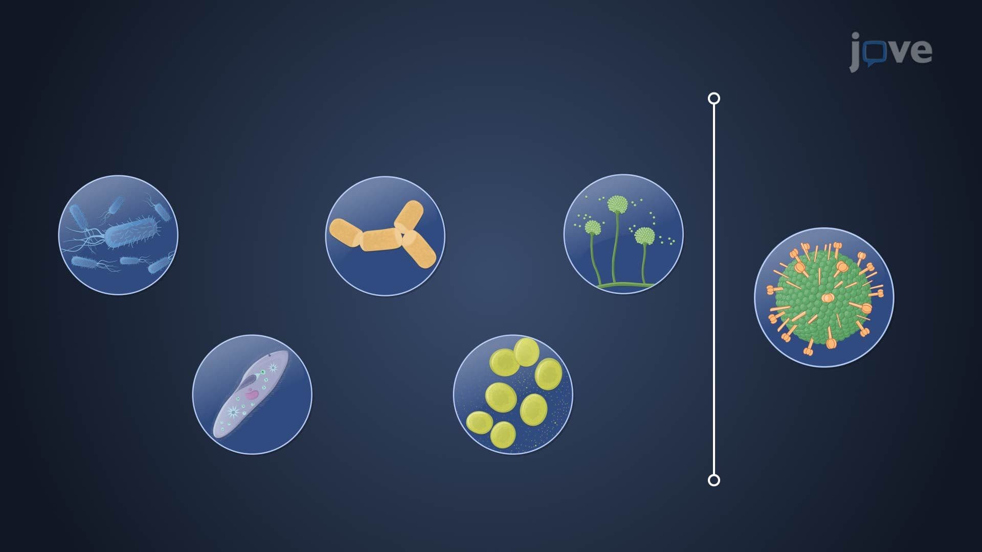
The simple staining technique represents one of the most essential skills in microbiology, serving as the foundation for bacterial identification in laboratories across the United States. Unlike complex differential staining methods such as Gram staining, this technique employs a single dye to create contrast between microbial cells and their background, making cellular morphology clearly visible under light microscopy.
The Science Behind Simple Staining Chemistry
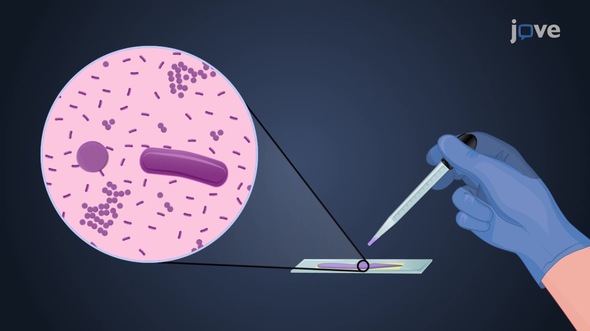
The effectiveness of simple staining relies on fundamental electrostatic principles. Basic dyes contain positively charged chromophores that exhibit strong affinity for negatively charged cellular components, particularly nucleic acids and peptidoglycan in bacterial cell walls. Common dyes used in US laboratories include crystal violet (producing deep purple coloration), methylene blue (creating bright blue cells), and safranin (yielding pink-red specimens). This electrostatic attraction ensures uniform dye binding across the entire cell surface.
Step-by-Step Laboratory Protocol
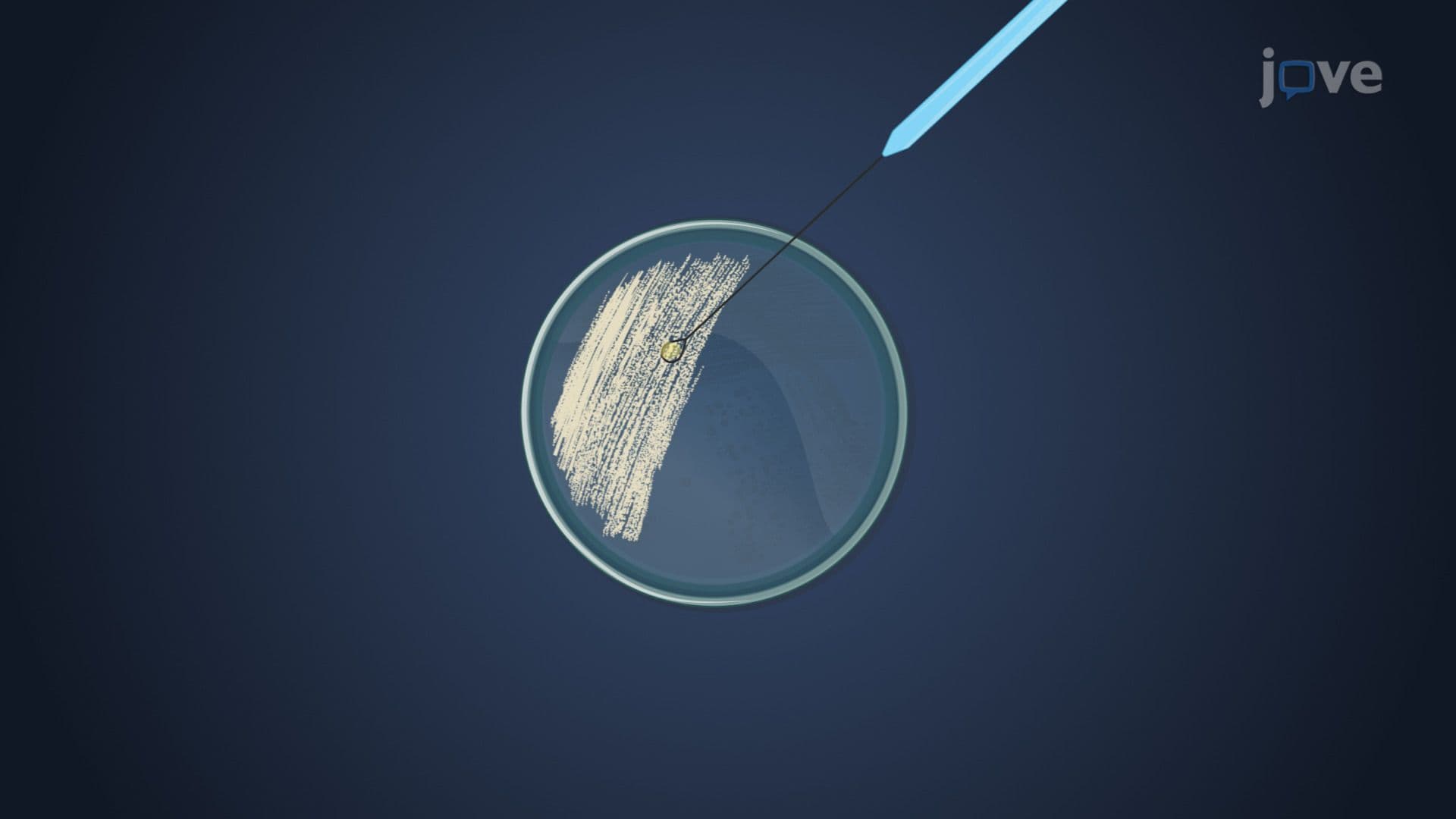
The simple staining procedure follows a standardized protocol taught in biology courses from high school AP Biology through undergraduate microbiology. First, technicians create a thin bacterial smear on a clean glass slide, allowing proper air-drying to prevent cell distortion. Heat fixation follows, typically performed by passing the slide through a Bunsen burner flame three times. The staining phase involves flooding the slide with basic dye for 60-120 seconds, followed by gentle water rinsing and careful blotting with absorbent paper.
Clinical and Educational Applications
In clinical settings, simple staining serves as a preliminary diagnostic tool in hospitals like Mayo Clinic and Cleveland Clinic, helping medical technologists quickly assess bacterial load and morphology in patient samples. For students preparing for standardized exams including the MCAT, AP Biology, and college microbiology courses, mastering this technique demonstrates understanding of fundamental laboratory skills. The technique frequently appears in practical laboratory exams and serves as preparation for more advanced staining procedures used in clinical microbiology certification programs.
Frequently Asked Questions
Simple staining technique is a basic microscopy method that uses a single dye to make microorganisms visible by creating contrast against a clear background. Unlike differential staining methods such as Gram staining, simple staining doesn't distinguish between different types of bacteria but rather highlights their size, shape, and arrangement for basic morphological analysis.
The three most frequently used basic dyes in US laboratories are crystal violet (producing deep purple cells), methylene blue (creating bright blue coloration), and safranin (yielding pink-red specimens). These dyes are chosen for their strong positive charge, which ensures effective binding to negatively charged bacterial cell components through electrostatic attraction.
The MCAT frequently includes questions about staining principles, particularly the electrostatic interactions between charged dyes and cellular components. AP Biology exams may test understanding of laboratory techniques and their applications in identifying microbial morphology. Students should focus on understanding the underlying chemistry and practical applications rather than memorizing specific protocols.
General biology, microbiology, and cell biology courses at most US colleges include hands-on simple staining laboratories. Pre-med students encounter this technique in introductory biology sequences, while microbiology majors study it as preparation for advanced diagnostic methods used in clinical and research settings.
Clinical laboratories use simple staining as a preliminary diagnostic tool to quickly assess bacterial morphology in patient samples, helping technologists determine cell shape, size, and arrangement before proceeding with more specific identification methods. This rapid screening helps prioritize samples and guide subsequent testing protocols in busy hospital environments.
No advanced chemistry background is required, the technique relies on basic electrostatic attraction principles taught in high school chemistry. Understanding that oppositely charged particles attract each other is sufficient to grasp why positively charged dyes bind to negatively charged bacterial cells, making this technique accessible to students at all levels.
Practice the step-by-step protocol until it becomes automatic: smear preparation, air-drying, heat fixation, staining, rinsing, and observation. Focus on understanding the purpose of each step rather than just memorizing the sequence, and review common troubleshooting scenarios like over-staining or inadequate heat fixation.
Progress to differential staining methods like Gram staining, acid-fast staining, and spore staining, which provide more specific information about bacterial characteristics. These advanced techniques build upon simple staining principles while adding complexity for detailed microbial identification and classification.
Related Micro-courses

13 Concepts
26 Concepts
13 Concepts