Loading subject page…
- Microbiology
- Introduction to Microbiology
- Three-Dimensional Microscopy in Microbiology
Micro-course: Introduction to Microbiology
15,419 views
Learn key concepts and earn your certificate.

Three-Dimensional Microscopy in Microbiology

00:00
01:28
Video Summary: What Is Three Dimensional Microscopy
Did you know that scientists at the CDC use three dimensional microscopy to study deadly pathogens like anthrax bacteria in unprecedented detail? Three dimensional microscopy techniques, including Differential Interference Contrast (DIC) and Confocal Scanning Laser Microscopy (CSLM), revolutionize how researchers visualize cellular structures by creating detailed, layered images that reveal internal organization. These advanced imaging methods allow scientists at institutions like Johns Hopkins to examine living cells without damaging them, making breakthrough discoveries in cancer research and infectious disease studies. Watch the full video on JoVE Coach to master this concept with expert-led visuals and step-by-step explanations.
- Understand how three dimensional microscopy creates detailed, layered images of cellular structures using advanced optical techniques
- Identify the key differences between DIC and CSLM methods and their specific applications in biological research
- Analyze how polarized light splitting in DIC microscopy generates pseudo-3D images based on refractive index variations
- Compare the advantages of unstained live-cell imaging versus traditional staining methods for cellular observation
- Evaluate the role of laser scanning and pinhole apertures in eliminating out-of-focus light for clearer imaging
- Apply knowledge of fluorescent dye enhancement to understand how CSLM improves visualization of cellular components
- Recognize the importance of computational image stacking in creating high-resolution three-dimensional reconstructions
- Explore real-world applications of these techniques in studying bacterial biofilms and complex cellular interactions
Understanding Three Dimensional Microscopy Fundamentals
Three dimensional microscopy represents a revolutionary advancement in cellular imaging that goes far beyond traditional flat microscopy. Unlike conventional light microscopy that produces two-dimensional images, these sophisticated techniques create detailed, layered representations of cellular structures that scientists can examine from multiple angles and depths. This technology has transformed research at leading US institutions like Harvard Medical School and the National Institutes of Health, where researchers study everything from cancer cell behavior to infectious disease mechanisms.
The fundamental principle behind three-dimensional imaging lies in capturing information from multiple focal planes within a specimen. Rather than seeing just the surface features, scientists can now peer deep into cells, tissues, and even complex biological communities like bacterial biofilms that cause hospital-acquired infections.
Differential Interference Contrast (DIC) Microscopy: Seeing the Invisible
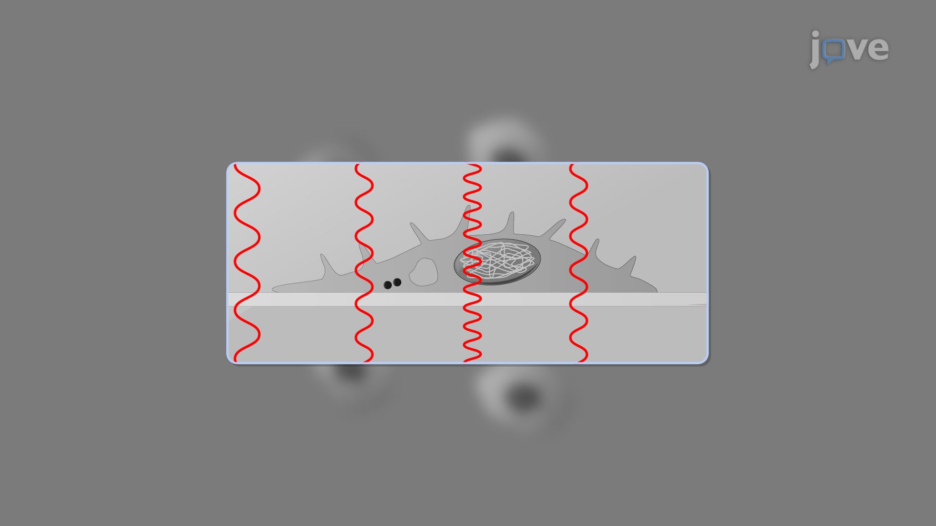
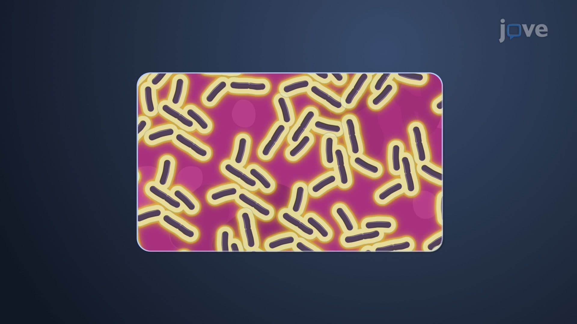
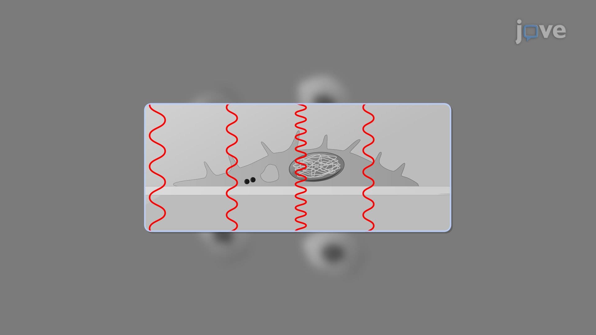
DIC microscopy employs an ingenious optical trick that makes transparent cellular structures visible without any staining or chemical treatment. The technique splits polarized light into two beams that travel slightly different paths through the specimen. When these beams recombine, they create interference patterns that highlight variations in the cell's refractive index-essentially revealing density differences within the specimen.
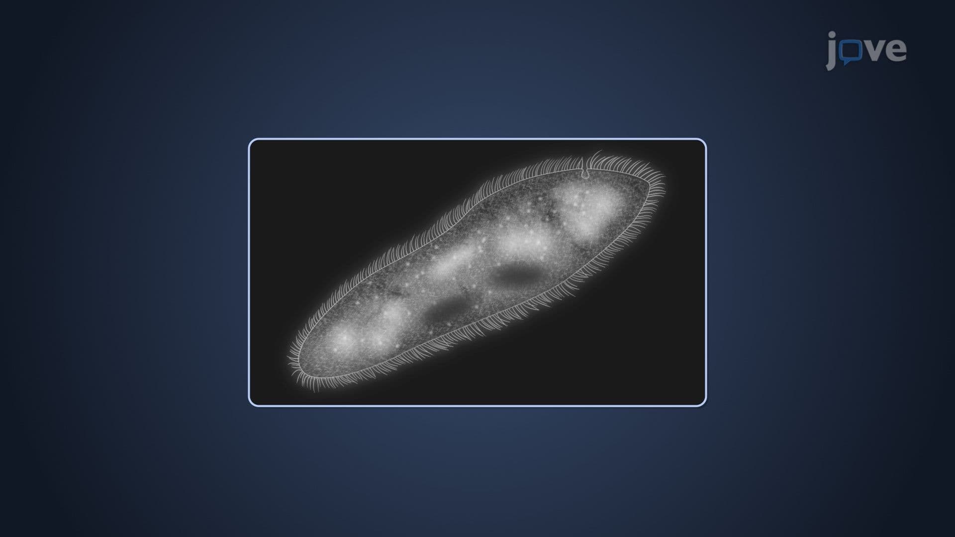
This method proves invaluable for studying living cells because it preserves their natural state. Researchers at the Mayo Clinic use DIC to observe cellular processes like mitosis in real-time, tracking how chromosomes move during cell division without introducing potentially harmful dyes or fixatives. The pseudo-three-dimensional appearance created by DIC makes cellular organelles like nuclei, vacuoles, and inclusion bodies stand out with remarkable clarity.
Confocal Scanning Laser Microscopy: Layer-by-Layer Precision
CSLM takes three-dimensional imaging to the next level by using laser light and sophisticated optical components to create true 3D reconstructions. The key innovation lies in its pinhole aperture system, which blocks out-of-focus light that would otherwise blur the image. By scanning the specimen point-by-point with a focused laser beam, CSLM builds up incredibly detailed images one focal plane at a time.
The real magic happens during computational processing, where specialized software stacks these individual planes to create comprehensive three-dimensional models. Researchers at Stanford University use this technique to study neural networks in brain tissue, revealing how individual neurons connect and communicate across different tissue layers.
Real-World Applications and Exam Relevance
Students preparing for the MCAT, AP Biology exams, or college-level cell biology courses will encounter three-dimensional microscopy in contexts ranging from basic cell structure identification to advanced research methodology questions. Understanding these techniques helps explain how scientists make discoveries about cellular processes that appear in textbooks and research papers. For instance, studies of biofilm formation in medical devices-a major concern in US hospitals-rely heavily on confocal microscopy to understand how bacteria organize themselves into complex, treatment-resistant communities.
Frequently Asked Questions
Three dimensional microscopy creates detailed, layered images of specimens that reveal internal structures and depth, unlike traditional microscopy that only shows flat, surface-level views. These advanced techniques use specialized optical systems to capture information from multiple focal planes within a specimen. The result is comprehensive 3D visualizations that scientists can examine from different angles and depths, providing crucial insights into cellular organization and biological processes.
Yes, understanding advanced microscopy techniques is essential for MCAT success and frequently appears in AP Biology questions about experimental design and cellular analysis. These exams often test your ability to interpret microscopy data and explain how different imaging methods provide specific types of information. Questions may ask you to compare imaging techniques, analyze research methodology, or explain how scientists use these tools to study cellular processes and disease mechanisms.
Most introductory biology and cell biology courses at US universities include hands-on microscopy labs where students learn to operate DIC and confocal systems. Advanced courses in microbiology, developmental biology, and biomedical research methods extensively cover these techniques as essential research tools. Students often analyze published research papers that use three-dimensional microscopy data and may complete projects involving image analysis and interpretation.
US hospitals use confocal microscopy to study antibiotic-resistant bacterial infections, while cancer research centers like MD Anderson employ these techniques to understand tumor cell behavior and test new treatments. The CDC relies on advanced microscopy for infectious disease surveillance, and pharmaceutical companies use 3D imaging to evaluate drug effectiveness at the cellular level. Medical device companies also use these methods to study biofilm formation on implants and catheters.
While the underlying optics involve complex physics, you can understand the basic principles and applications with standard high school biology and chemistry knowledge. The key concepts focus on how light interacts with specimens and how computers process multiple images to create 3D reconstructions. Most biology courses emphasize practical applications and interpretation rather than detailed optical calculations, making these concepts accessible to students with diverse academic backgrounds.
Focus on understanding the key differences between DIC and CSLM techniques, including their specific advantages and applications. Practice interpreting microscopy images and explaining how each method reveals different types of cellular information. Create comparison charts showing when scientists would choose each technique, and study real research examples that demonstrate practical applications in medical and biological research.
Mastering these concepts provides essential foundation knowledge for upper-level courses in cell biology, microbiology, and biomedical research methods. Understanding advanced imaging techniques helps students critically evaluate scientific literature and design their own research projects. These skills prove valuable for students pursuing careers in medicine, research, biotechnology, or graduate studies in biological sciences.
Build on this foundation by exploring electron microscopy techniques, fluorescence labeling methods, and digital image analysis software used in biological research. Advanced topics include super-resolution microscopy, live-cell imaging techniques, and quantitative image analysis methods that complement three-dimensional imaging capabilities.
Related Micro-courses

13 Concepts
26 Concepts
13 Concepts