Loading subject page…

- Molecular Biology
- Cell Division
- Spindle Assembly
Micro-course: Cell Division
101,001 views
Learn key concepts and earn your certificate.



Spindle Assembly


00:00
02:50
Video Summary: What Is Spindle Assembly
Every time your skin cells replace themselves or your muscles grow stronger, millions of cells must divide perfectly-but how do chromosomes know exactly where to go? Spindle assembly is the cellular process that builds the molecular machinery responsible for splitting chromosomes during cell division. For instance, when cancer researchers at Johns Hopkins study why tumor cells divide uncontrollably, they often focus on spindle assembly defects. This intricate process involves microtubules forming a bipolar structure that ensures each daughter cell receives the correct genetic material. Watch the full video on JoVE Coach to master this concept with expert-led visuals and step-by-step explanations.
- Understand what spindle assembly is and why this process is essential for accurate chromosome segregation during eukaryotic cell division
- Identify the three main types of microtubules formed during spindle assembly: interpolar, kinetochore, and astral microtubules
- Analyze how centrosomes and acentrosomal mechanisms contribute to spindle formation in different cellular contexts
- Compare the roles of various motor proteins (dynein, kinesin-5, kinesin-14, chromokinesins) in spindle positioning and stabilization
- Evaluate how nuclear envelope breakdown coordinates with microtubule nucleation during mitosis
- Apply knowledge of spindle assembly defects to understand cancer cell biology and genetic disorders
- Recognize the molecular mechanisms that ensure balanced forces determine final spindle length and chromosome positioning
- Explore how Ran-GTP signaling promotes localized microtubule stabilization around mitotic chromosomes
Understanding Spindle Assembly Fundamentals
Spindle assembly represents one of cell biology's most precisely orchestrated events, where cells construct a bipolar microtubule array to ensure accurate chromosome distribution. This process becomes critical when studying genetic diseases-for example, researchers at Memorial Sloan Kettering have identified spindle assembly defects in hereditary cancer syndromes affecting American families.
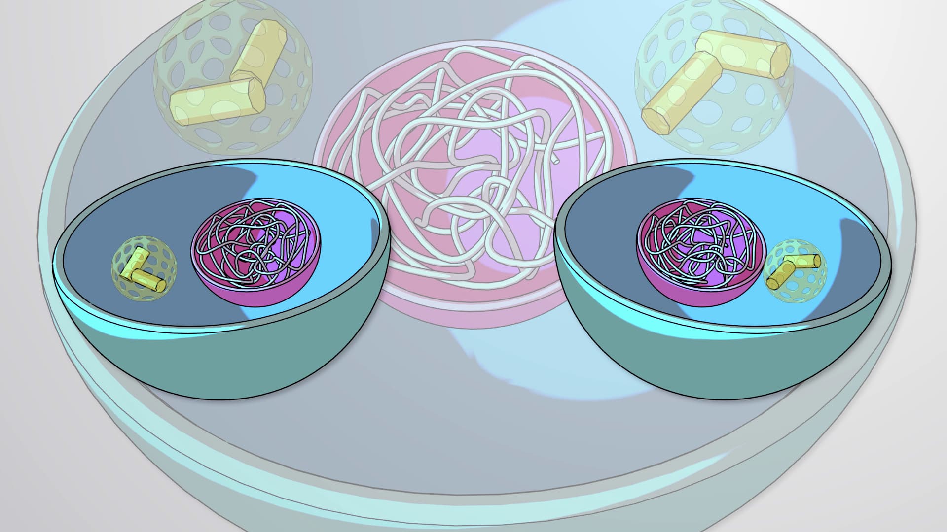
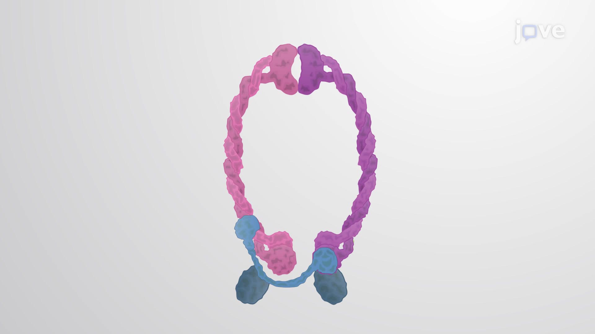
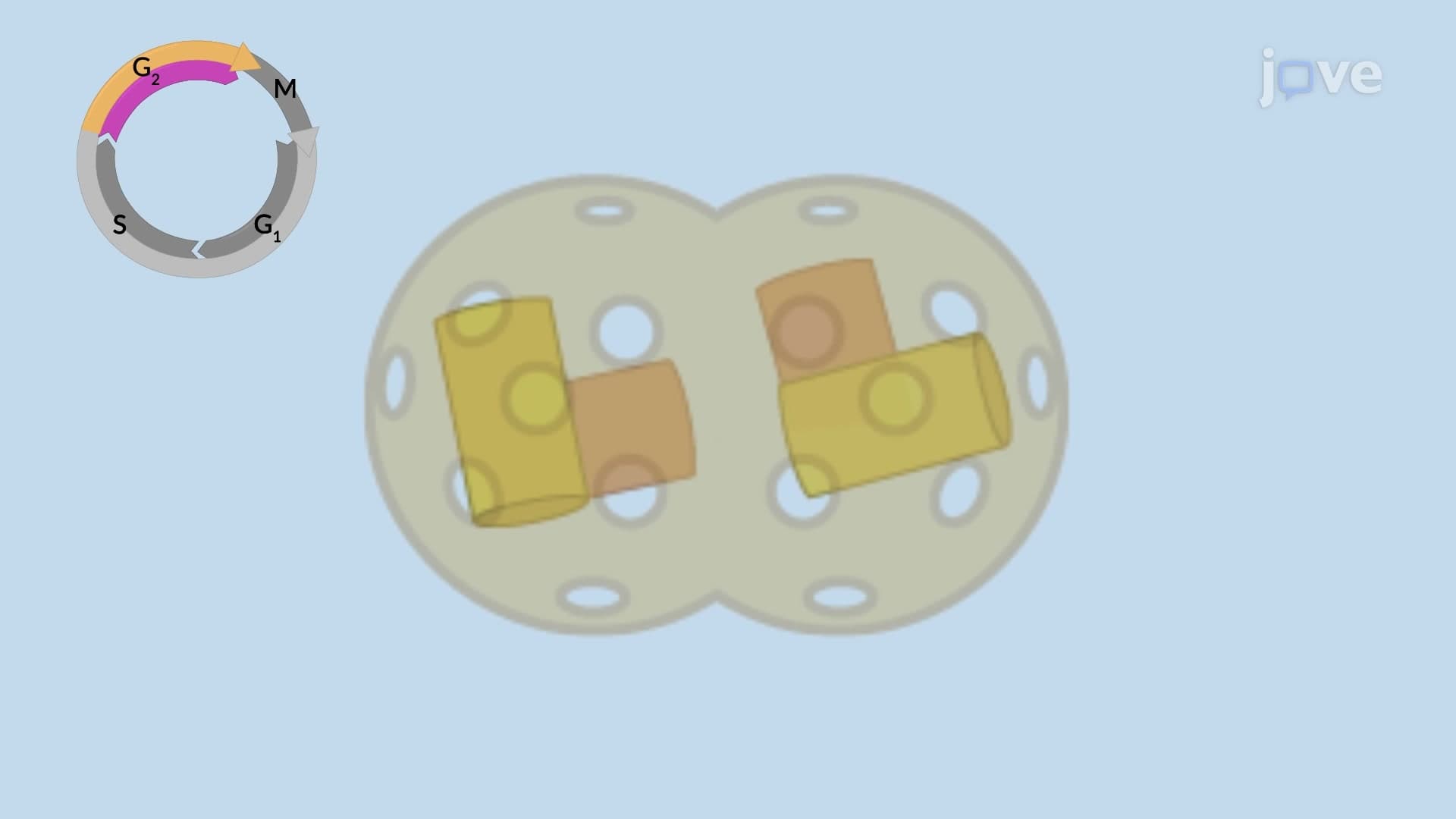
The assembly process begins when duplicated centrosomes position themselves at opposite cell poles. Think of centrosomes as molecular construction sites that anchor microtubules, the cellular "cables" responsible for chromosome movement. This positioning creates the foundation for the entire spindle apparatus, much like how construction crews establish anchor points before building a suspension bridge.
Microtubule Organization and Nuclear Changes
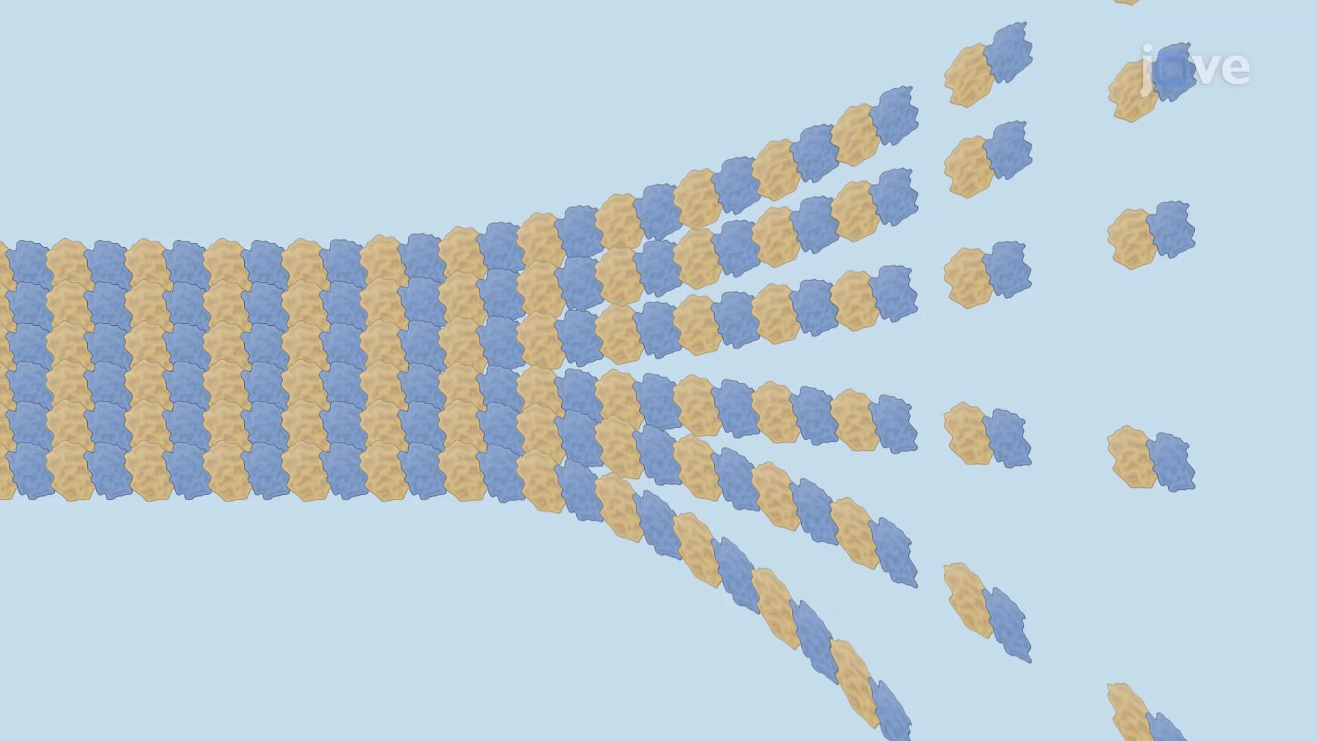
During spindle assembly, cells coordinate two major events: microtubule nucleation and nuclear envelope breakdown. M-Cdk enzymes-master regulators of cell division-phosphorylate nuclear envelope components, essentially dismantling the barrier between nucleus and cytoplasm. This breakdown exposes condensed chromosomes to the newly formed microtubule network.
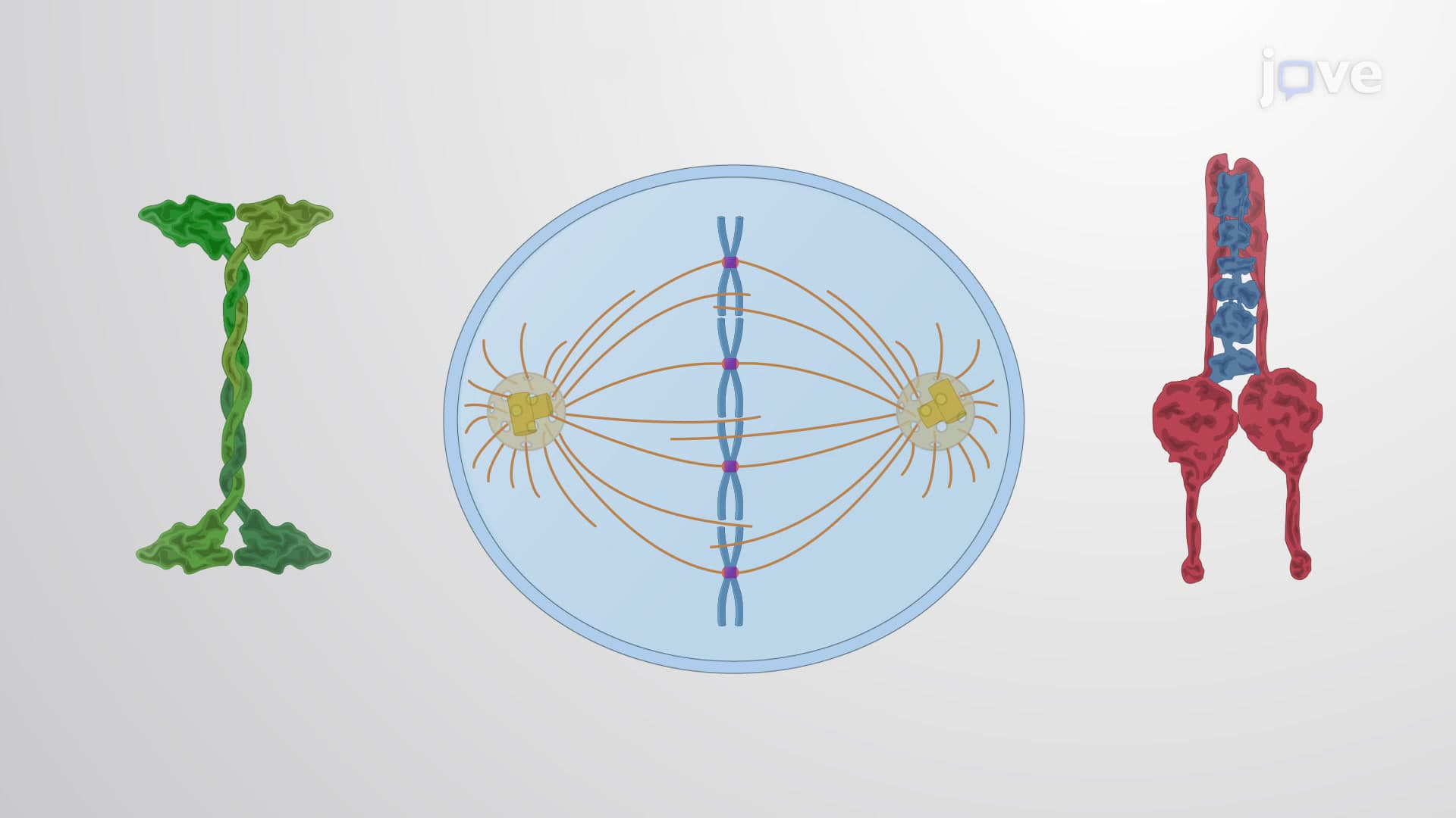
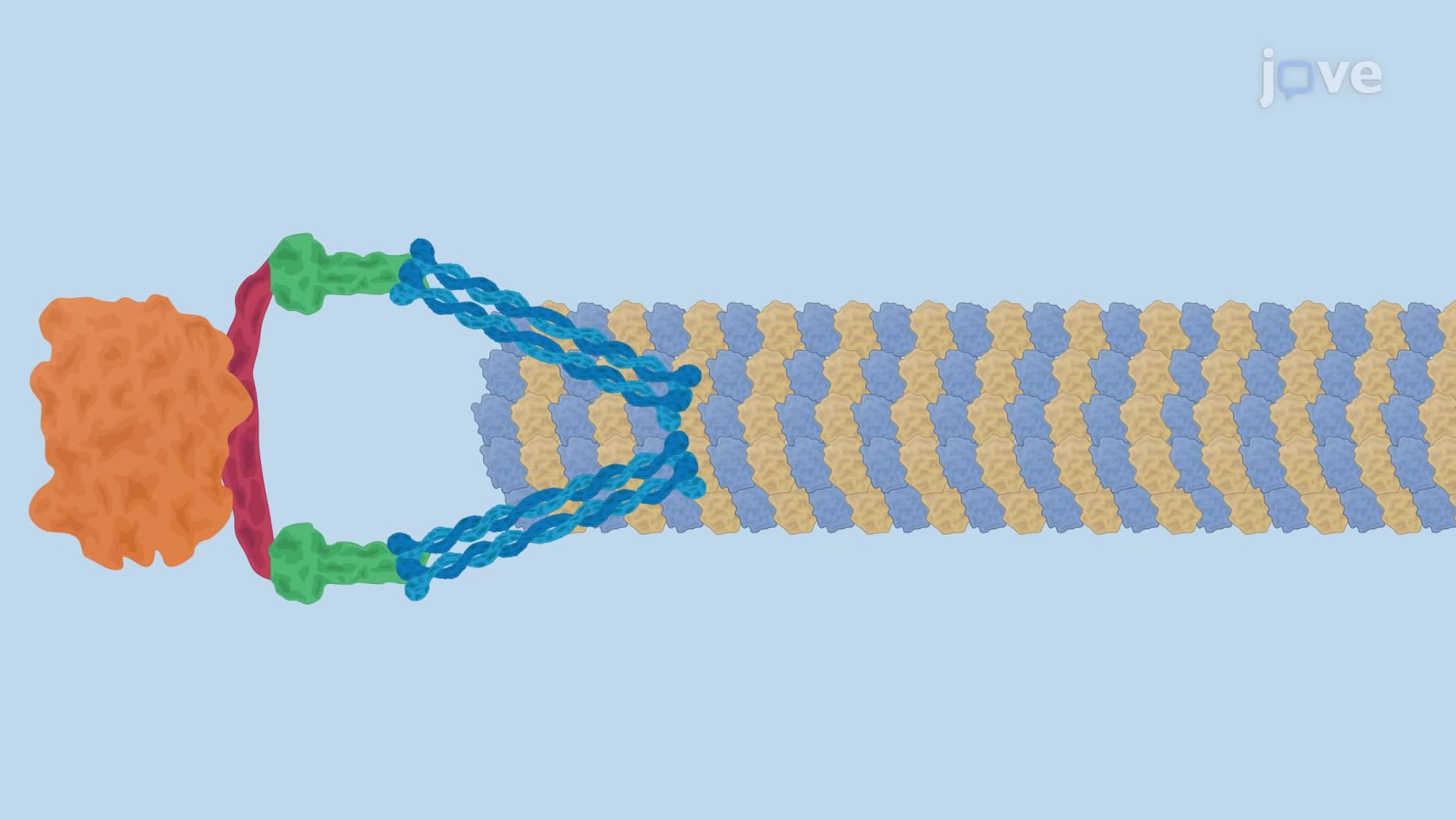
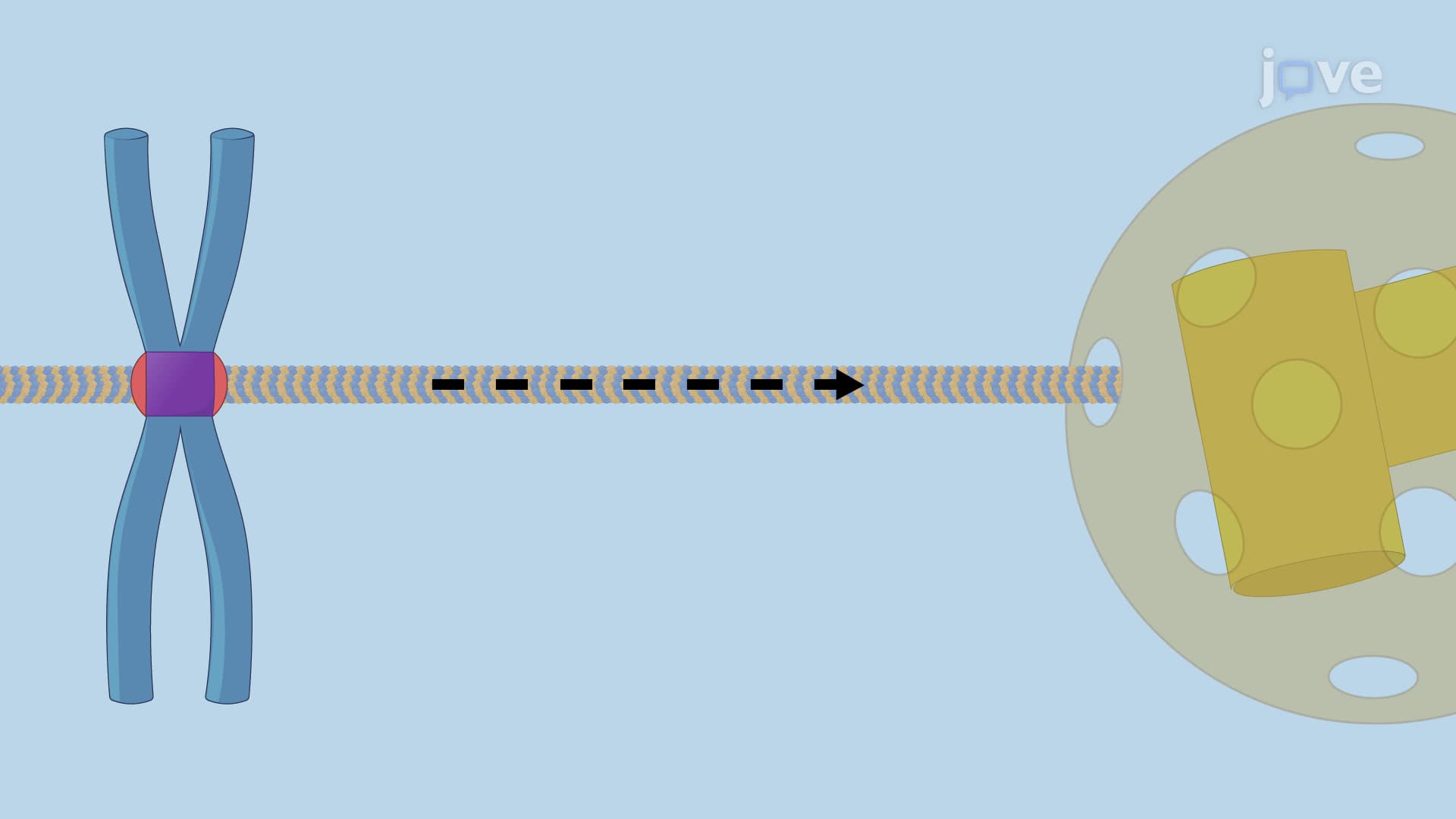
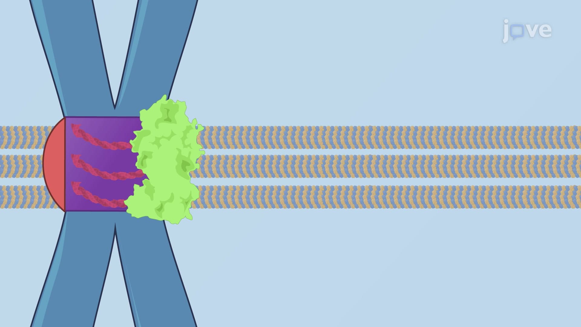
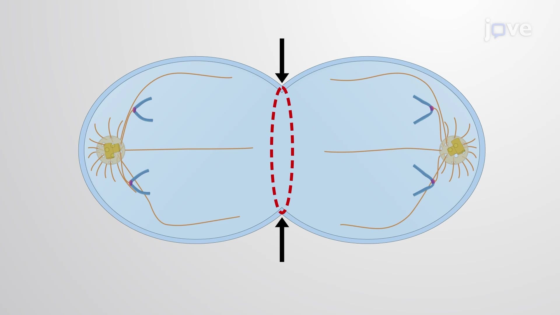
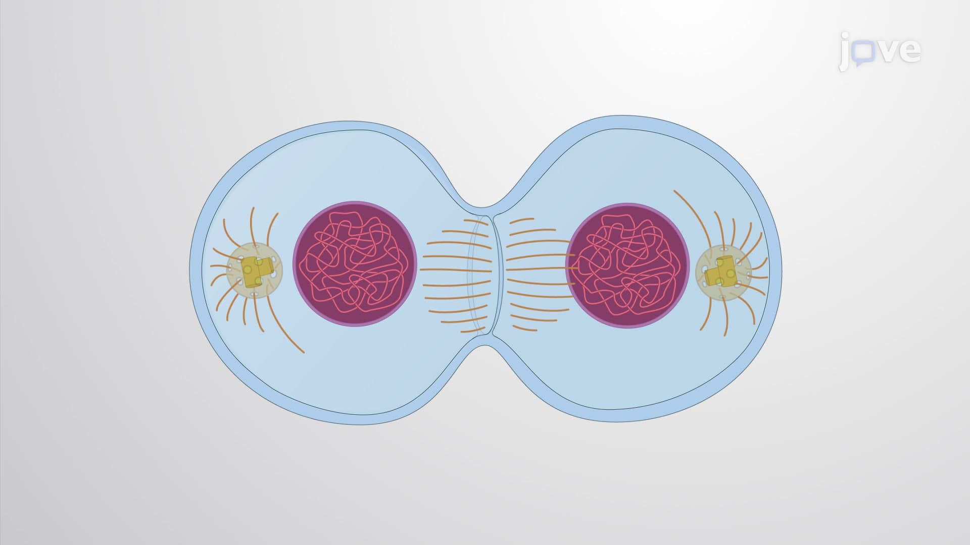
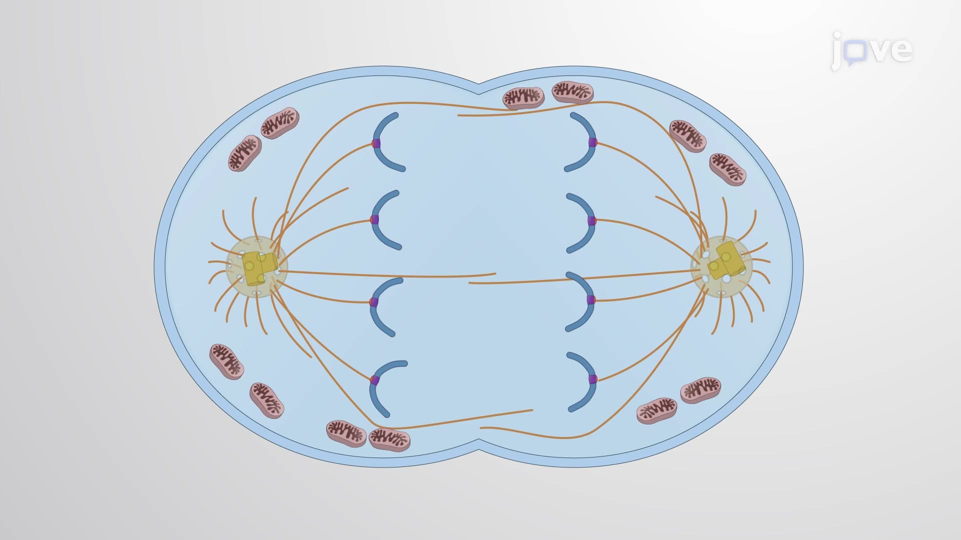
The resulting microtubule array consists of three distinct populations. Interpolar microtubules from opposite poles create overlapping zones at the cell center. Kinetochore microtubules directly attach to chromosome attachment sites, while astral microtubules contact the cell membrane to position the entire apparatus. Students preparing for AP Biology exams should focus on how these three microtubule types work together-this concept frequently appears in free-response questions about cell division.
Motor Protein Coordination and Force Balance
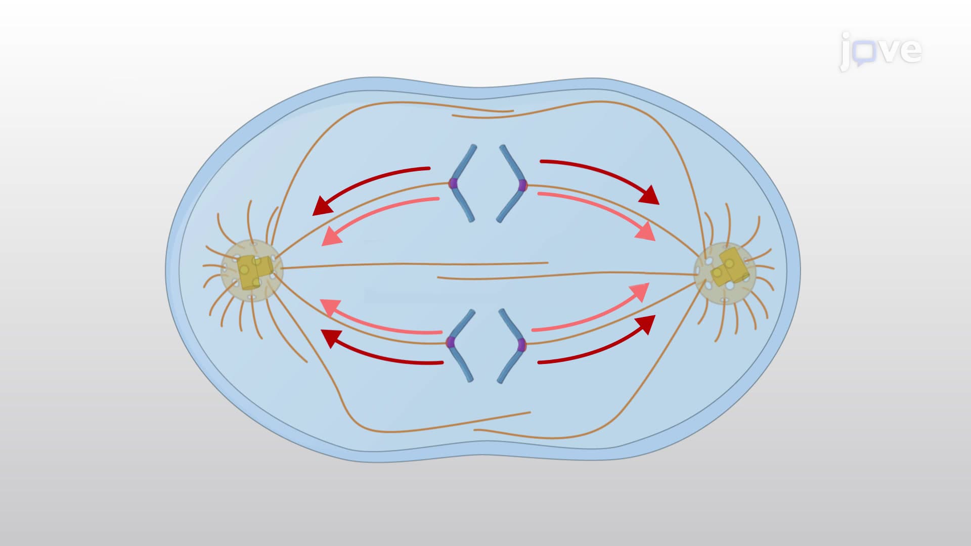
Multiple motor proteins fine-tune spindle assembly through opposing forces. Dynein motors pull spindle poles toward cell edges, while kinesin-5 pushes poles apart by sliding interpolar microtubules. Meanwhile, kinesin-14 generates tension pulling poles together, and chromokinesins push chromosomes away from poles. This force balance determines final spindle dimensions-a concept that appears regularly on MCAT Cell Biology sections.
Alternative Assembly Mechanisms
Not all cells rely solely on centrosomes for spindle assembly. In acentrosomal systems, mitotic chromosomes activate Ran-GTP, which releases microtubule-stabilizing factors locally. This mechanism ensures spindle formation even when centrosomes malfunction, highlighting the redundancy built into this essential process. Understanding both centrosomal and acentrosomal mechanisms helps students tackle complex scenarios in college cell biology courses and prepares them for advanced study in developmental biology or cancer research.
Frequently Asked Questions
Spindle assembly is the process where cells build a bipolar microtubule structure that separates chromosomes during division. This ensures each daughter cell receives exactly the right genetic material, preventing cancer-causing chromosome imbalances. Without proper spindle assembly, cells would distribute chromosomes randomly, leading to genetic disorders or cell death.
AP Biology frequently tests spindle assembly through diagrams showing mitotic phases and questions about chromosome movement mechanisms. Students should memorize the three microtubule types and understand how motor proteins create balanced forces. Practice drawing spindle diagrams and labeling centrosomes, kinetochores, and microtubule arrays for maximum exam success.
The MCAT emphasizes understanding how spindle defects relate to cancer biology and genetic diseases. Focus on motor protein functions, centrosome roles, and how spindle checkpoint mechanisms prevent errors. Questions often connect spindle assembly to pharmaceutical targets, since many cancer drugs disrupt microtubule function to stop tumor cell division.
Spindle assembly defects contribute to many cancers seen in US hospitals, including breast, lung, and colon cancers. For example, researchers at MD Anderson have identified spindle checkpoint mutations in families with hereditary cancer syndromes. These defects allow cancer cells to survive with abnormal chromosome numbers, promoting tumor growth and drug resistance.
Not at all-spindle assembly builds logically on basic cell biology concepts you already know. Start with understanding that cells need to divide chromosomes equally, then learn how microtubules accomplish this task. Focus on the big picture first: centrosomes anchor cables that pull chromosomes apart, with motor proteins providing fine-tuned control.
Create labeled diagrams showing all three microtubule types and practice drawing spindle formation stages. Use flashcards for motor protein functions and make concept maps connecting spindle assembly to cancer biology. Many students find success studying this topic alongside mitosis phases, since spindle assembly occurs during prophase and metaphase transitions.
Dive deeper into cell cycle checkpoints, particularly the spindle checkpoint that monitors chromosome attachment. Study cancer biology to understand how spindle-targeting drugs work, or explore developmental biology to see how spindle orientation affects tissue formation. These advanced topics build directly on spindle assembly fundamentals.
Related Micro-courses