Loading subject page…


89,755 views

Video Summary: What Is Peritoneum
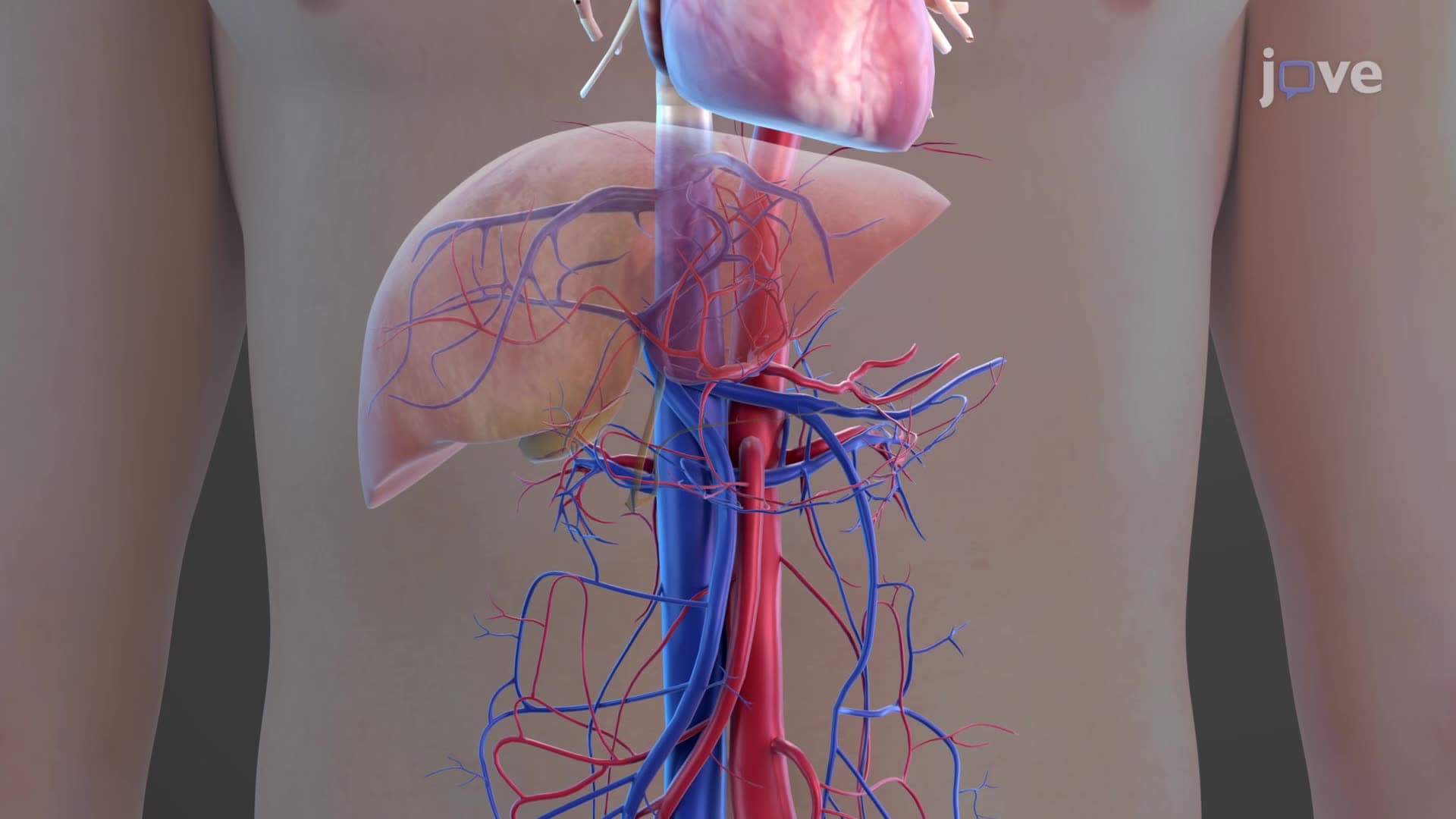
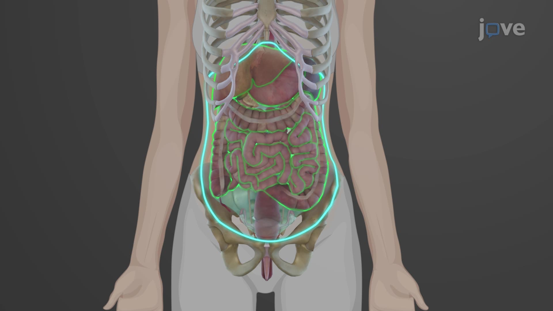
Did you know that your abdomen contains nearly two square meters of protective membrane-roughly the size of a ping-pong table? This peritoneum anatomy biology masterpiece lines your entire abdominal cavity, creating a sophisticated barrier system that protects vital organs like those affected in appendicitis cases treated daily in US emergency rooms. The peritoneum functions as your body's largest serous membrane, featuring complex folds that support digestive organs and facilitate crucial physiological processes. Watch the full video on JoVE Coach to master this concept with expert-led visuals and step-by-step explanations.
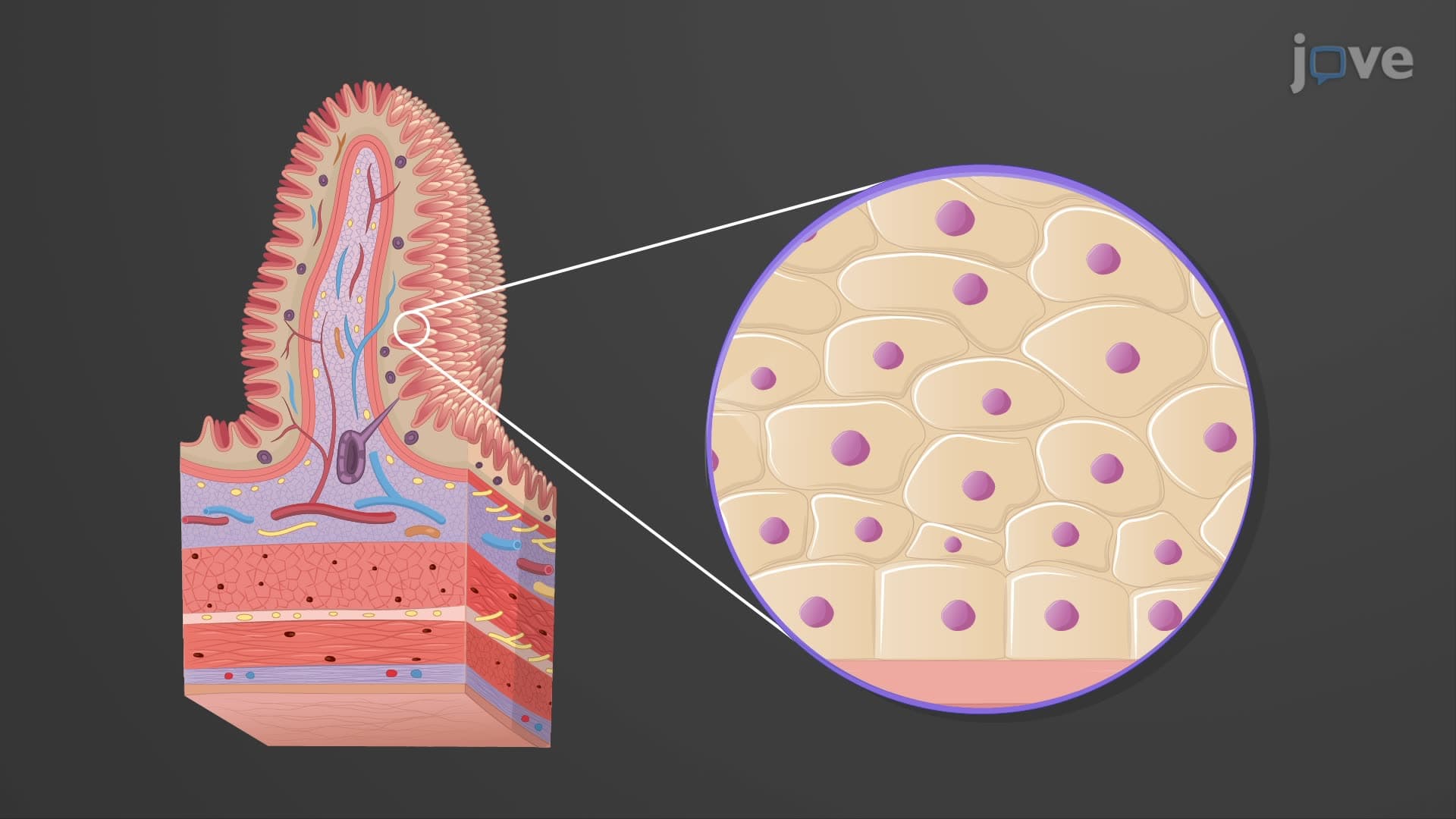
The peritoneum represents one of anatomy's most elegant protective systems-a continuous, glistening membrane that transforms the potentially chaotic abdominal space into an organized, protected environment. This peritoneal membrane explained simply is a two-layered serous membrane that creates both a protective barrier and a sophisticated support network for your digestive organs.
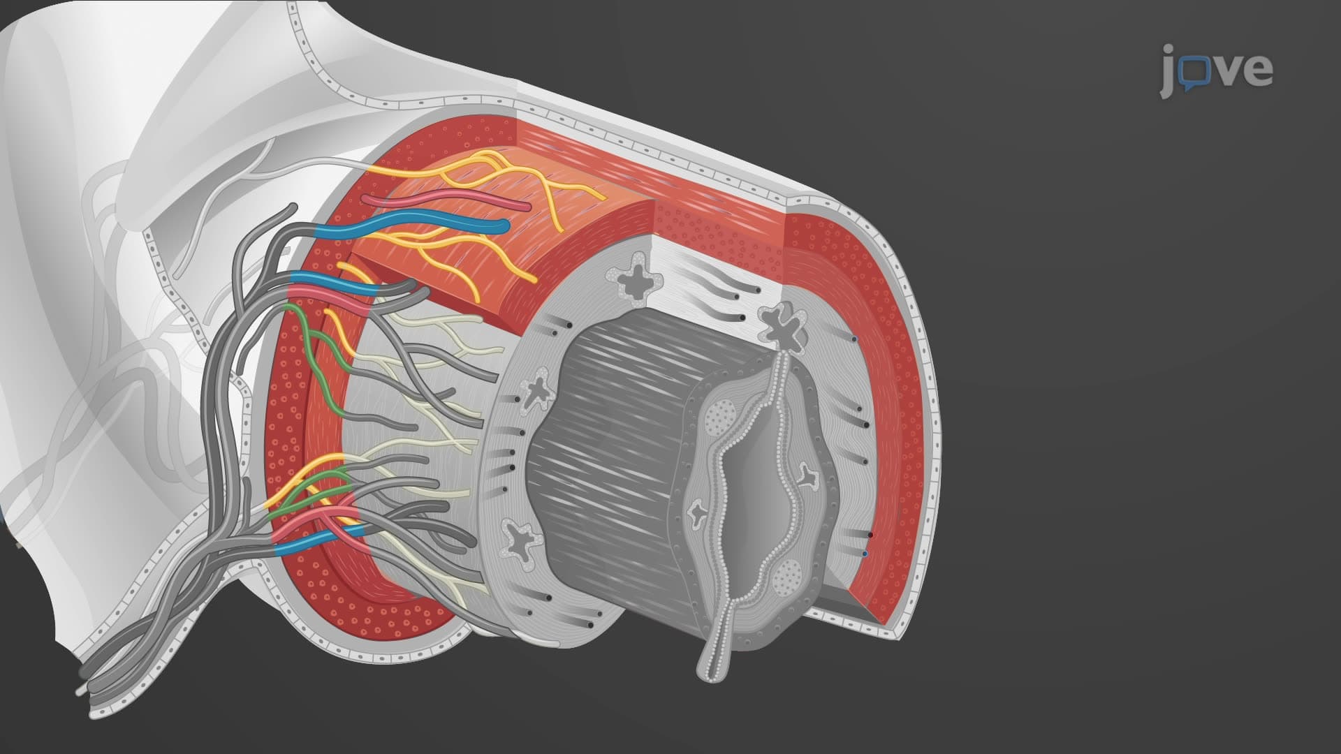
Understanding visceral parietal peritoneum differences is crucial for mastering digestive anatomy. The parietal peritoneum lines the inner abdominal wall, diaphragm, and pelvis, while the visceral peritoneum directly covers individual organs like the stomach, liver, and intestines. Between these layers lies the peritoneal cavity-a potential space containing approximately 50-100 milliliters of peritoneal cavity fluid that acts like natural lubricant, allowing organs to glide smoothly during breathing, digestion, and movement.
This arrangement appears frequently on MCAT questions and AP Biology exams, where students must differentiate between intraperitoneal organs (completely covered by visceral peritoneum) and retroperitoneal organs (only partially covered, lying behind the peritoneum).
The mesentery omentum peritoneum system creates five major folds that serve as both structural supports and highways for blood vessels, lymphatics, and nerves. The greater omentum-often called the "abdominal policeman"-drapes over intestines like a protective apron, rich in infection-fighting immune cells and fat storage. In cases of appendicitis, this structure often walls off infection, preventing widespread peritonitis.
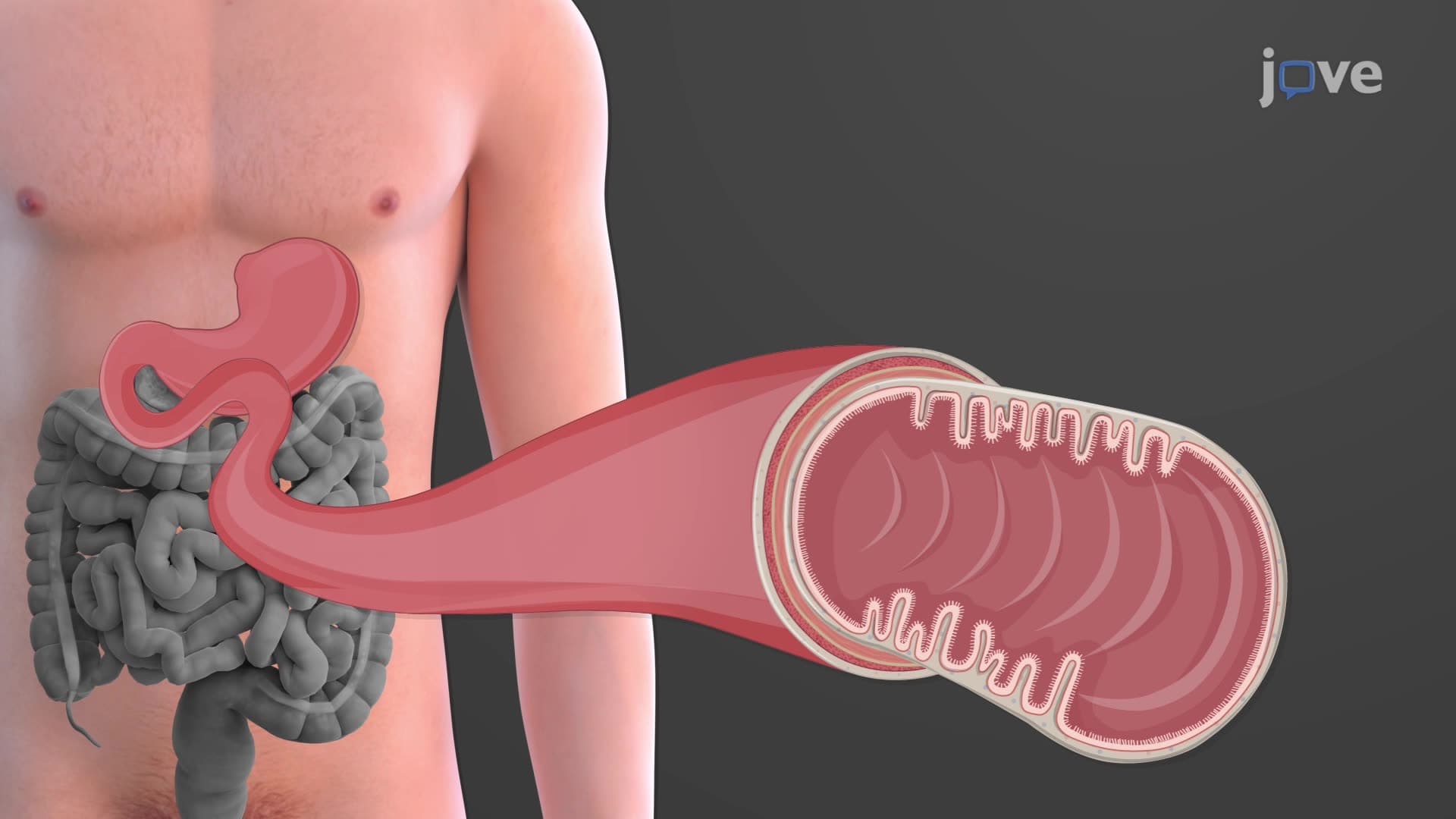
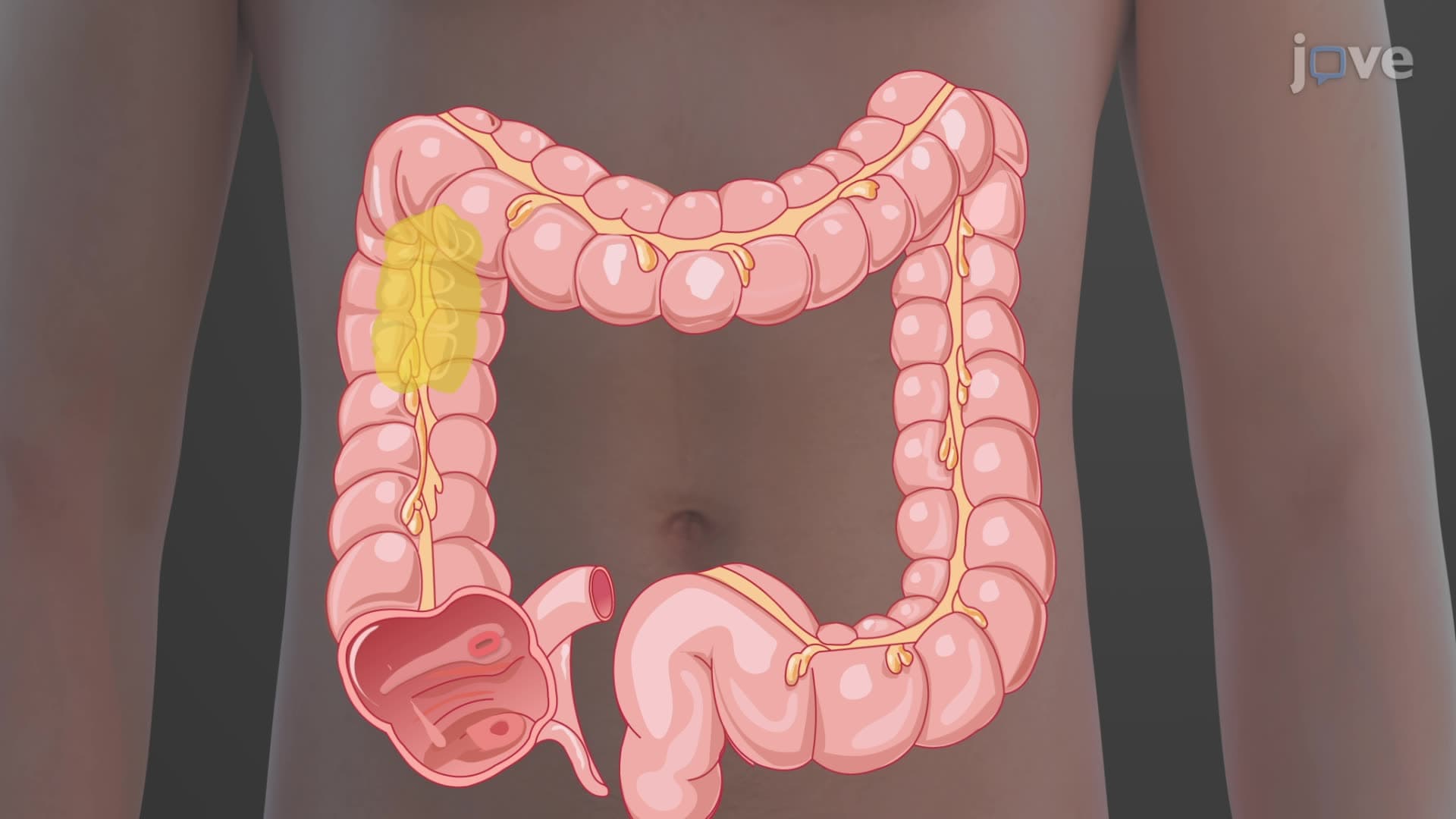
The mesentery, a fan-shaped peritoneal fold, suspends the small intestine from the posterior abdominal wall, while the mesocolon performs similar functions for the large intestine. The falciform ligament connects the liver to the anterior abdominal wall, and the lesser omentum links the stomach and duodenum to the liver.
Understanding peritoneum anatomy structure proves essential for recognizing conditions like peritonitis (inflammation of the peritoneum), commonly seen in US emergency departments following appendix rupture or bowel perforation. Medical students studying for the USMLE frequently encounter questions about peritoneal reflections and their clinical significance in surgical procedures and disease progression.
Related Micro-courses