Loading video…


 40 Concepts
 89.8K views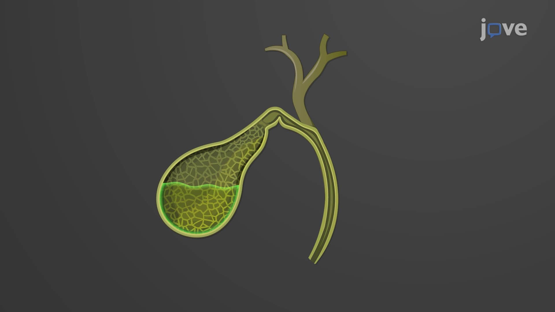

Video Summary: What Is Gallbladder
Did you know that your gallbladder can concentrate bile up to 20 times its original strength, making it one of the body's most efficient storage organs? Understanding gallbladder anatomy biology reveals how this small, kiwi-sized organ plays a crucial role in fat digestion. When you eat a bacon cheeseburger at your local diner, your gallbladder contracts to release concentrated bile, helping break down those fats for absorption. Watch the full video on JoVE Coach to master this concept with expert-led visuals and step-by-step explanations.
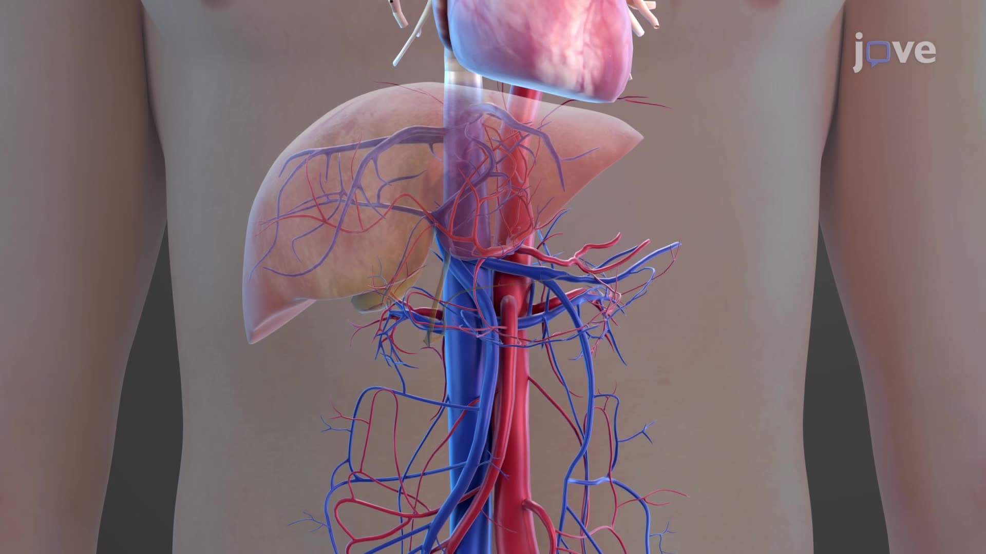
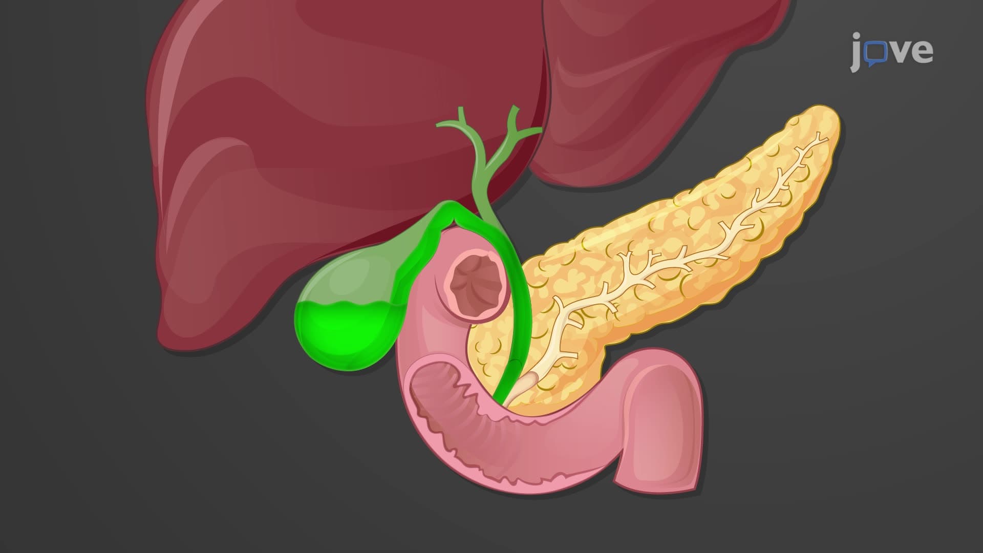
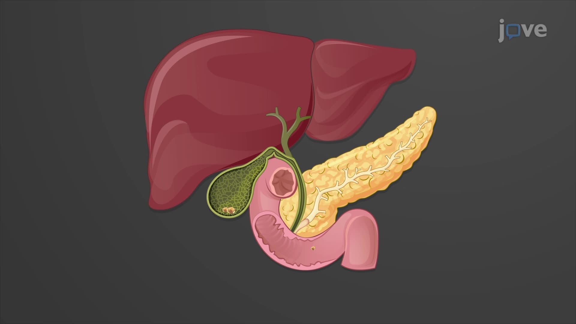
The gallbladder structure function explained begins with understanding its role as a specialized storage and concentration facility for bile. This remarkable organ demonstrates how evolution has optimized space and efficiency in human anatomy. Located in a depression called the gallbladder fossa on the liver's inferior surface, this muscular sac measures approximately 10 centimeters in length-roughly equivalent to a large kiwi fruit.
What is the gallbladder in digestive anatomy becomes clearer when examining its three distinct regions. The fundus represents the rounded, blind end that extends slightly beyond the liver's edge. The body forms the central portion, while the neck tapers toward the cystic duct gallbladder connection. This anatomical organization isn't arbitrary-each region serves specific functions in bile storage and release.
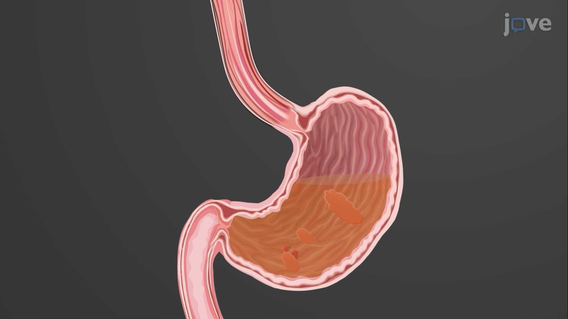
The internal architecture features honeycomb-like mucosal folds called rugae, similar to those found in the stomach. These folds allow dramatic expansion as bile storage gallbladder capacity increases from about 30-50 milliliters when empty to potentially 300 milliliters when fully distended. Students preparing for the MCAT should note that this expandability is crucial for accommodating varying bile volumes between meals.
Bile concentration gallbladder activity represents one of the most efficient concentration processes in human physiology. The gallbladder can concentrate bile 5-20 times its original strength by actively reabsorbing water and electrolytes while retaining bile salts, cholesterol, and bilirubin. This concentration process is particularly important for AP Biology students studying membrane transport mechanisms.
The release mechanism involves cholecystokinin CCK gallbladder stimulation. When fatty foods enter the duodenum-like the cheese and meat in a typical American burger-CCK hormone is released from duodenal cells. This hormone triggers smooth muscle contraction in the gallbladder wall, forcing concentrated bile through the cystic duct, into the common bile duct, and finally into the duodenum where it emulsifies fats.
Understanding gallbladder anatomy function becomes clinically significant when considering gallstone formation. When bile becomes supersaturated with cholesterol or bilirubin, these substances can crystallize into gallstones. The Western diet, common in the United States, with its high cholesterol and fat content, contributes to the prevalence of gallbladder disease affecting approximately 20 million Americans.
College students studying for pre-med requirements should understand that gallbladder dysfunction can significantly impact fat digestion and absorption, leading to deficiencies in fat-soluble vitamins (A, D, E, K). This connection frequently appears on USMLE Step 1 examinations, particularly in questions linking anatomical knowledge to physiological consequences.
Related Micro-courses