Loading video…


 12 Concepts
 628K views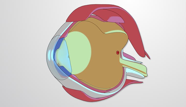

Video Summary: What Is Vision
Did you know that your brain processes visual information from both eyes even though you only see one unified image? Vision is the complex biological process that transforms light waves into meaningful visual perception, allowing us to navigate our world. When a baseball player at Yankee Stadium tracks a 95-mph fastball, their visual system is performing millions of calculations in milliseconds-from light detection in the retina to object recognition in the cerebral cortex. Watch the full video on JoVE Coach to master this concept with expert-led visuals and step-by-step explanations.
Vision represents one of the most sophisticated sensory systems in the human body, transforming electromagnetic radiation (light) into meaningful visual experiences. Unlike simple light detection, vision involves complex neural processing that allows us to recognize faces, read text, judge distances, and navigate three-dimensional space with remarkable precision.
The journey of visual processing begins when light reflects off objects and enters the eye through the cornea-the eye's transparent front layer that provides initial focusing power. After passing through the pupil (controlled by the iris), light reaches the crystalline lens, which fine-tunes focus to project sharp images onto the retina.
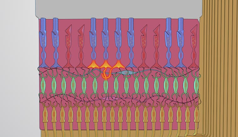
The retina contains two primary types of photoreceptors: rods and cones. Rods, numbering about 120 million per eye, excel in low-light conditions and detect motion, while cones (6-7 million per eye) provide color vision and sharp detail recognition. When light strikes these photoreceptors, they undergo a chemical change that alters neurotransmitter release, converting light energy into electrical signals.
Retinal ganglion cells collect information from multiple photoreceptors, performing initial processing like edge detection-a critical step that helps us distinguish objects from backgrounds. These cells send axons through the optic nerve, which partially crosses at the optic chiasm, ensuring each brain hemisphere receives input from both eyes for depth perception.
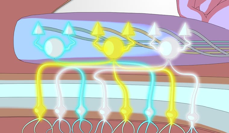
Most visual information reaches the thalamus (specifically the lateral geniculate nucleus), where different visual features undergo parallel processing. Color information follows one pathway while motion detection follows another-a principle crucial for understanding conditions like motion blindness, where patients can see stationary objects but cannot detect movement.
The primary visual cortex (V1) in the occipital lobe receives thalamic input and maintains a precise topographic map of visual space. Neurons here respond to specific orientations, spatial frequencies, and movement directions. This organization explains why damage to specific cortical areas can cause predictable visual field defects.
From V1, information flows to specialized cortical areas: the "what" pathway (ventral stream) identifies objects and faces, while the "where/how" pathway (dorsal stream) processes spatial location and movement. This dual-pathway concept frequently appears on AP Psychology exams and helps explain how we can recognize a friend's face while simultaneously tracking their hand gestures.
Understanding vision proves essential for pre-med students preparing for the MCAT, as visual system questions commonly test knowledge of neural pathways, photoreceptor function, and cortical organization. Medical students will later apply this knowledge when studying conditions like macular degeneration, glaucoma, and cortical blindness.
Related Micro-courses