Loading video…


 12 Concepts
 83.2K views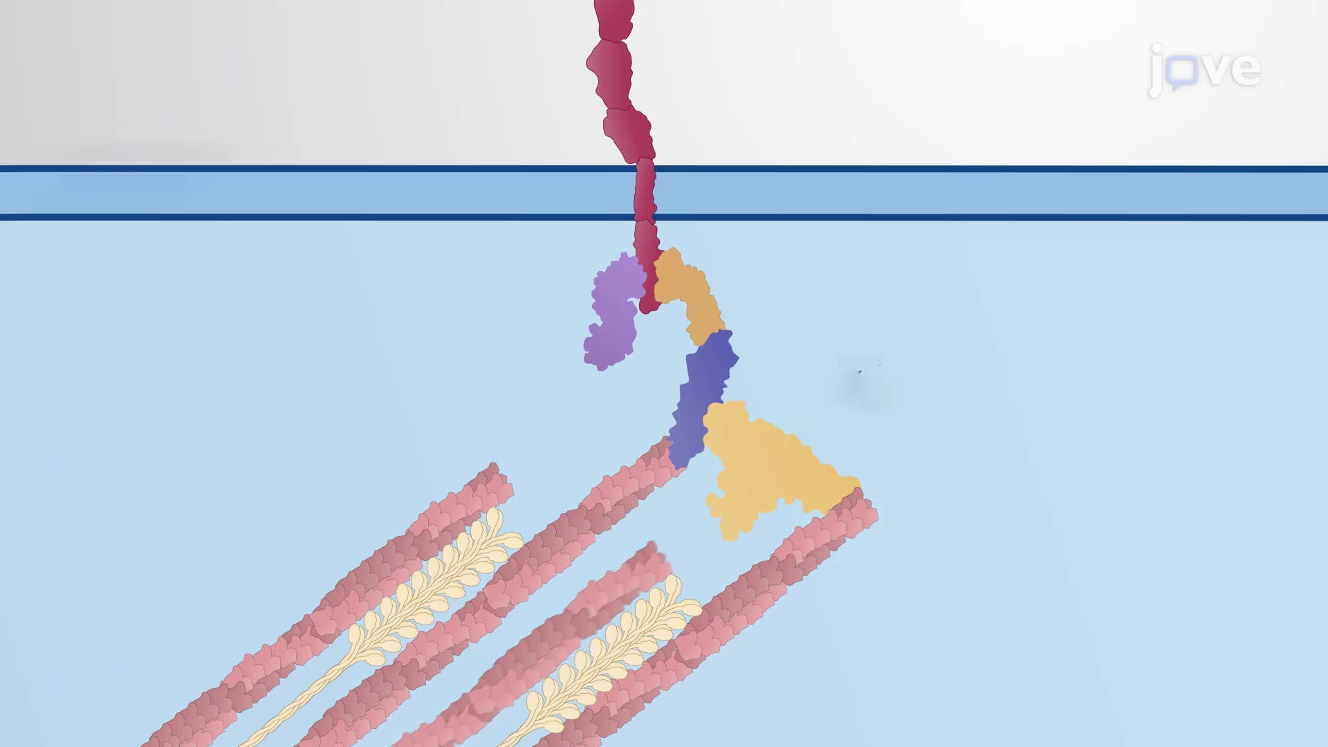

Video Summary: Tension Response at Adherens Junctions Explained
Ever wonder how your skin stays intact when you stretch or how your intestinal lining withstands constant movement? The tension response at adherens junctions is the cellular mechanism that keeps epithelial tissues strong under mechanical stress. When dermatologists treat stretch marks or gastroenterologists examine intestinal damage, they're observing the results of this fundamental process called mechanotransduction. Through specialized proteins like alpha-catenin and vinculin, cells detect mechanical forces and strengthen their connections accordingly. Watch the full video on JoVE Coach to master this concept with expert-led visuals and step-by-step explanations.
The tension response at adherens junctions represents one of biology's most elegant examples of cellular engineering. Unlike rigid structures, these intercellular connections function as dynamic, responsive networks that adapt to mechanical stress in real-time. This process is fundamental to maintaining tissue integrity in organs that experience constant physical forces.
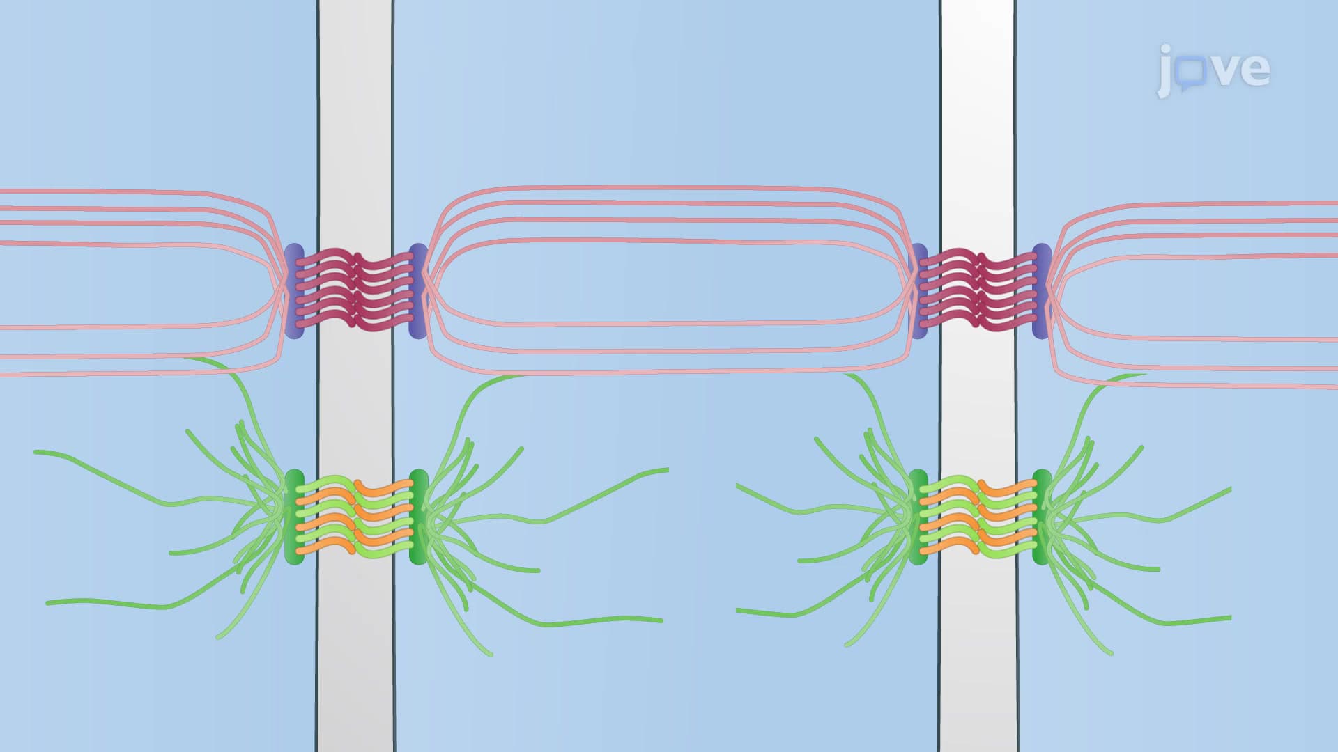
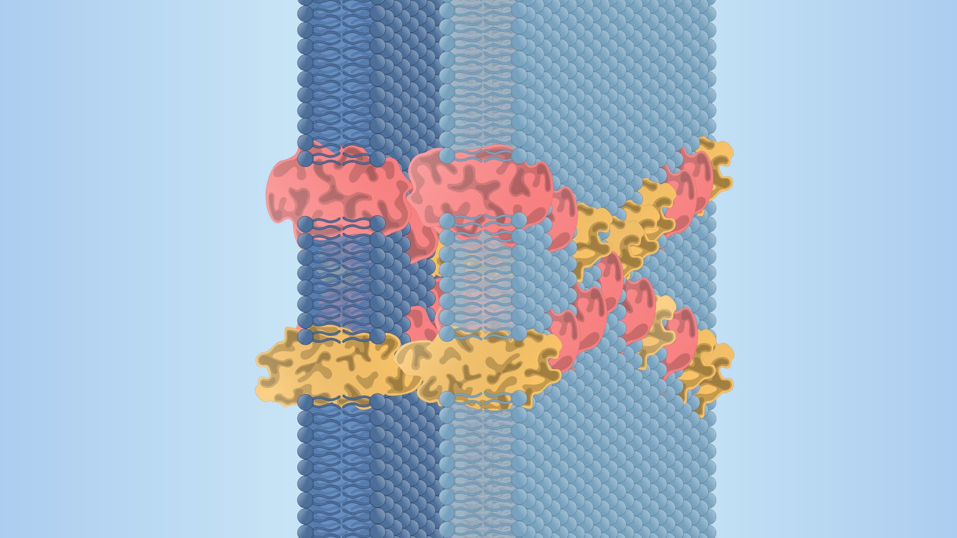
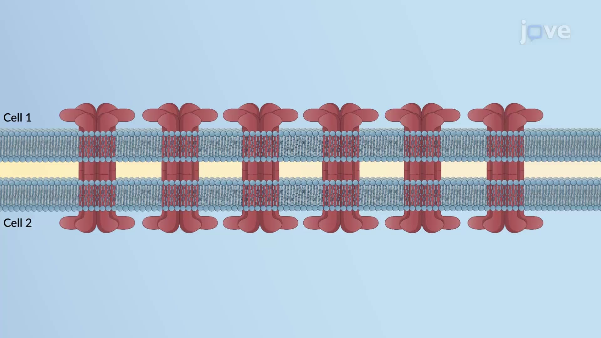
Mechanotransduction at adherens junctions operates through a sophisticated molecular cascade. When epithelial tissues-such as those lining the digestive tract or forming skin barriers-encounter stretching, compression, or shearing forces, specialized protein complexes respond immediately. The adherens junctions form what scientists call an "adhesion belt," a continuous zone of attachment that encircles each cell like a molecular corset.
This system becomes particularly important in clinical contexts. For example, when gastroenterologists study inflammatory bowel disease, they examine how compromised adherens junctions contribute to intestinal barrier dysfunction. Similarly, dermatologists investigating skin elasticity and aging focus on how mechanical stress responses change over time.
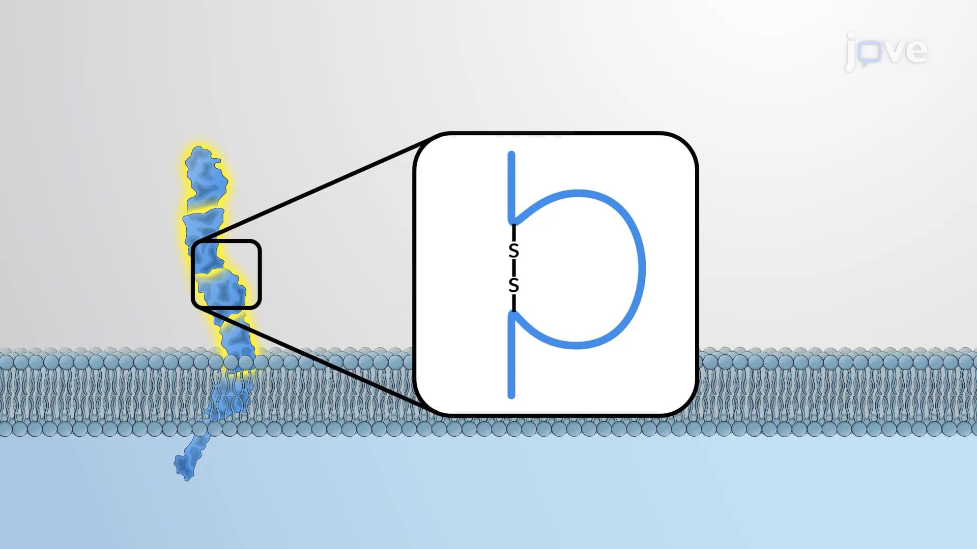
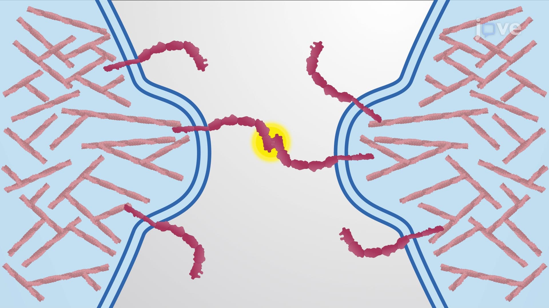
Alpha-catenin serves as the primary mechanosensor in this system, functioning like a biological strain gauge. Under normal conditions, alpha-catenin maintains a compact, folded structure. However, when contractile forces increase-such as during muscle contraction or tissue stretching-the protein undergoes conformational changes that expose hidden binding sites. This unfolding mechanism is so precise that researchers use it as a model for designing synthetic materials that respond to mechanical stress.
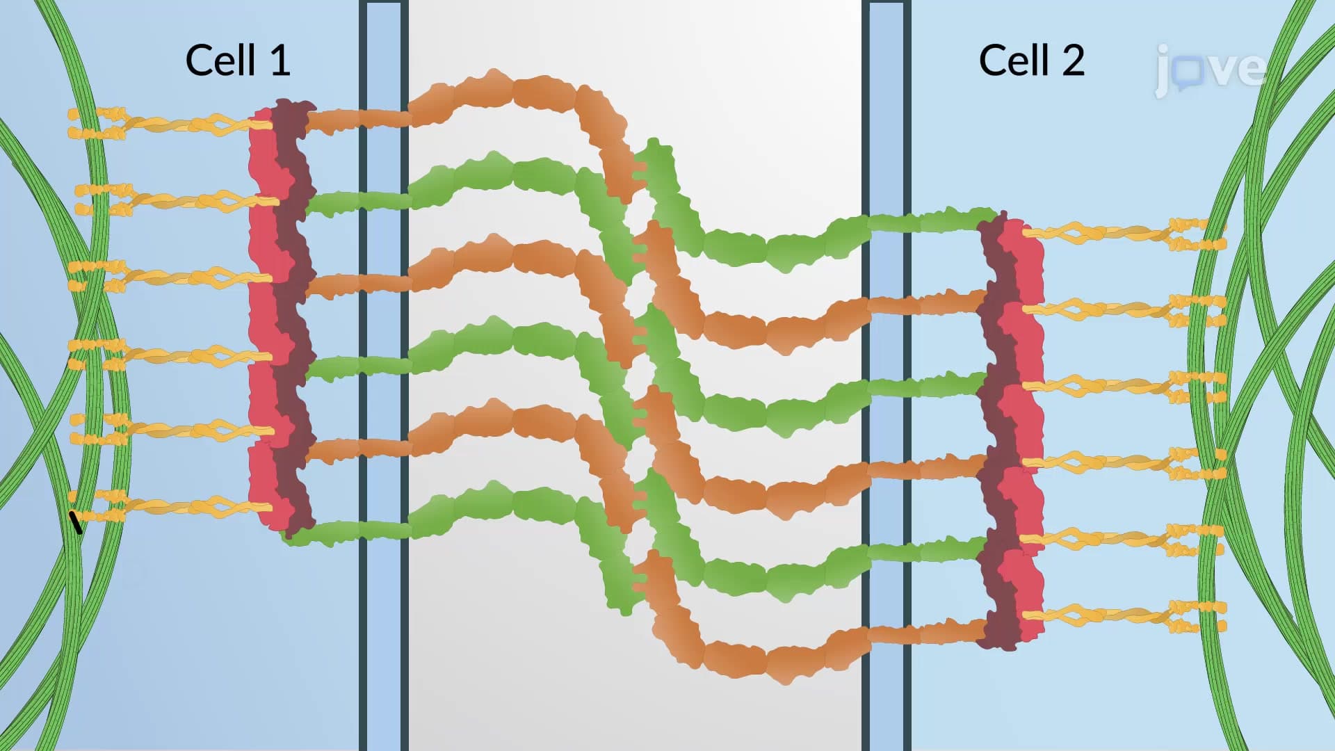
The interaction between unfolded alpha-catenin and vinculin exemplifies cellular communication at its finest. Vinculin normally exists in an inactive, closed conformation within the cytoplasm. Upon binding to the newly exposed sites on alpha-catenin, vinculin transforms into its active, open form. This activated vinculin then recruits additional actin filaments, effectively reinforcing the junction and distributing mechanical loads across neighboring cells.
This mechanism appears frequently on standardized exams, particularly the MCAT and AP Biology tests, where students must demonstrate understanding of protein conformational changes and cellular responses to environmental stimuli. College biochemistry and cell biology courses also emphasize these concepts when covering cytoskeletal dynamics and tissue mechanics.
Related Micro-courses