Loading video…


 12 Concepts
 83.2K views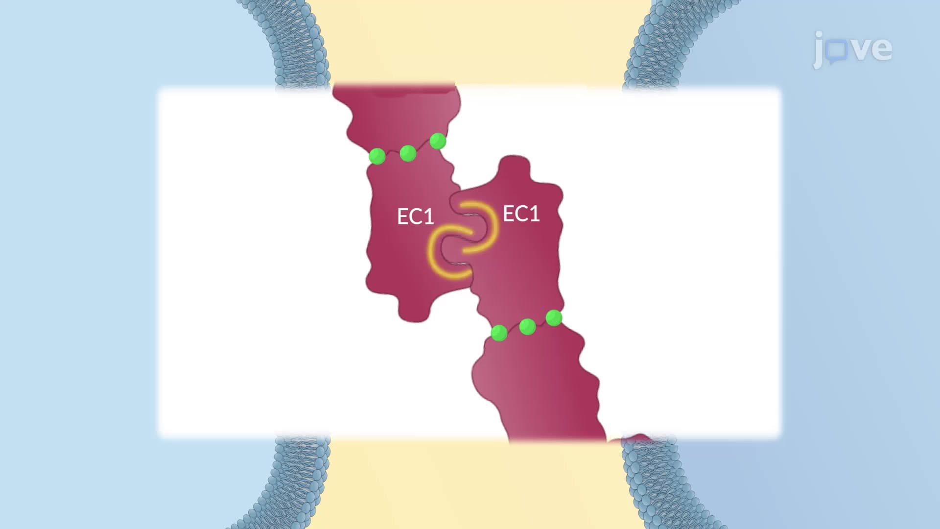

Video Summary: What Is Structure of Cadherins
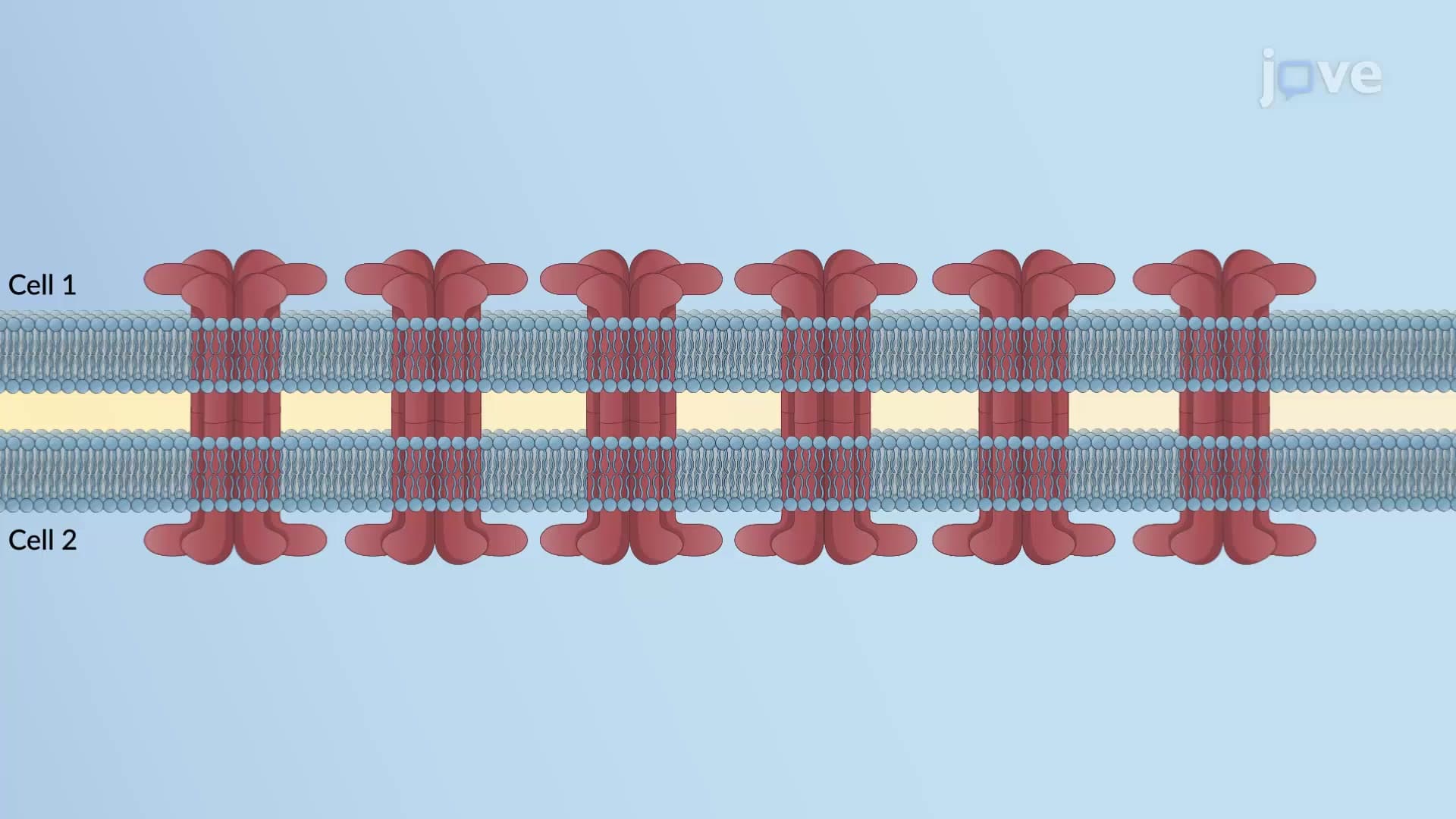
Did you know that without tiny molecular "handshakes" between cells, your skin would literally fall apart? The structure of cadherins consists of specialized cell adhesion molecules that act like cellular velcro, holding our tissues together through calcium-dependent binding mechanisms. These proteins are essential in conditions like pemphigus, where patients at Johns Hopkins Hospital experience painful skin blistering when cadherin function fails. Each cadherin molecule features three main structural components: extracellular domains for cell-to-cell binding, a transmembrane region, and an intracellular tail for signaling. Watch the full video on JoVE Coach to master this concept with expert-led visuals and step-by-step explanations.
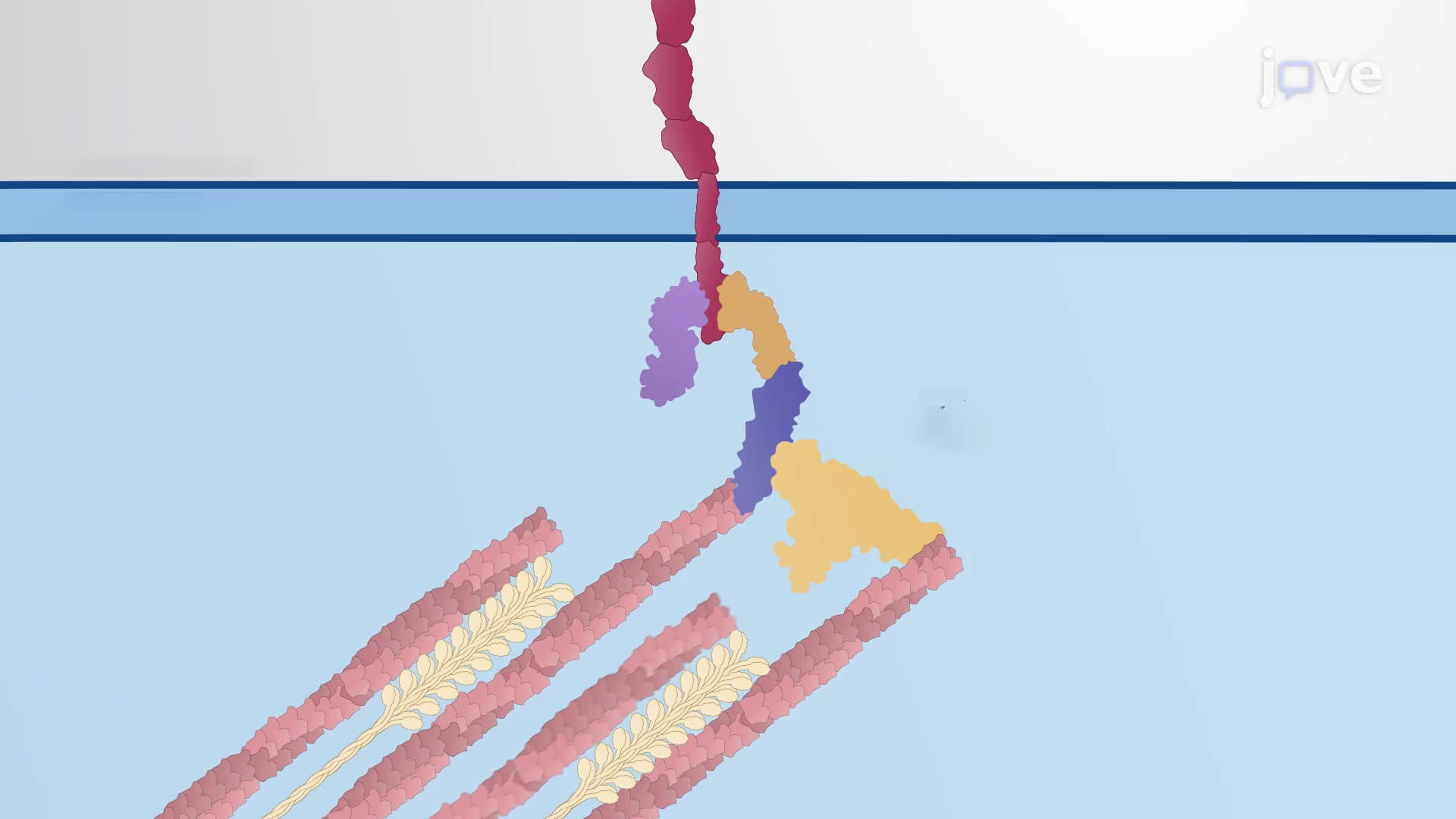
The structure of cadherins represents one of biology's most elegant solutions to cellular organization. These transmembrane proteins function as the primary architects of cell-cell adhesion, with over 100 different types identified in humans. Each cadherin molecule spans the cell membrane once, creating a bridge between the cell's interior and exterior environments.
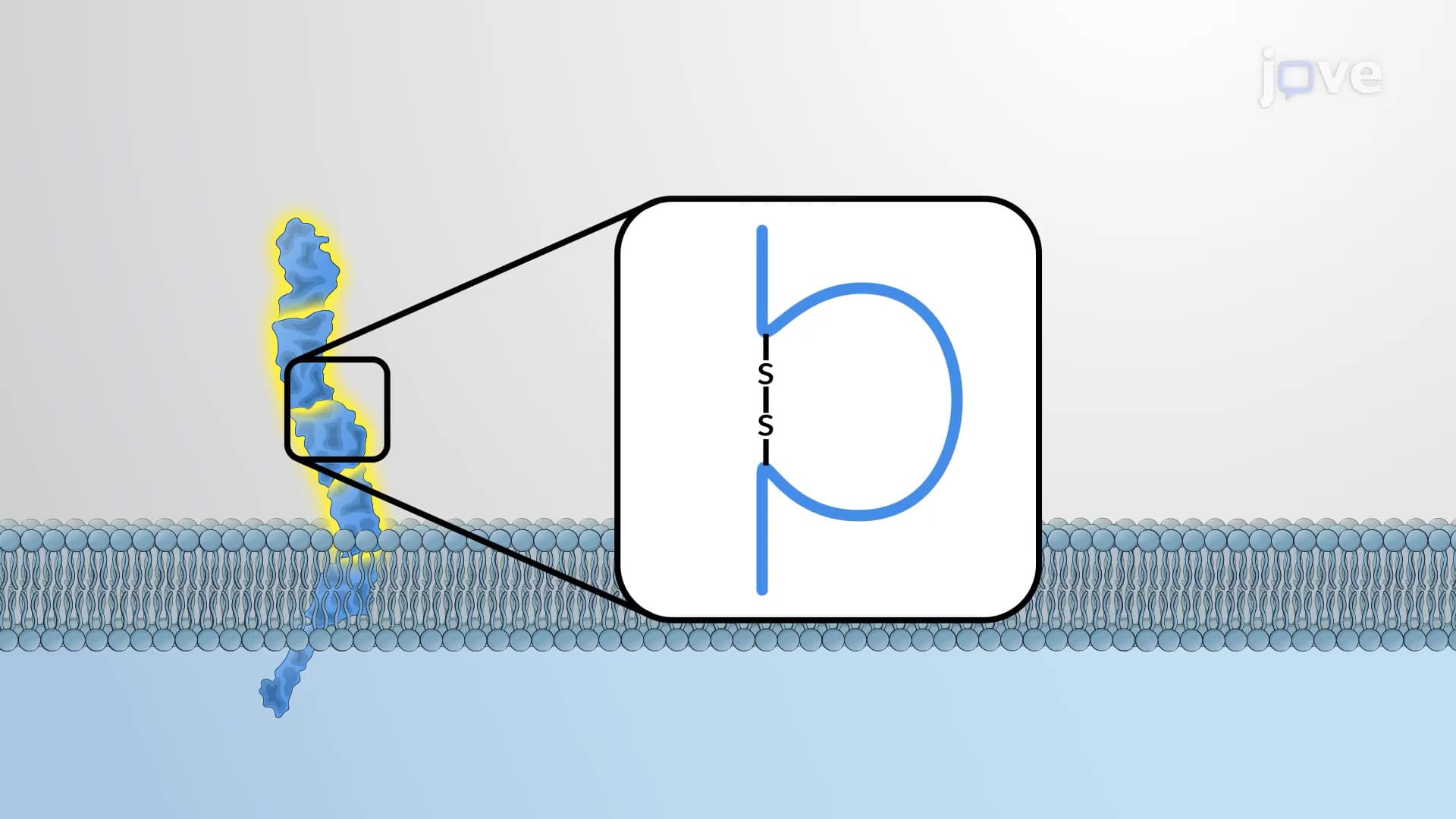
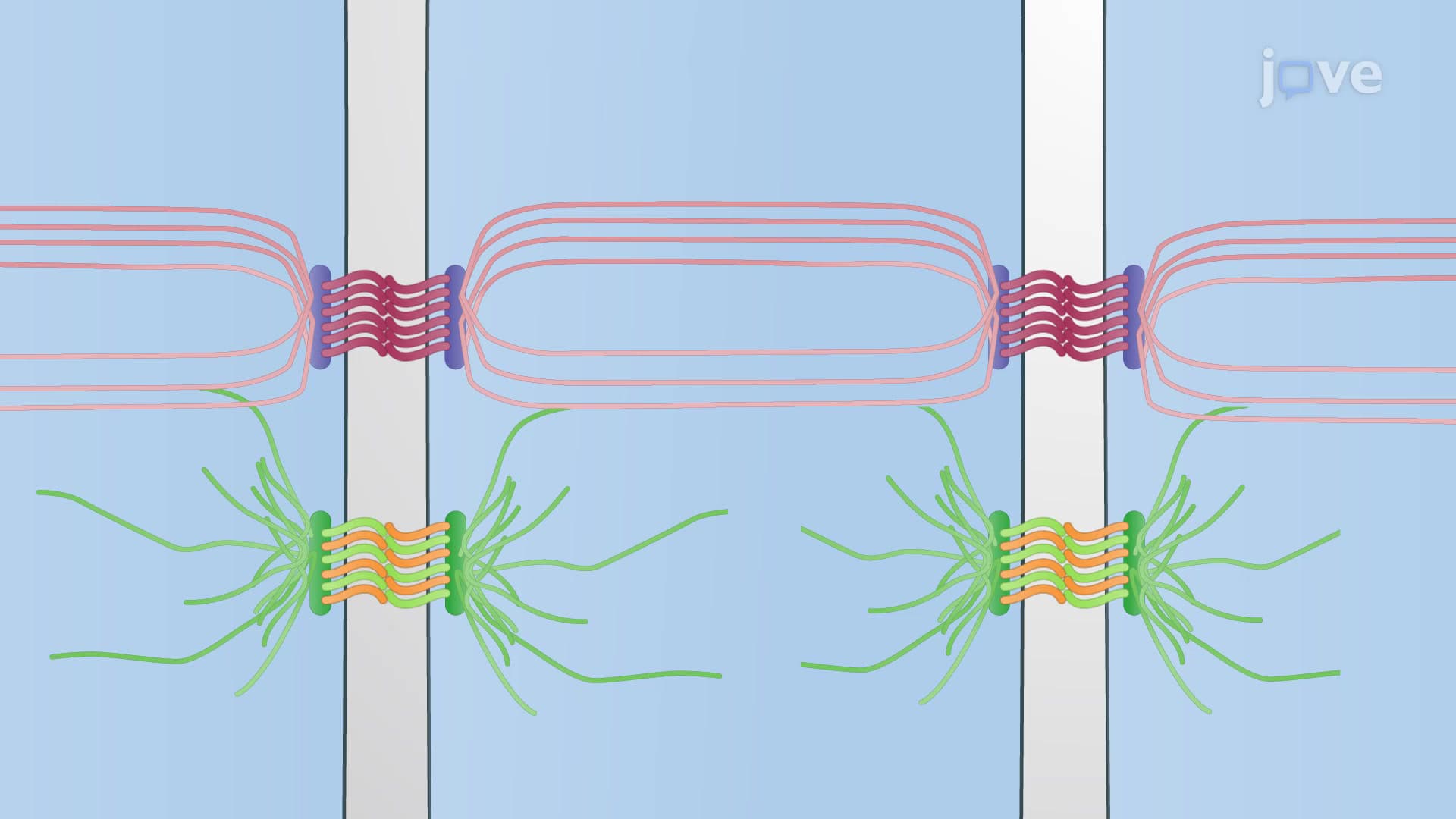
Cadherins exhibit a modular three-part architecture. The extracellular region contains multiple cadherin domains (typically 5 in classical cadherins), each approximately 110 amino acids long. These domains fold into beta-sandwich structures connected by flexible linker regions containing three calcium-binding sites each. The single transmembrane domain anchors the protein within the lipid bilayer, while the cytoplasmic tail interacts with intracellular proteins like beta-catenin and alpha-catenin, connecting to the actin cytoskeleton.
This organization proves crucial for AP Biology students studying cell communication, as it demonstrates how protein structure directly relates to function. College biochemistry courses at institutions like UCLA and MIT emphasize how this modular design allows for both specificity and strength in cellular adhesion.
The presence of calcium ions transforms cadherins from flexible, non-functional molecules into rigid, adhesion-competent structures. Each interdomain hinge contains three calcium-binding pockets that, when occupied, lock adjacent domains into proper orientation. This calcium dependence explains why EDTA treatment (which chelates calcium) causes tissues to dissociate-a principle used in laboratory cell culture techniques taught in universities across the US.
Medical students preparing for the MCAT encounter this concept when studying tissue development and cancer metastasis, where calcium availability directly impacts cell migration patterns.
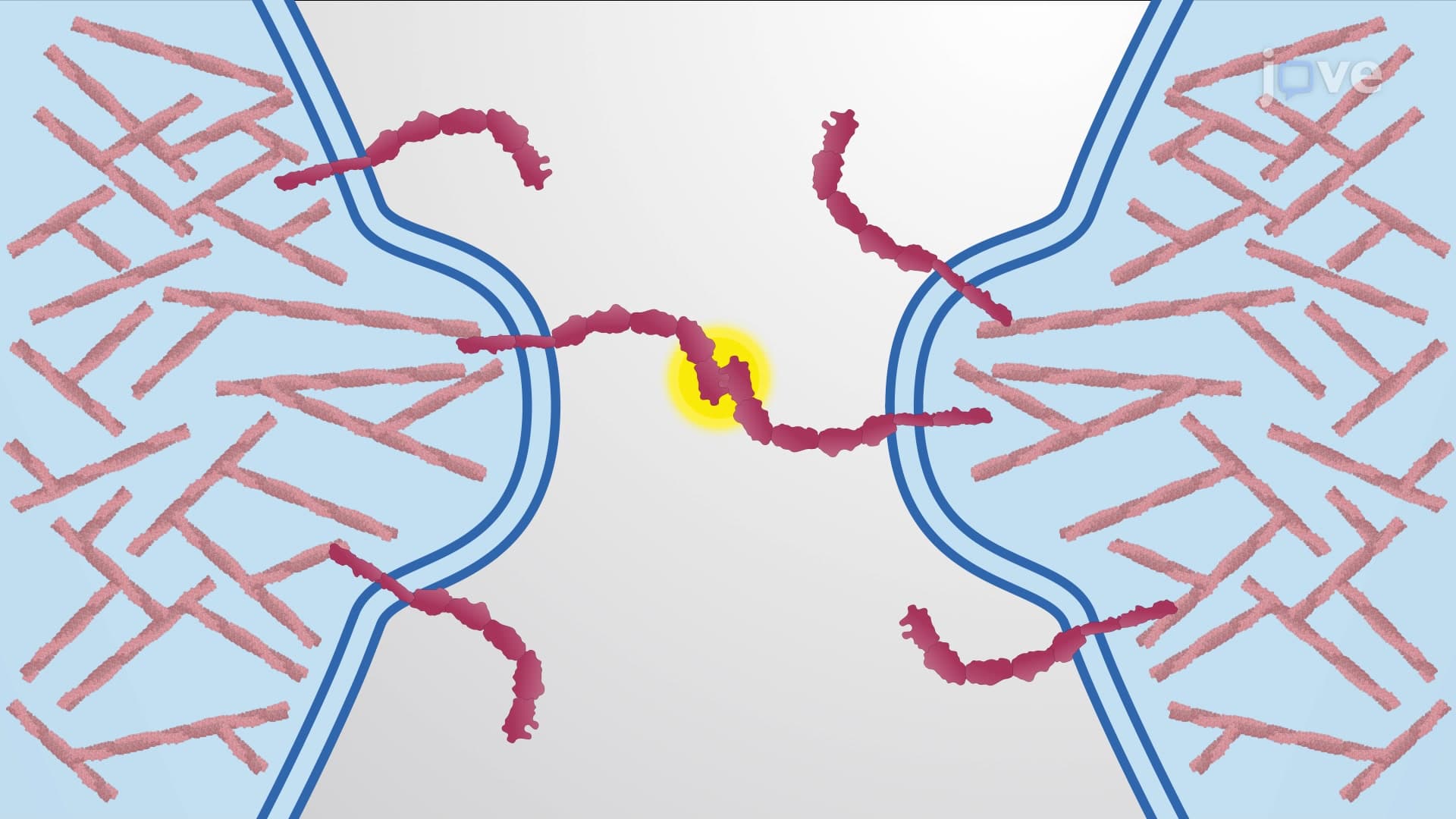
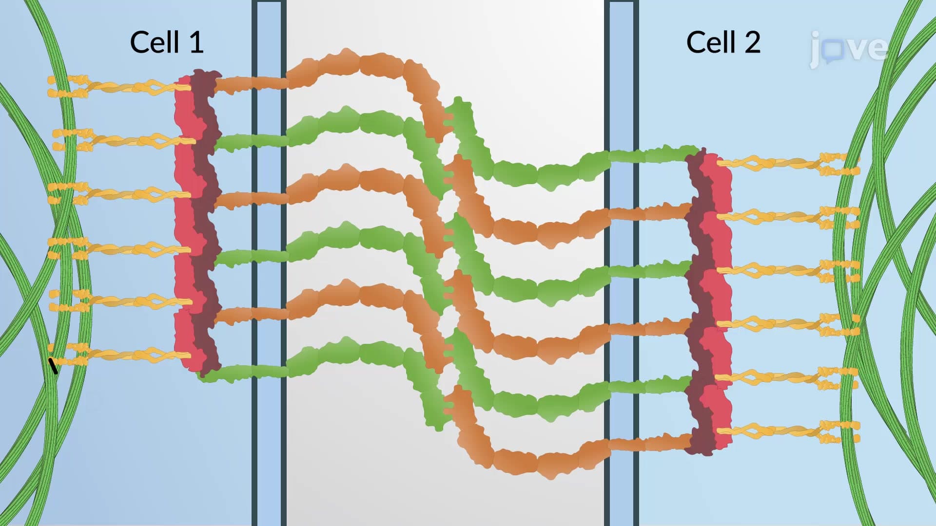
The N-terminal EC1 domain contains the primary adhesive interface, featuring a distinctive knob-and-pocket structure. During trans-binding, the knob from one cell's cadherin inserts into the pocket of another cell's cadherin of the same type. This mechanism ensures homophilic binding-E-cadherins bind preferentially to other E-cadherins, N-cadherins to N-cadherins, and so forth.
This selectivity underlies tissue organization during embryonic development and maintains tissue boundaries in adults. Students studying for AP Biology exams often encounter this concept in developmental biology contexts, particularly when examining how neural crest cells migrate during vertebrate development.
Related Micro-courses