Loading subject page…

- Anatomy and Physiology
- Digestive System
- Blood Supply to the Digestive System
Micro-course: Digestive System
89,747 views
Learn key concepts and earn your certificate.


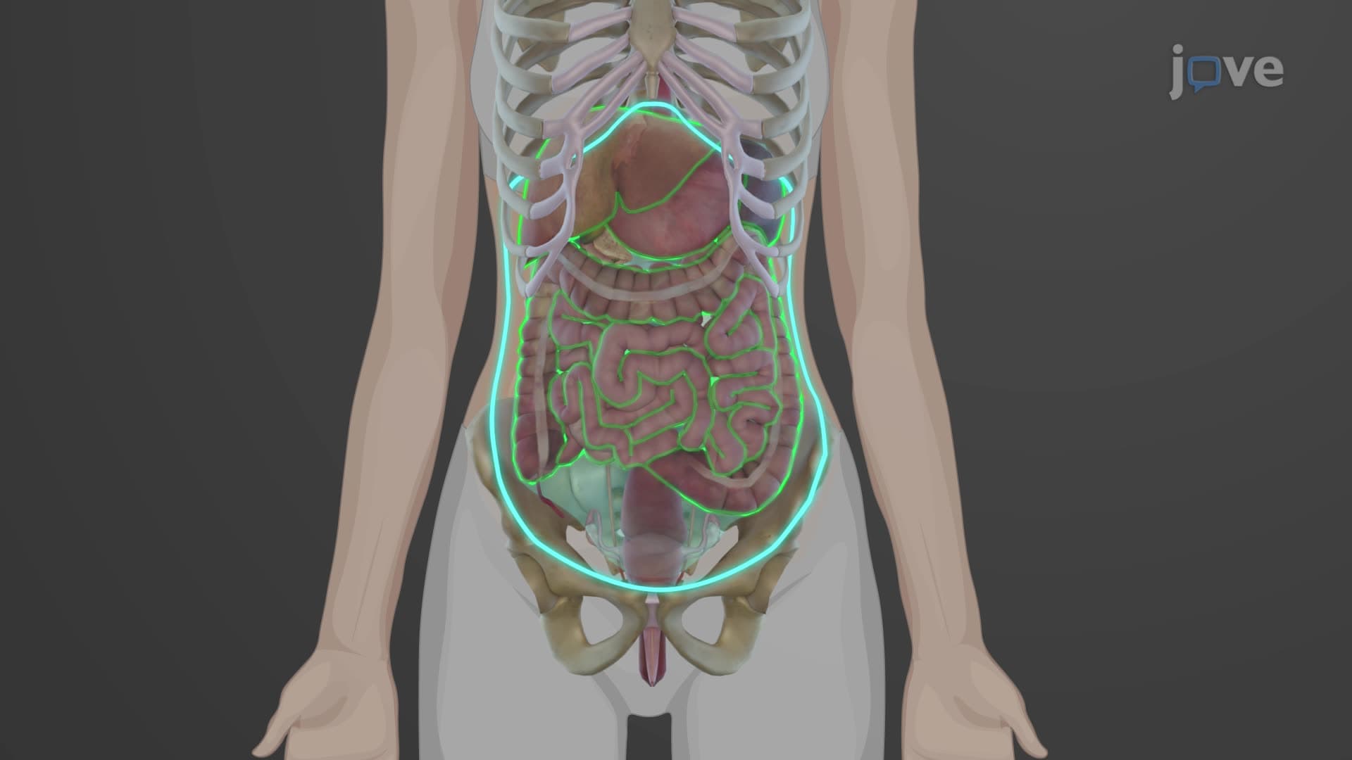
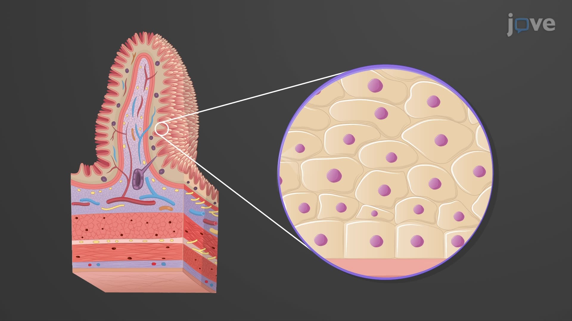
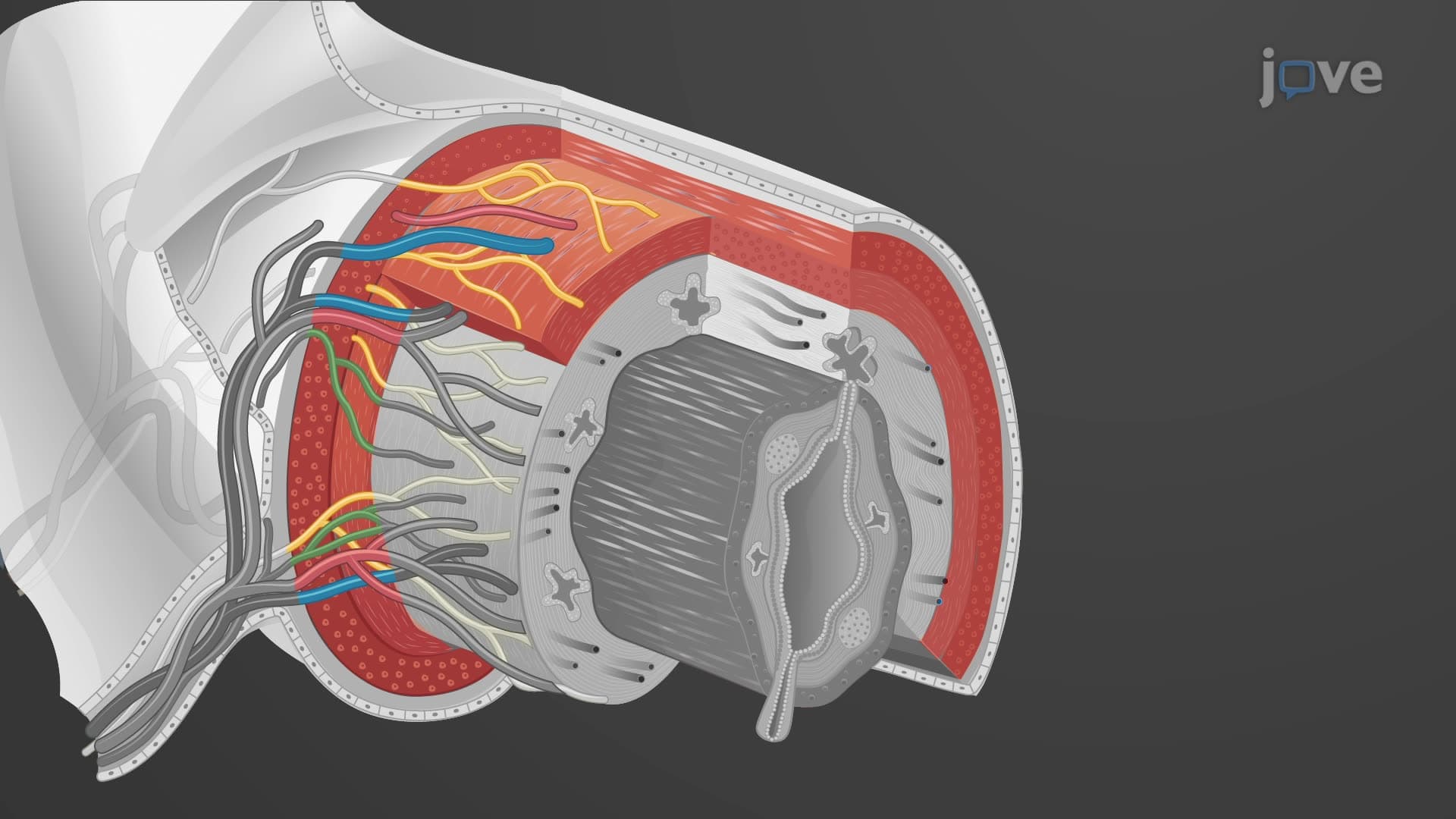

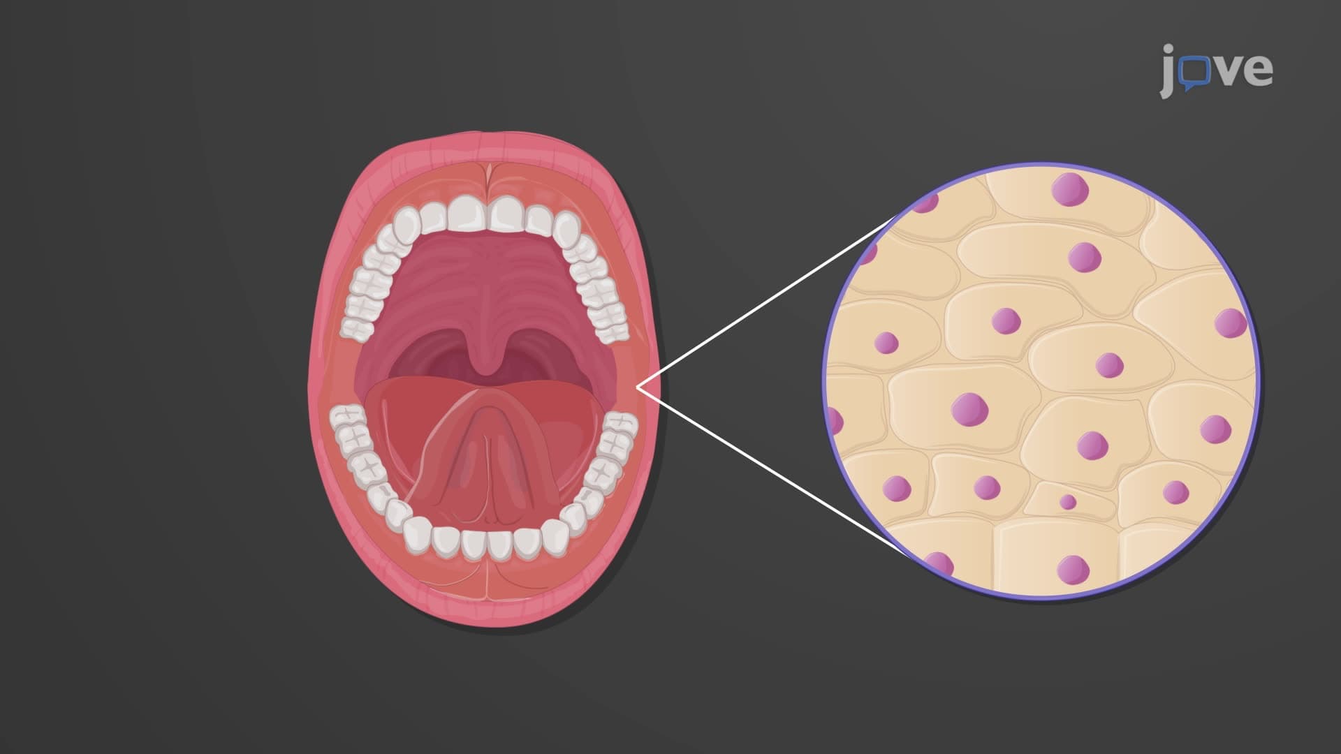

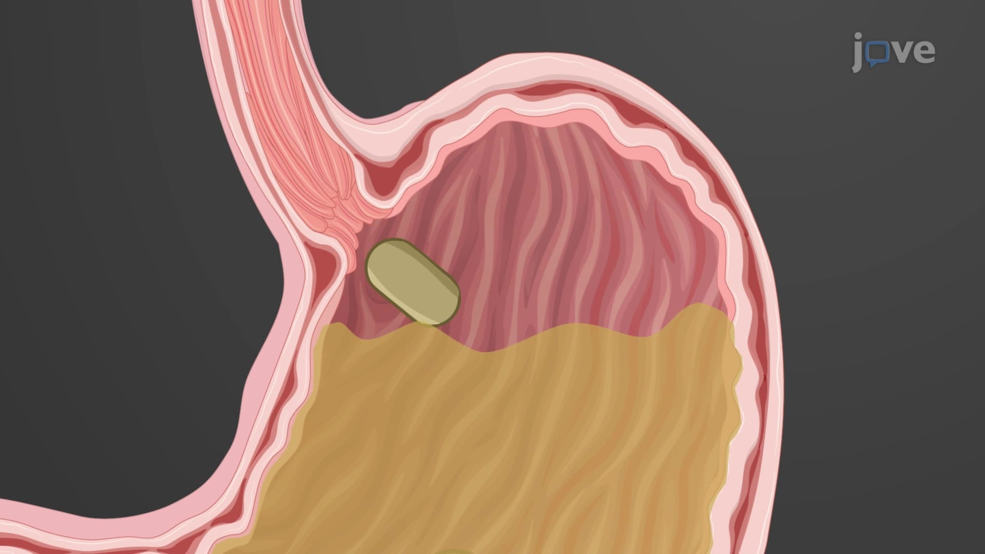
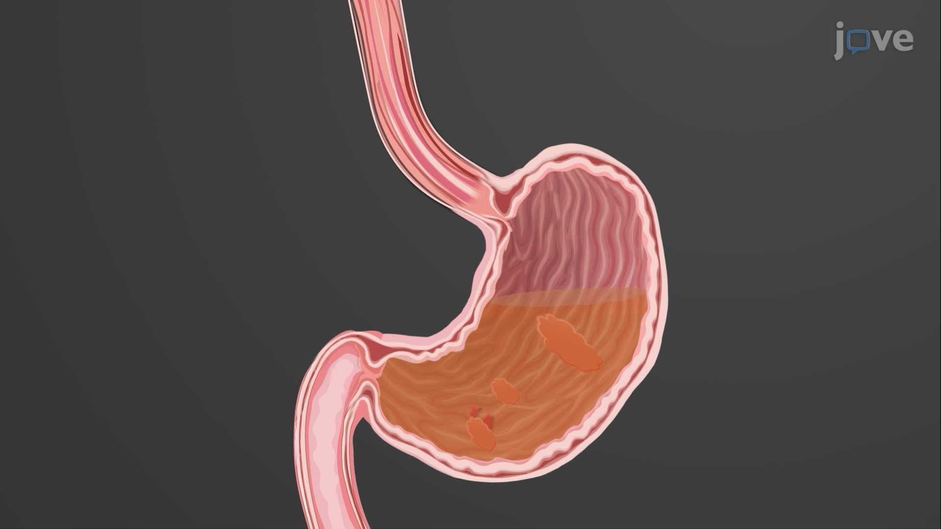

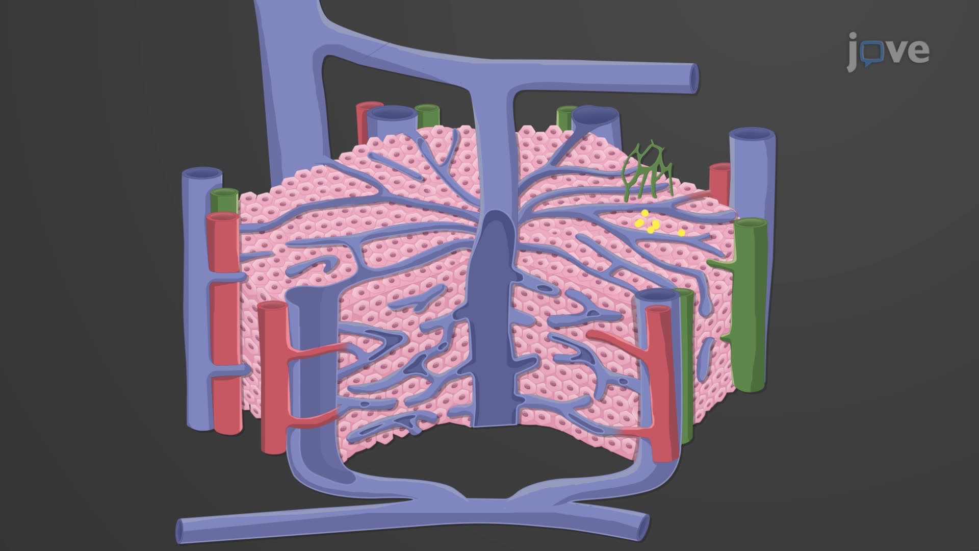
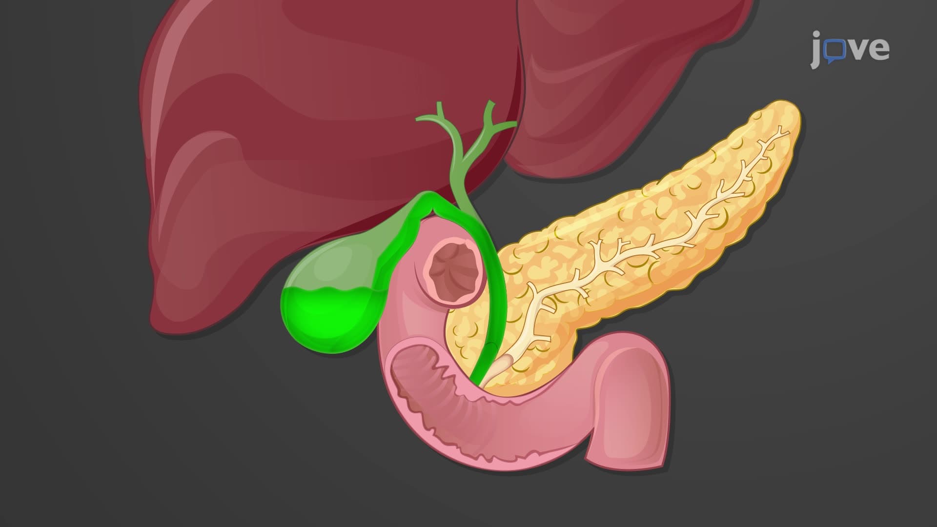
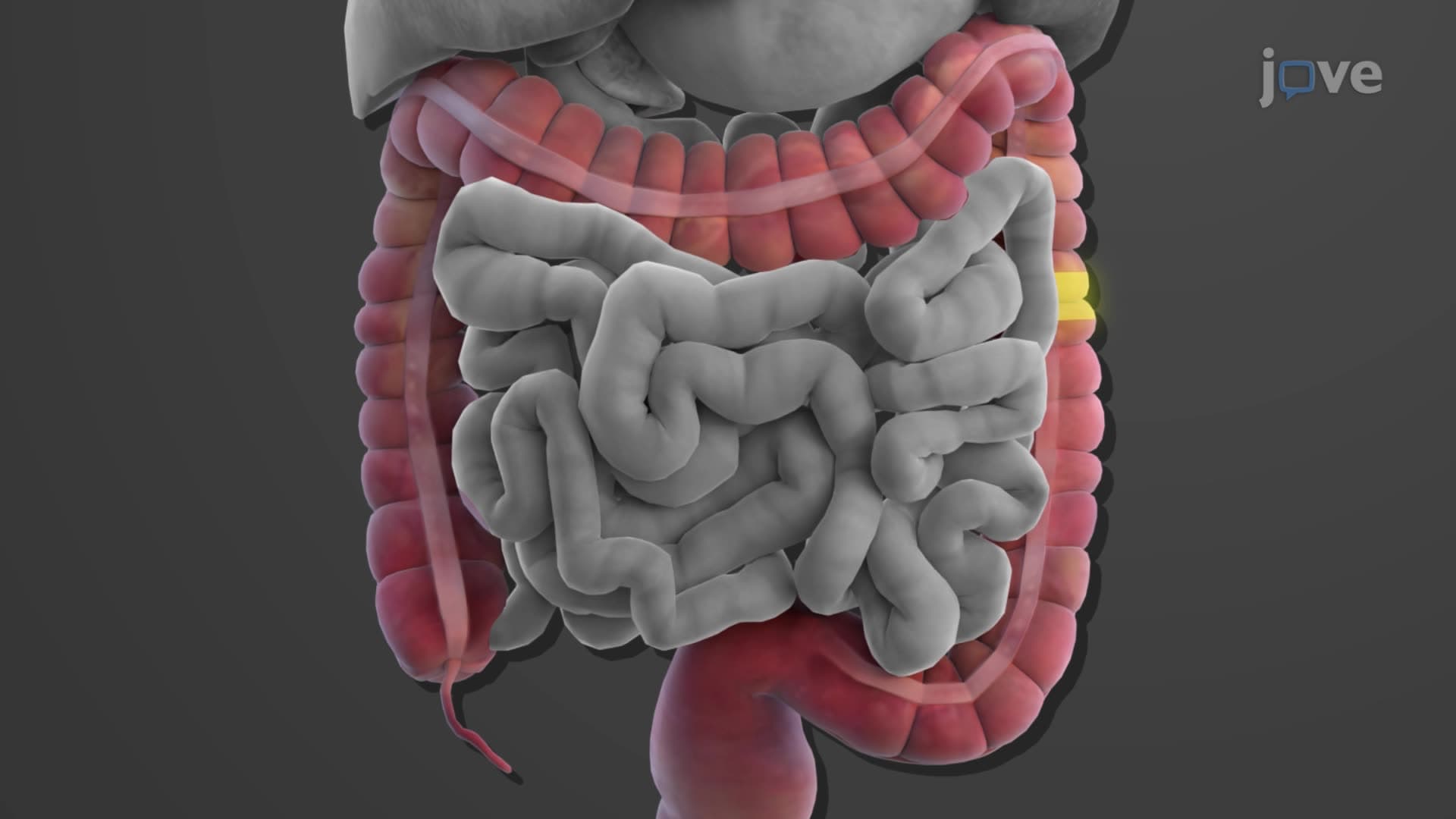
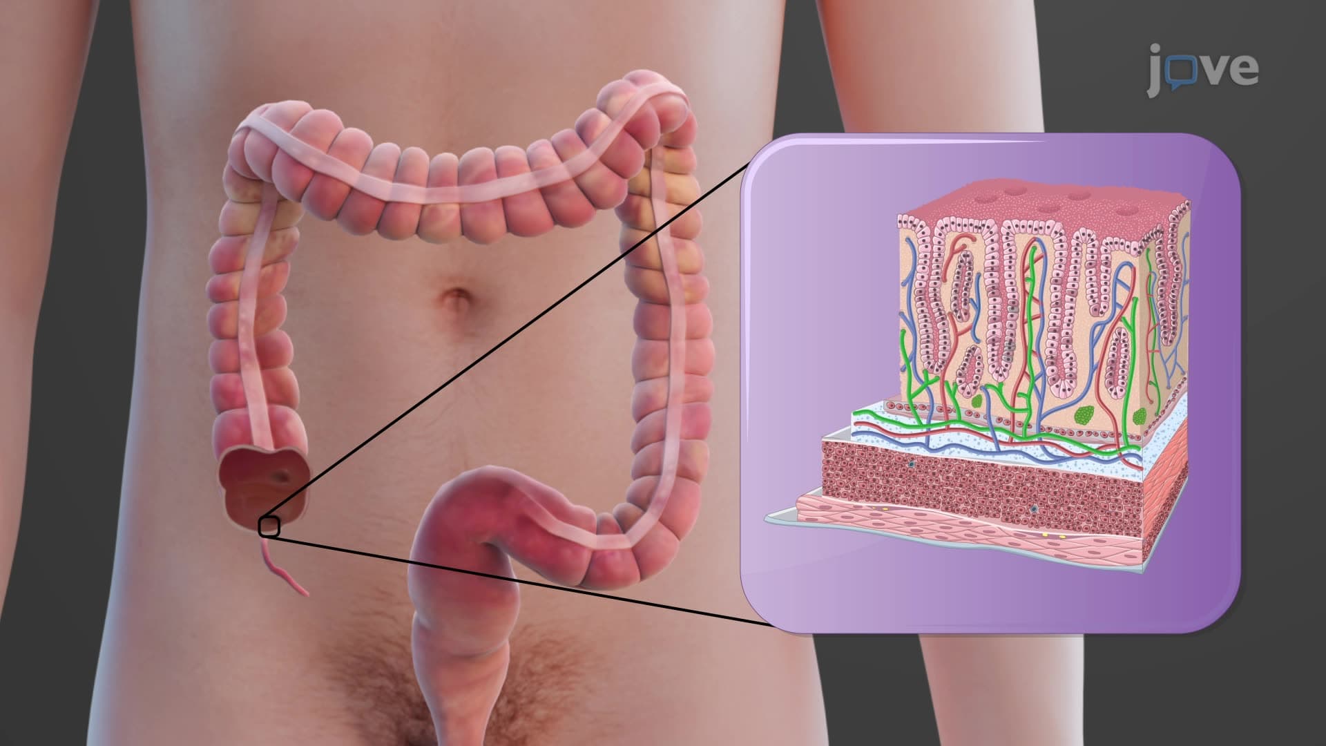
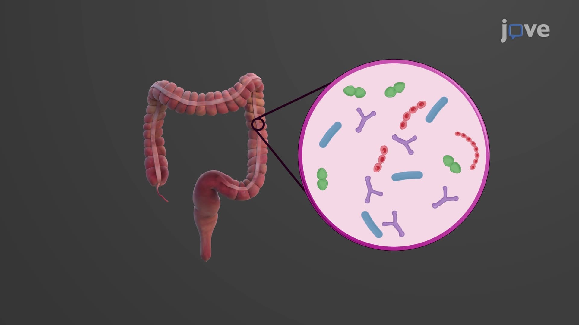
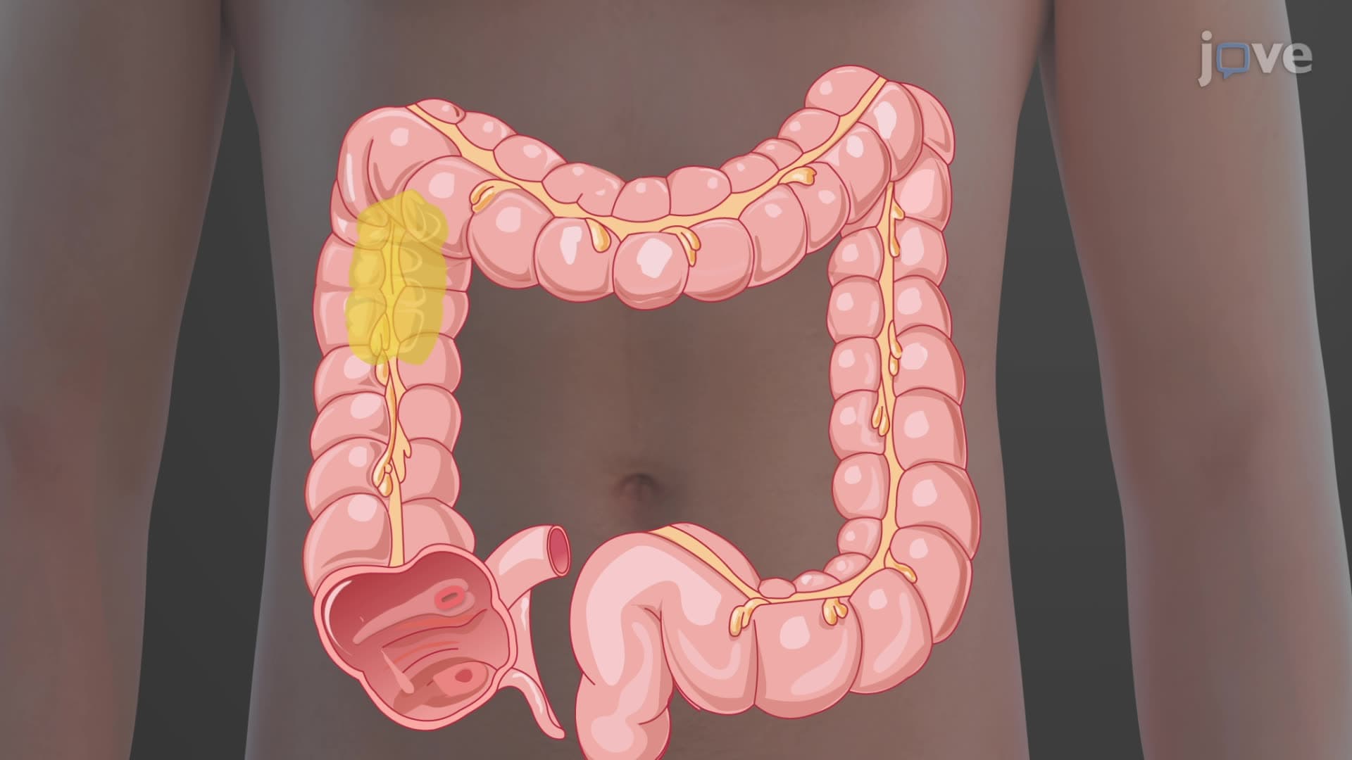
Blood Supply to the Digestive System

00:00
01:16
Video Summary: Blood Supply to the Digestive System Explained
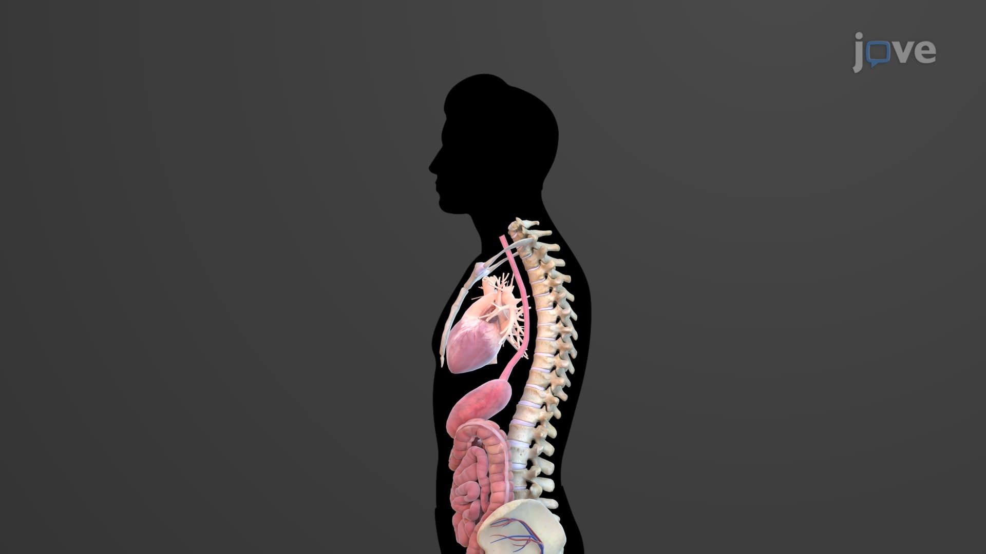
Ever wonder how your stomach gets the oxygen it needs during a Thanksgiving feast? The blood supply digestive system relies on a complex network called splanchnic circulation, featuring major arteries like the celiac trunk and mesenteric vessels that deliver oxygenated blood to every organ from your esophagus to your colon. This intricate vascular system ensures organs like the liver and pancreas receive adequate perfusion even during heavy metabolic demands. Watch the full video on JoVE Coach to master this concept with expert-led visuals and step-by-step explanations.
- Understand the Blood Supply To The Digestive System Explained through the splanchnic circulation and its three major arterial branches
- Identify the celiac trunk's three main branches (left gastric, splenic, and common hepatic arteries) and their target organs
- Analyze how the superior mesenteric artery supplies the small intestine and proximal large intestine structures
- Compare the drainage patterns of the hepatic portal system versus systemic venous return
- Evaluate the functional significance of the inferior mesenteric artery in supplying the distal colon
- Apply knowledge of venous drainage patterns, including portal and systemic circulation differences
- Recognize clinical implications of vascular anatomy in digestive system pathology
- Explore the relationship between arterial supply and venous drainage in maintaining GI tract homeostasis
Understanding Blood Supply To The Digestive System Explained
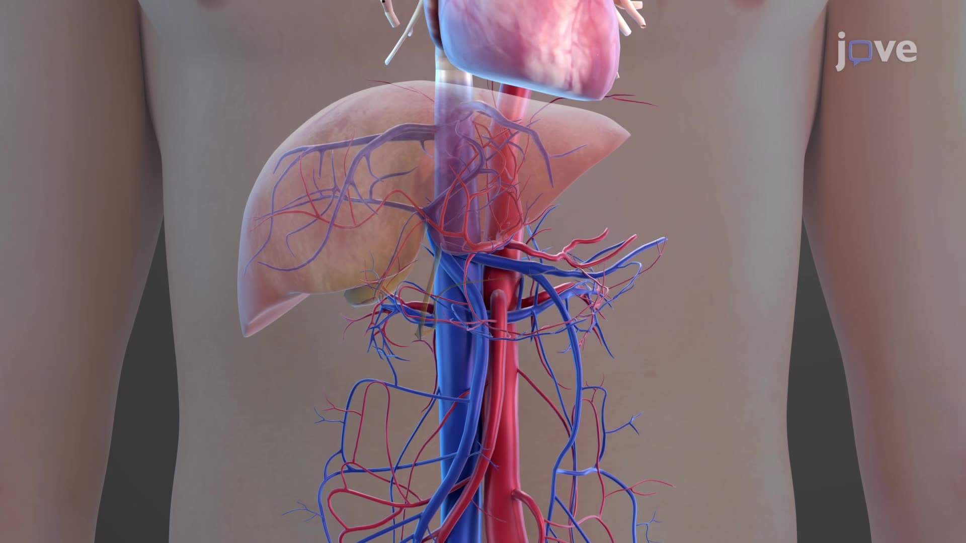
The digestive system's blood supply represents one of the most metabolically demanding circulatory networks in the human body. The GI blood supply anatomy explained centers on splanchnic circulation, which divides digestive organs into three distinct vascular territories. This organization reflects evolutionary efficiency, ensuring each region receives adequate perfusion while maintaining functional independence.
The Celiac Artery System and Upper GI Perfusion
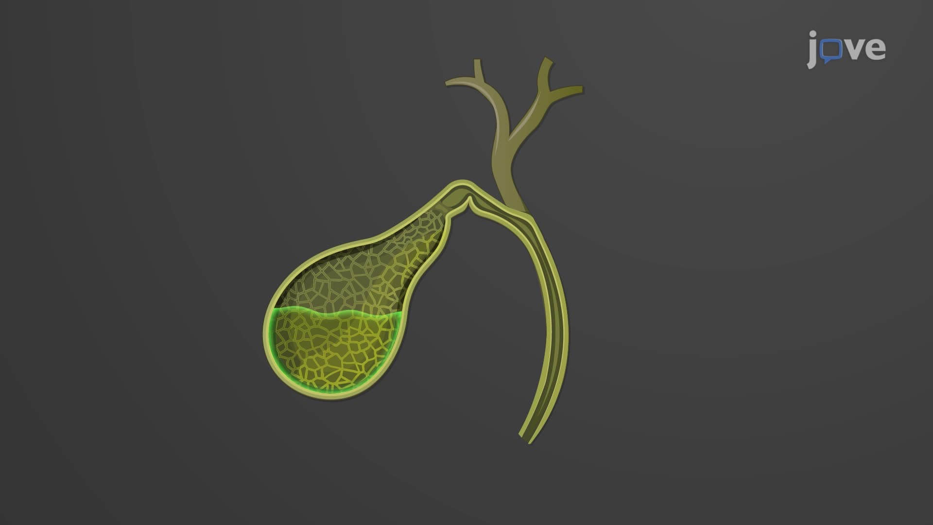
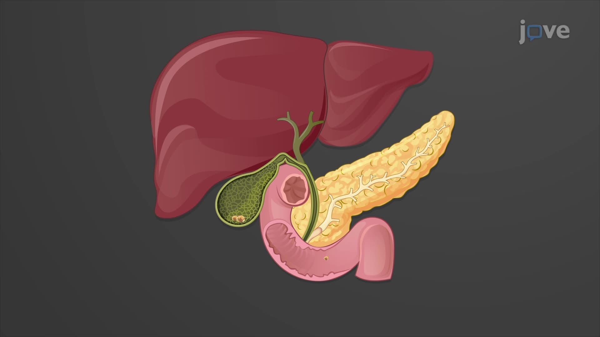
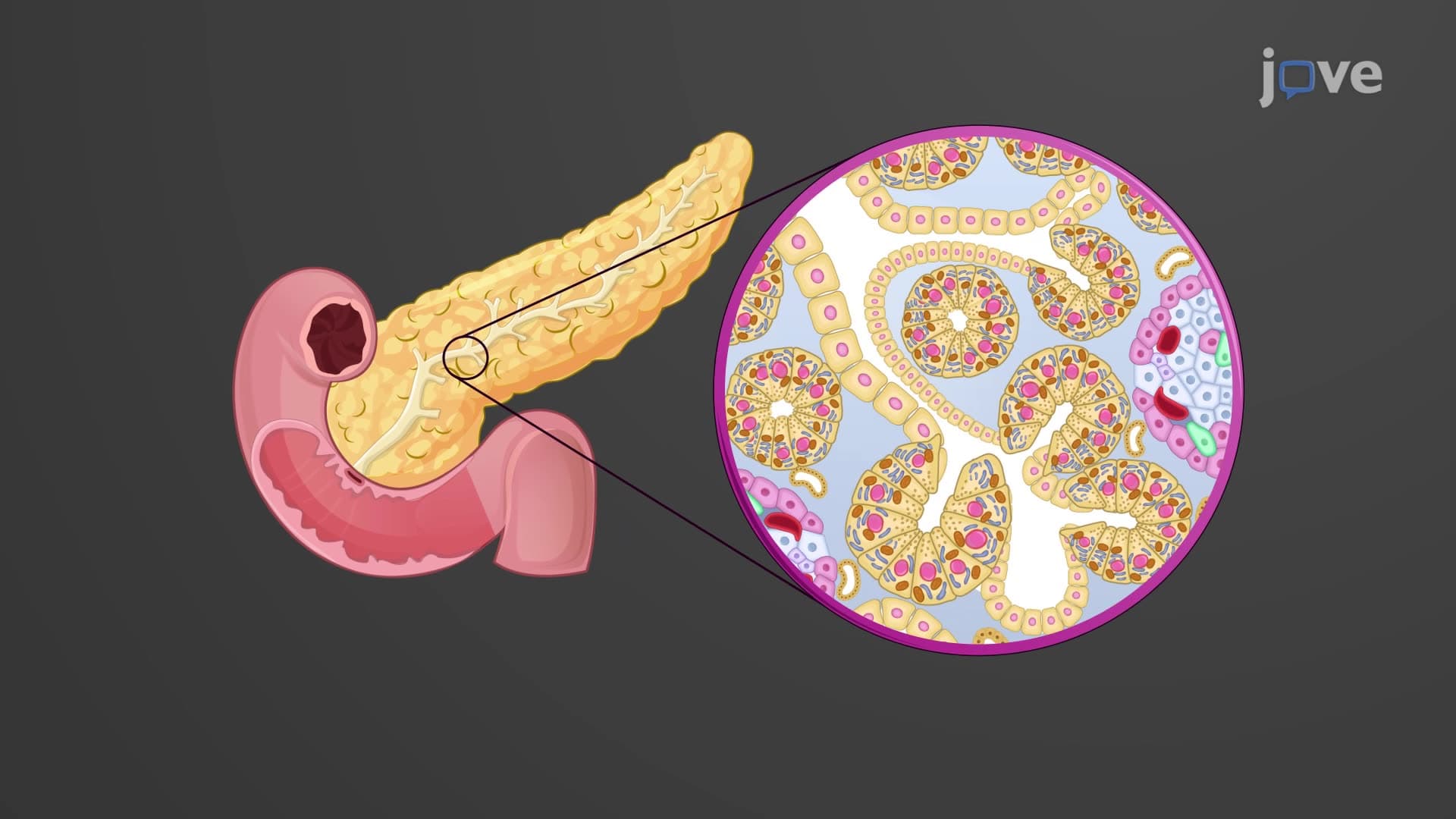
The celiac artery GI supply serves as the primary vascular highway for upper digestive organs. Originating at the T12 vertebral level, the celiac trunk immediately trifurcates into three essential branches. The left gastric artery supplies the esophagus and lesser curvature of the stomach, while the splenic artery provides blood to the spleen, pancreas, and stomach's greater curvature. The common hepatic artery branches further, supplying the liver, gallbladder, and duodenum through the gastroduodenal artery.
This arrangement proves clinically significant in conditions like peptic ulcer disease, where bleeding often involves these arterial territories. Medical students preparing for the MCAT or USMLE frequently encounter questions about celiac trunk pathology, particularly in cases of median arcuate ligament syndrome affecting celiac artery compression.
Superior and Inferior Mesenteric Circulation
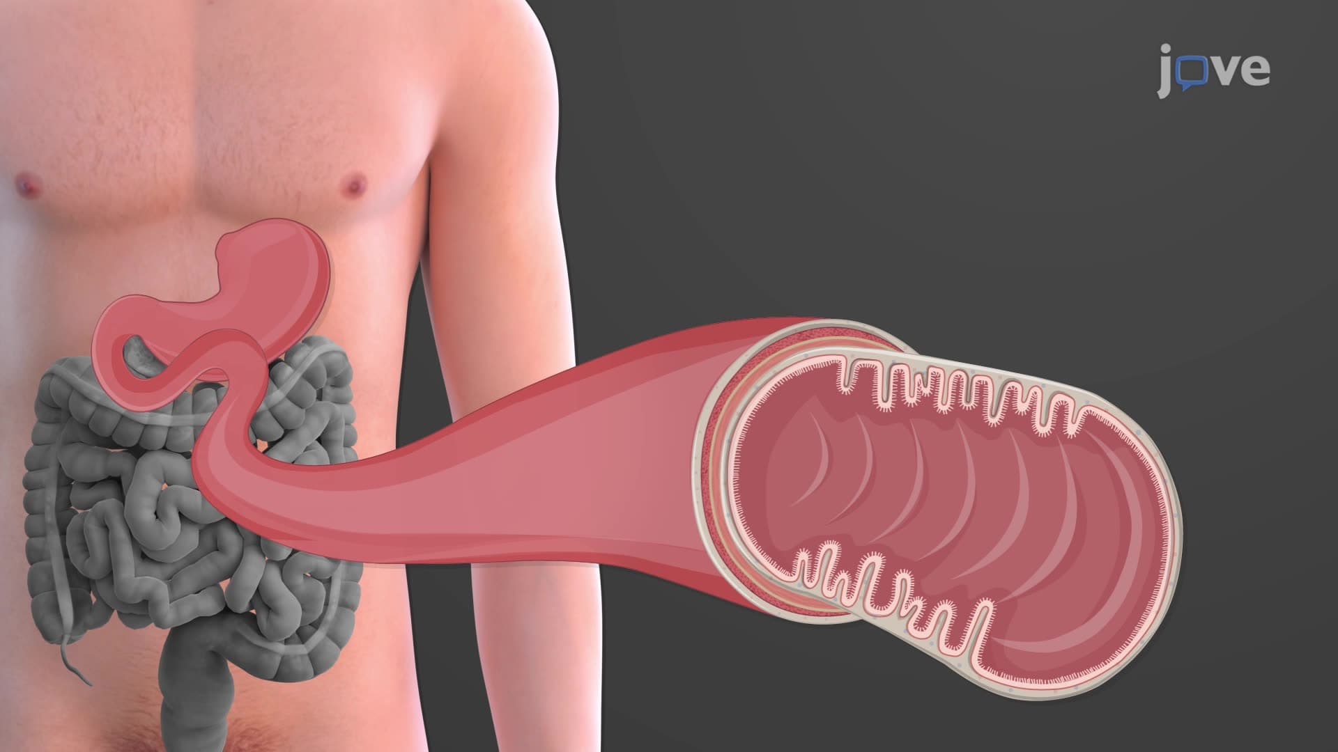
The superior inferior mesenteric arterial system demonstrates remarkable anatomical precision. The superior mesenteric artery (SMA) emerges at L1, supplying the entire small intestine through jejunal and ileal branches, plus the ascending colon and proximal transverse colon via the ileocolic and right colic arteries. This vessel's extensive territory makes it vulnerable to embolic events, creating medical emergencies requiring immediate intervention.
The inferior mesenteric artery (IMA) serves the remaining large intestine through left colic, sigmoid, and superior rectal arteries. Understanding this distribution proves essential for AP Biology students studying digestive physiology and college anatomy courses covering abdominal vascularization.
Portal Venous System and Hepatic Circulation
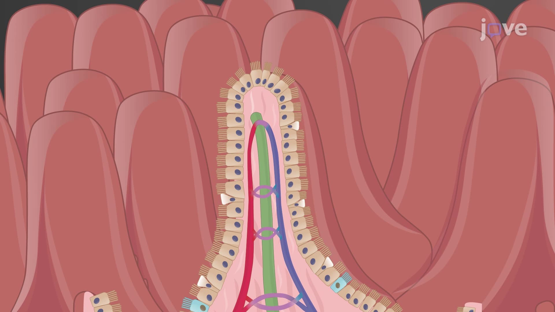
Hepatic portal blood flow represents a unique circulatory arrangement where venous blood from digestive organs passes through liver sinusoids before returning to systemic circulation. The superior mesenteric vein joins the splenic vein, forming the hepatic portal vein that processes nutrient-rich blood from intestinal absorption. This GI venous portal drainage system allows the liver to metabolize absorbed nutrients, detoxify substances, and regulate glucose homeostasis before blood reaches systemic circulation.
Frequently Asked Questions
The blood supply to the digestive system operates through splanchnic circulation, featuring three major arterial systems: the celiac trunk, superior mesenteric artery, and inferior mesenteric artery. These vessels deliver oxygenated blood to all GI organs, while venous drainage occurs through both portal and systemic circulation pathways. This dual system ensures efficient nutrient processing through the liver while maintaining adequate organ perfusion.
MCAT and USMLE questions typically focus on arterial territories, portal circulation pathways, and clinical correlations with GI pathology. Students must identify celiac trunk branches, understand mesenteric artery distribution patterns, and recognize portal versus systemic venous drainage. Practice questions often involve abdominal pain scenarios requiring vascular anatomy knowledge for diagnosis.
AP Biology exams emphasize the relationship between blood supply and digestive function, particularly nutrient absorption and transport. Students should understand how portal circulation supports liver metabolism, arterial supply meets metabolic demands during digestion, and vascular regulation affects digestive efficiency. Questions often connect blood flow to homeostatic mechanisms.
Mesenteric ischemia affects approximately 60,000-70,000 Americans annually, often involving superior mesenteric artery occlusion. Portal hypertension from liver disease creates complications like esophageal varices, while celiac artery compression syndrome causes chronic abdominal pain. Understanding vascular anatomy helps medical professionals diagnose these conditions through imaging and clinical correlation.
Basic knowledge of cardiovascular anatomy and digestive organ location provides sufficient foundation for understanding GI blood supply. High school biology students can grasp these concepts by focusing on major vessel names, general distribution patterns, and functional relationships. Visual aids and systematic organization help students master this material without overwhelming detail.
Create systematic charts organizing arteries by territory (celiac = upper GI, SMA = small bowel + right colon, IMA = left colon), use mnemonics for celiac trunk branches, and practice with unlabeled diagrams. Focus on clinical correlations and pathology examples to reinforce anatomical relationships. Spaced repetition with active recall proves more effective than passive reading.
Advanced students should investigate collateral circulation pathways, autonomic regulation of splanchnic blood flow, and pathophysiology of portal hypertension. Explore imaging techniques like CT angiography for vascular assessment, surgical considerations in GI procedures, and pharmacological agents affecting digestive system circulation. These topics prepare students for advanced medical coursework and clinical applications.
Related Micro-courses
25 Concepts

25 Concepts

20 Concepts