Loading subject page…

Micro-course: Digestive System
89,747 views
Learn key concepts and earn your certificate.


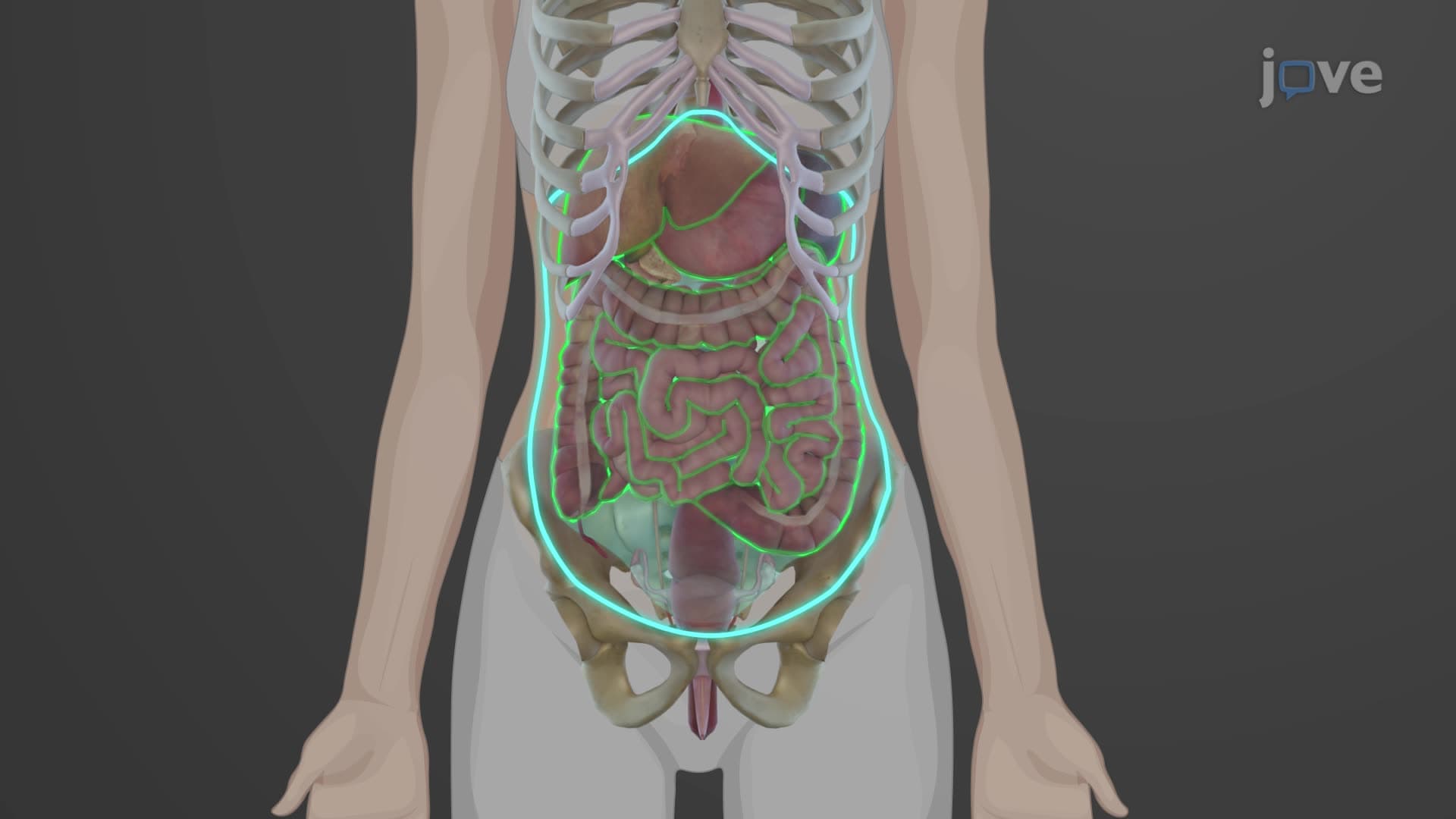
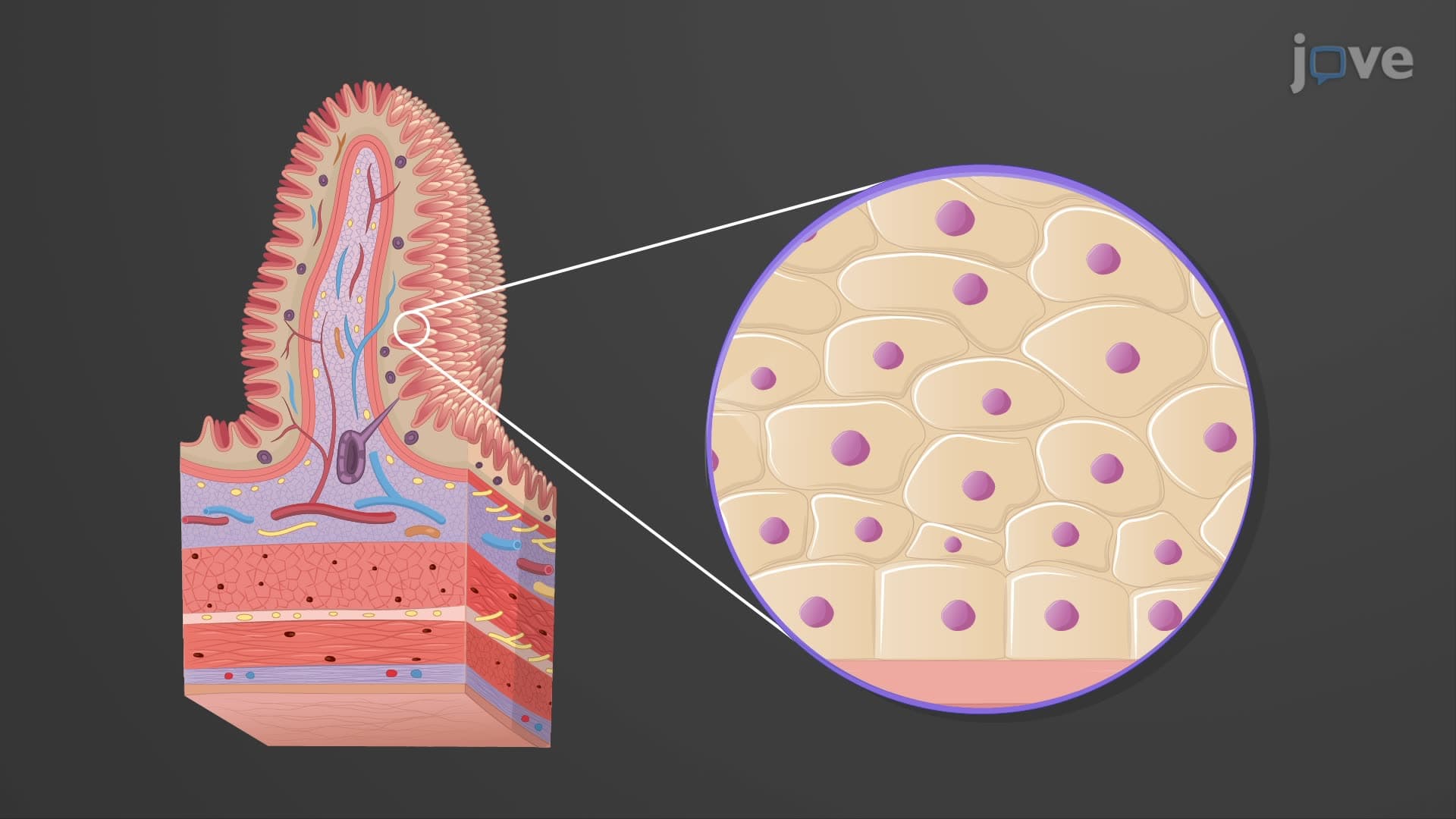
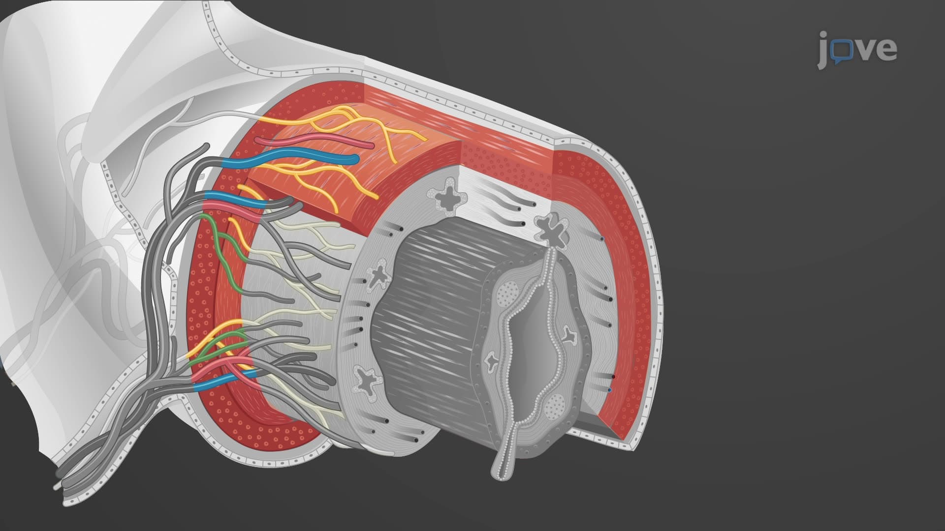

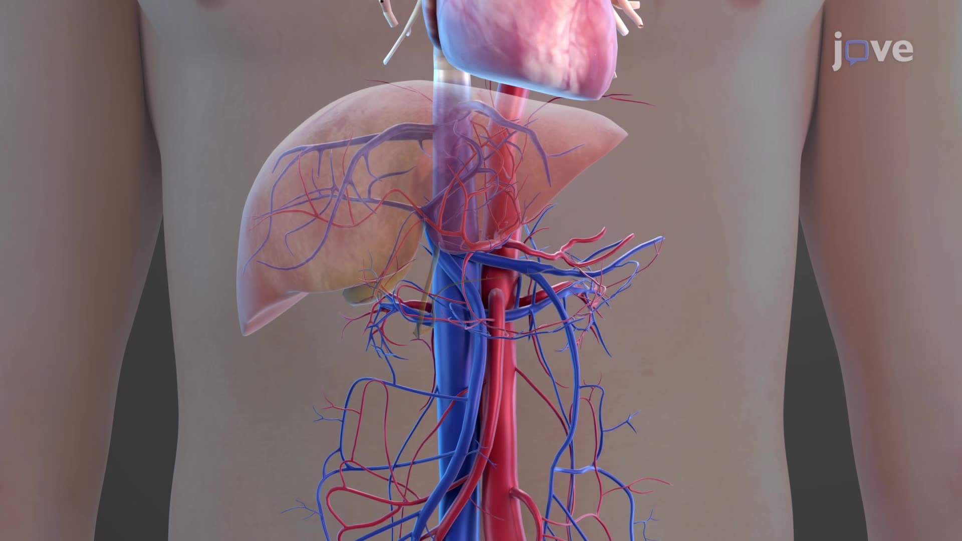

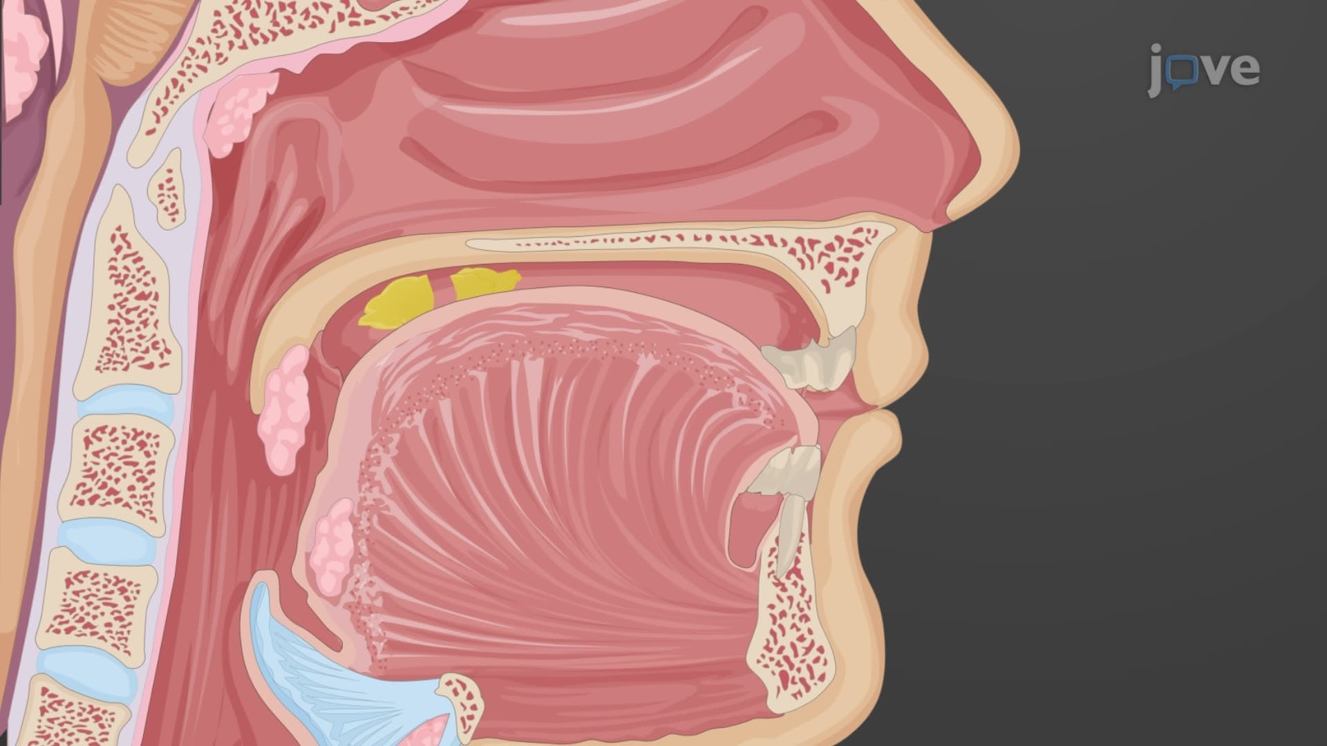
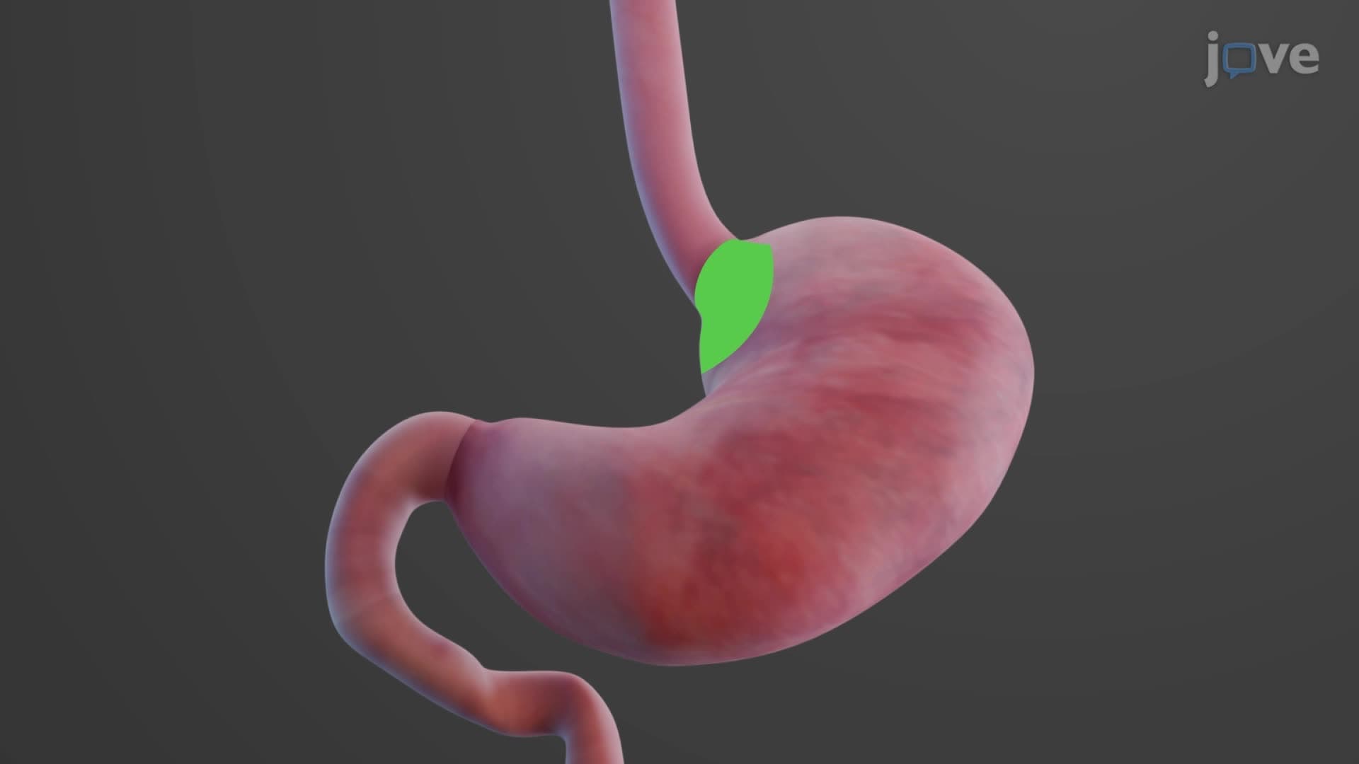
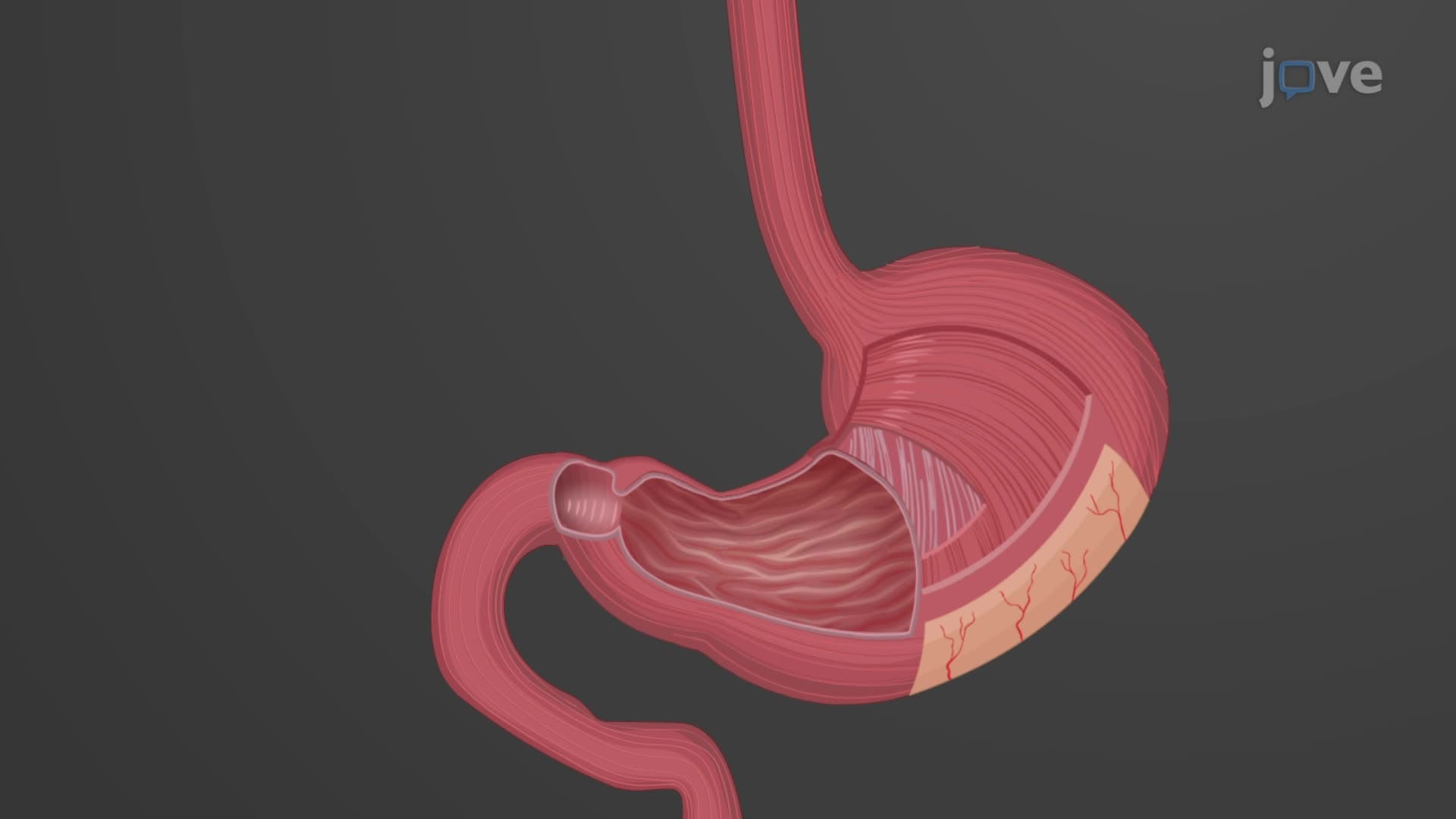
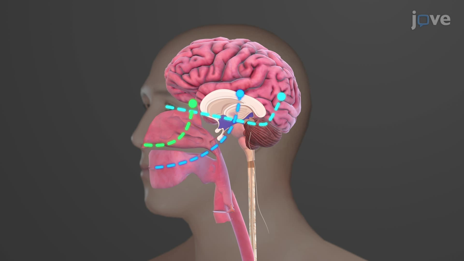
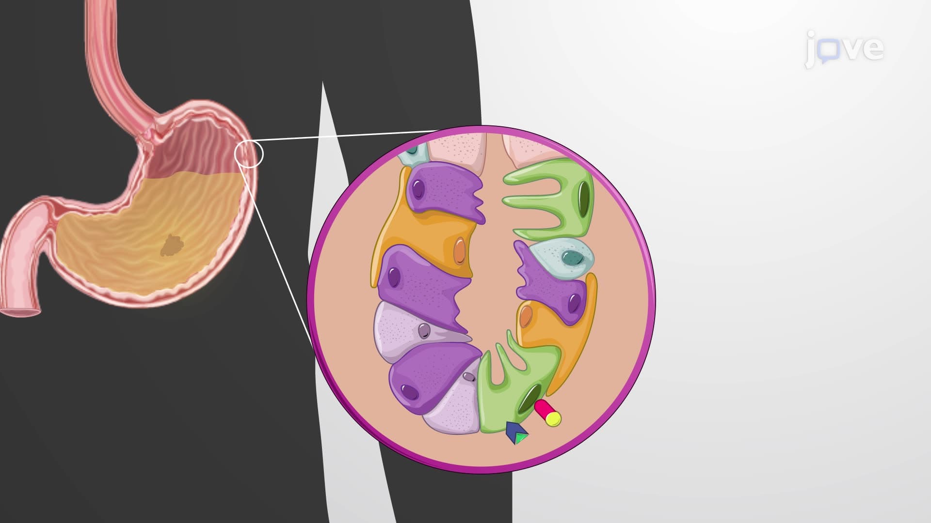
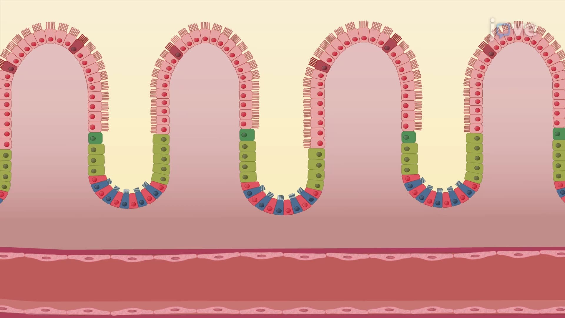
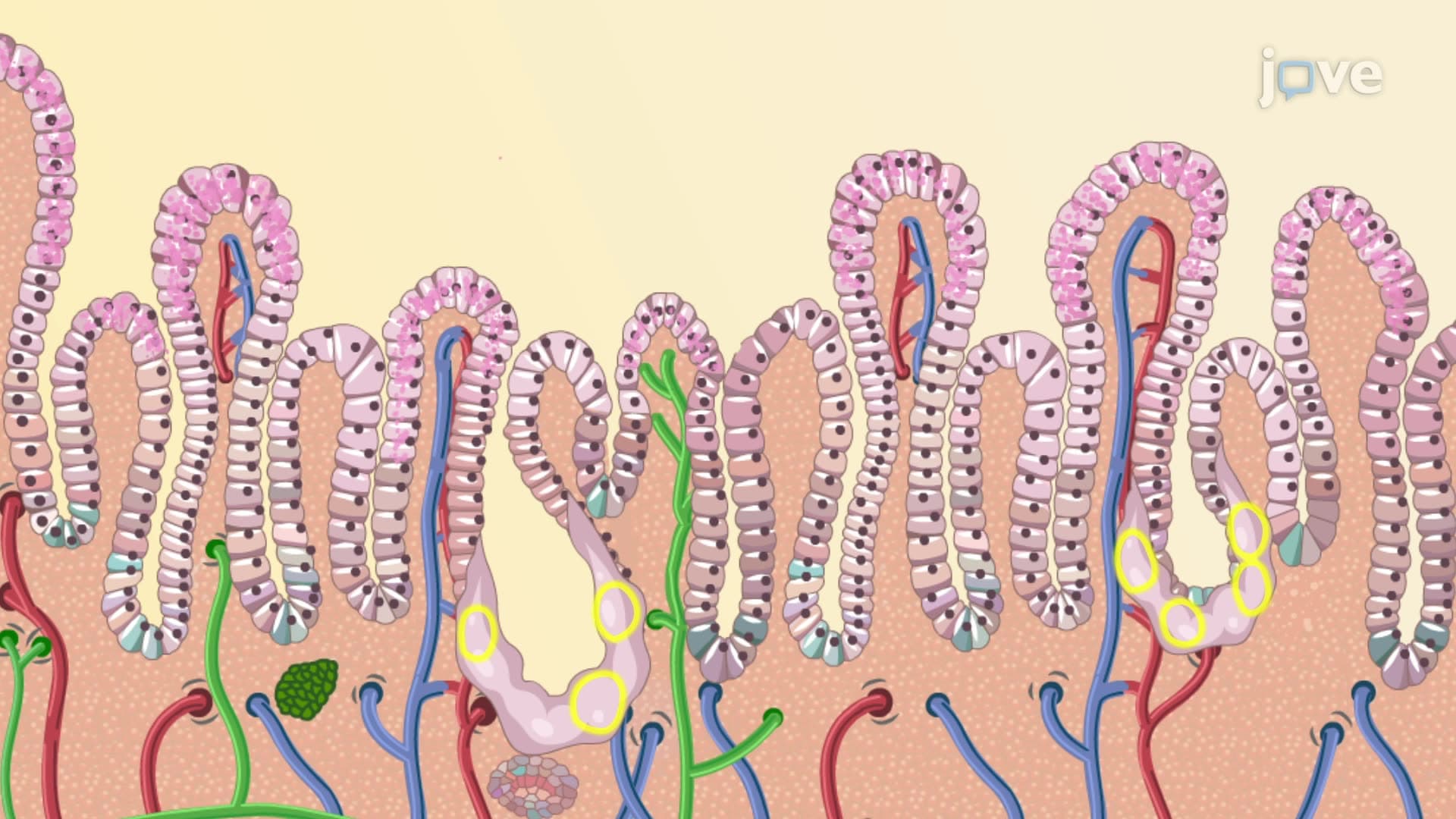
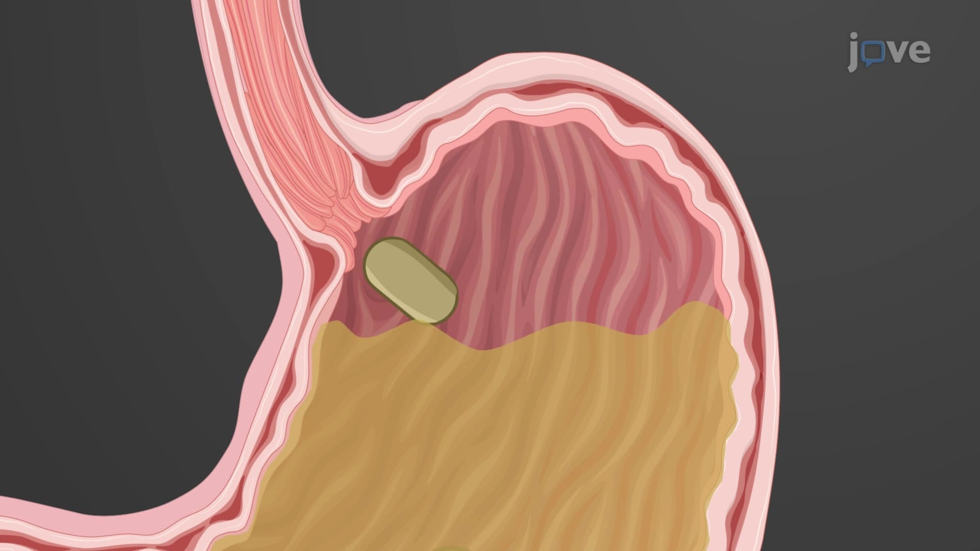
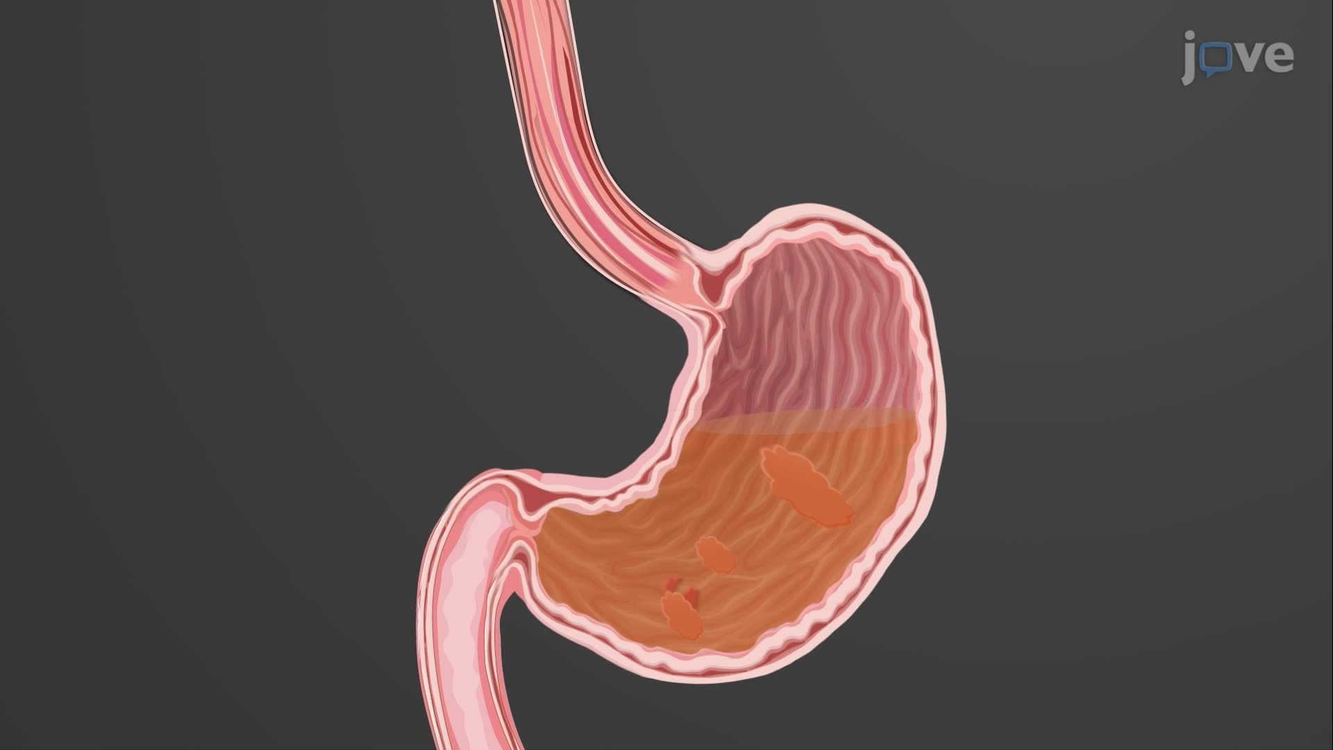

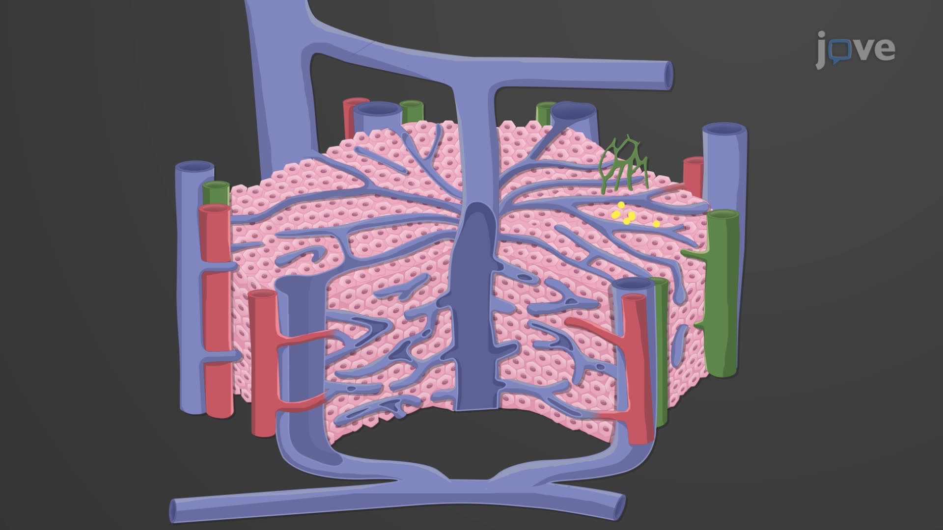
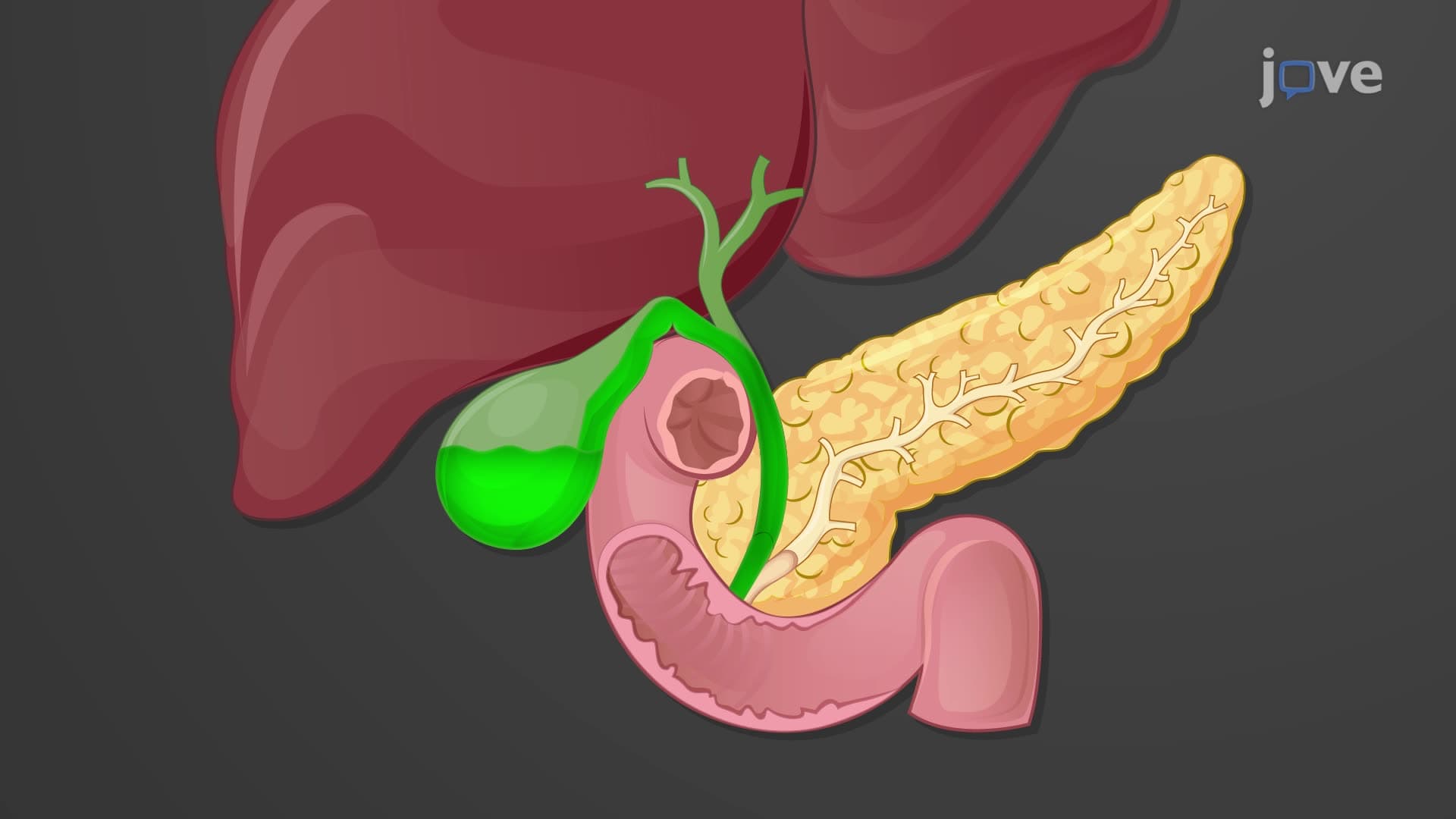
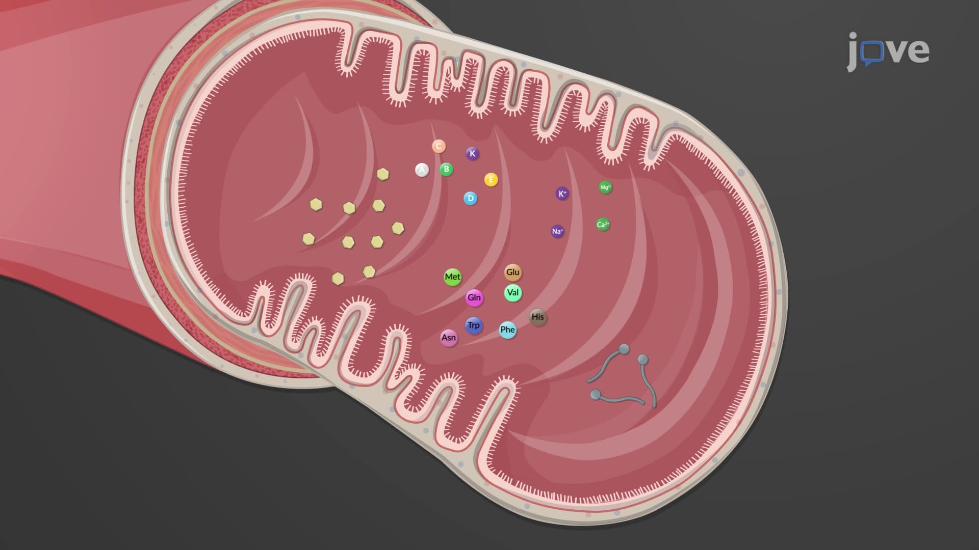
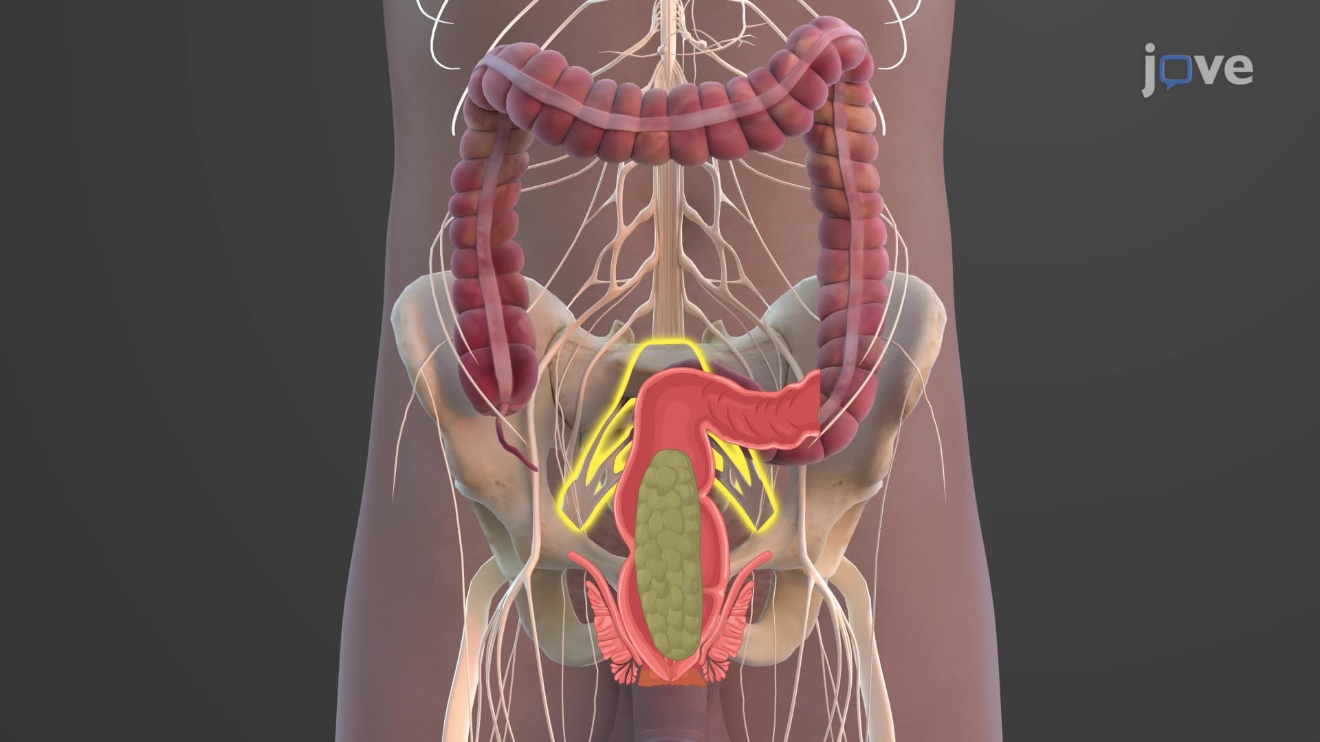
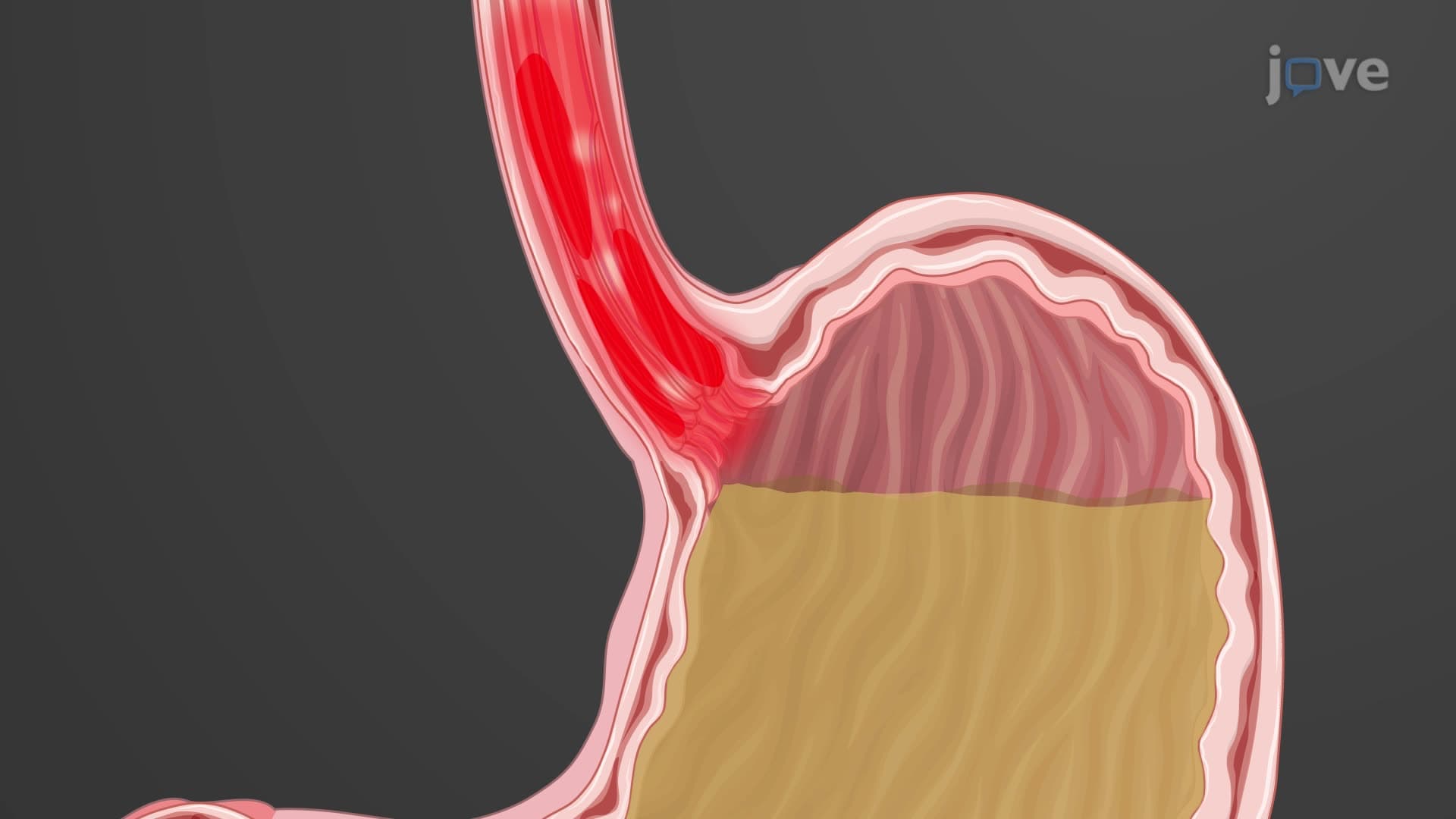
Esophagus

00:00
01:24
Video Summary: What Is Esophagus
Ever wonder how a single gulp of water travels from your mouth to your stomach in just 6-10 seconds? The esophagus anatomy biology reveals this fascinating muscular tube that makes swallowing possible through coordinated contractions. When a Johns Hopkins Medical School student studies digestive physiology, they learn how this 25-centimeter conduit uses specialized sphincters and layered muscle tissue to transport everything from pizza to medicine safely into the stomach. Watch the full video on JoVE Coach to master this concept with expert-led visuals and step-by-step explanations.
- Understand what is esophagus structure and its role as a muscular conduit connecting the pharynx to the stomach
- Identify the four distinct layers of esophageal wall structure and their specific functions in food transport
- Analyze how the esophageal sphincter anatomy prevents gastric acid reflux while allowing food passage
- Compare skeletal and smooth muscle distribution throughout the esophagus location anatomy
- Evaluate the protective mechanisms of nonkeratinized stratified squamous epithelium in the esophageal lining
- Apply knowledge of peristalsis esophagus function to explain coordinated swallowing mechanics
- Recognize clinical significance of gastroesophageal junction anatomy in digestive health
- Explore how esophageal glands contribute to successful food bolus transport through mucus production
What is Esophagus: The Body's Food Highway
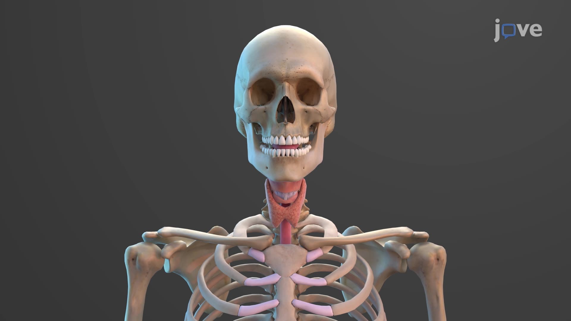
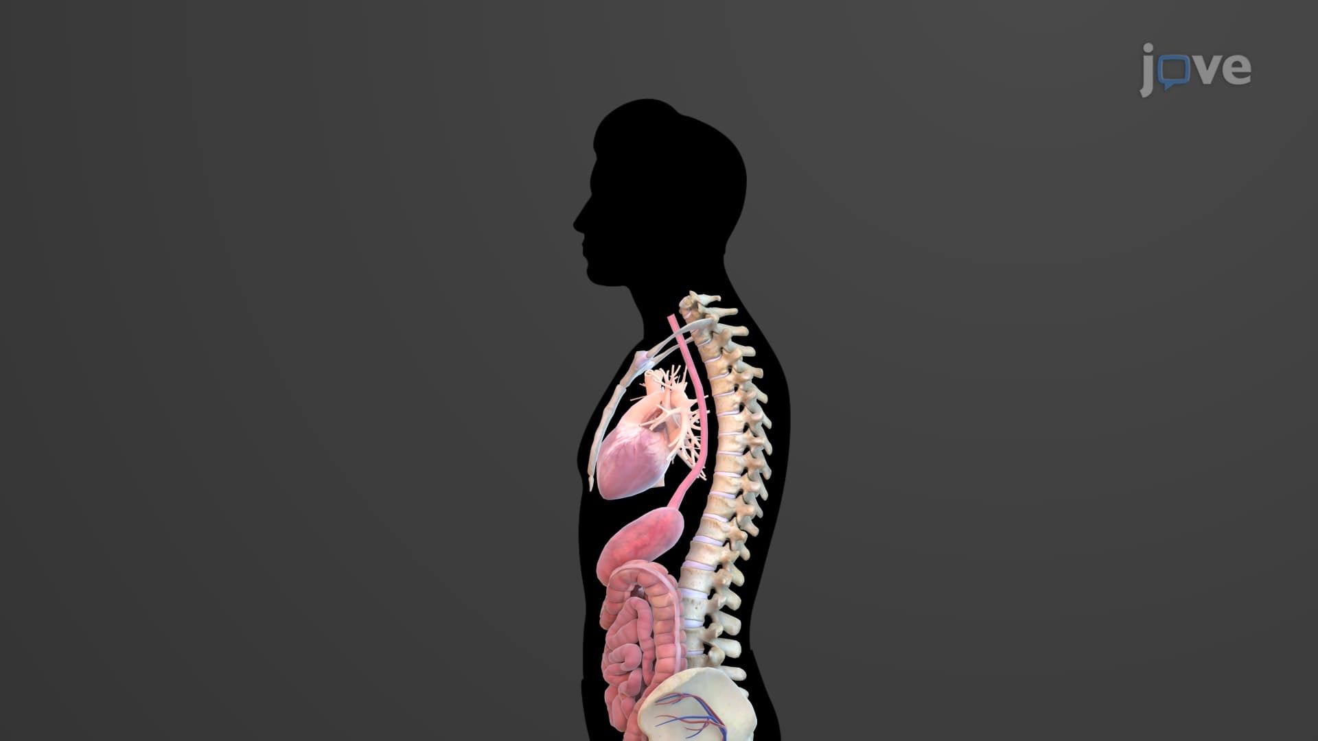
The esophagus serves as the body's primary food tube esophagus explained through its remarkable 25-centimeter muscular design. This collapsible conduit runs from the pharynx through the mediastinum, piercing the diaphragm at the esophageal hiatus before connecting to the stomach. Unlike rigid pipes, the esophagus remains flattened when empty, expanding only during active swallowing-a design that maximizes thoracic space while maintaining transport efficiency.
Medical students at universities like Stanford and Harvard learn that what is the esophagus in digestive anatomy extends beyond simple food transport. This organ demonstrates sophisticated neural control, coordinating with the central nervous system to execute precise swallowing sequences that prevent aspiration and ensure efficient gastric delivery.
Esophageal Wall Architecture and Function
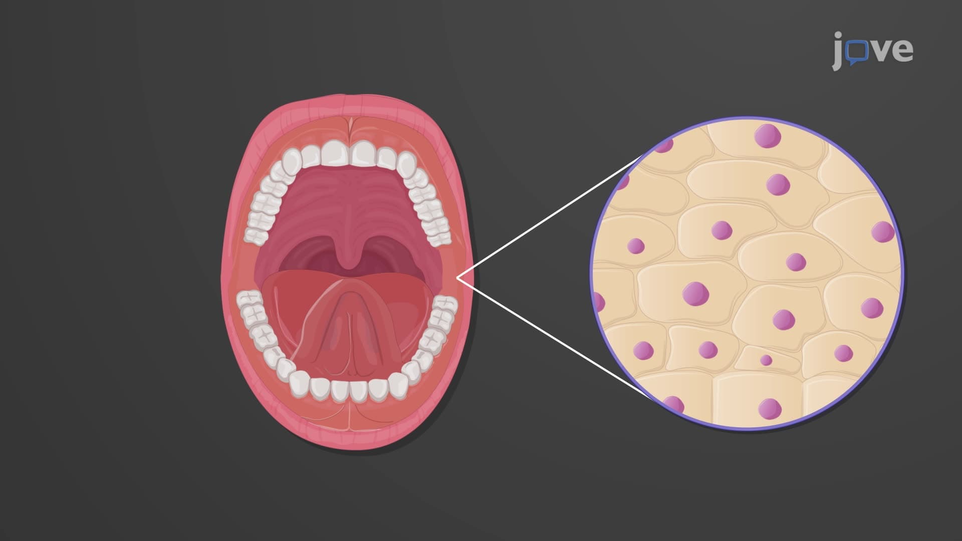
The esophageal wall structure comprises four distinct layers, each serving specialized functions. The mucosa features nonkeratinized stratified squamous epithelium-the same protective tissue found in the oral cavity-providing resistance against mechanical abrasion from food boluses. This epithelial choice reflects evolutionary adaptation to the esophagus's dual exposure to both food particles and potential gastric acid exposure.
The submucosa houses esophageal glands that secrete lubricating mucus, facilitating smooth food passage. These glands become clinically significant in conditions like Barrett's esophagus, frequently discussed in MCAT preparation courses and gastroenterology rotations at institutions like Mayo Clinic and Cleveland Clinic.
Muscular Control and Sphincter Dynamics
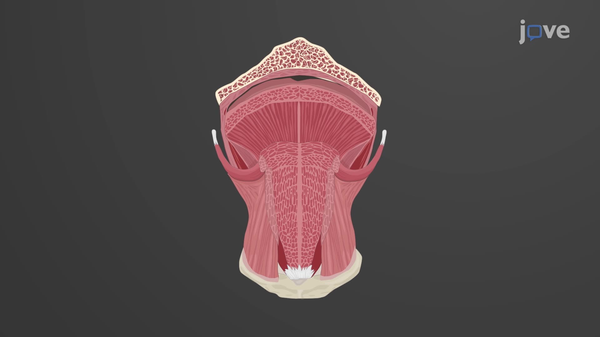
Peristalsis esophagus function relies on the muscularis externa's unique skeletal-to-smooth muscle transition. The upper third contains voluntary skeletal muscle, allowing conscious initiation of swallowing, while the lower two-thirds feature involuntary smooth muscle that continues peristaltic waves automatically.
The esophageal sphincter anatomy includes two critical structures: the upper esophageal sphincter (cricopharyngeus muscle) and the lower esophageal sphincter (gastroesophageal sphincter). The lower sphincter's function becomes particularly relevant in AP Biology courses when studying digestive disorders like gastroesophageal reflux disease (GERD), which affects over 20% of Americans according to the American College of Gastroenterology.
Clinical Applications in Medical Education
Understanding esophagus location anatomy proves essential for nursing students preparing for NCLEX examinations and medical students facing USMLE Step 1. The esophagus's relationship to surrounding structures-including the trachea, aortic arch, and left atrium-becomes critical knowledge for interpreting chest imaging and understanding potential surgical complications.
Frequently Asked Questions
The esophagus is a 25-centimeter muscular tube that transports food from the mouth to the stomach through coordinated muscle contractions called peristalsis. It's essential because it prevents food from entering the respiratory system while ensuring efficient delivery to the stomach for digestion. The esophagus also contains protective mechanisms that prevent stomach acid from damaging the throat and lungs.
MCAT questions often test esophageal sphincter function, peristalsis mechanisms, and tissue layer composition in digestive physiology passages. AP Biology exams typically include esophageal structure in broader digestive system questions, focusing on how muscular contractions coordinate food transport. Students should understand both the anatomical layers and physiological processes for comprehensive exam preparation.
HESI A2 and TEAS exams commonly test basic esophageal function, including peristalsis, sphincter locations, and the esophagus's role in preventing aspiration. Questions often focus on practical applications like patient positioning during feeding and recognizing signs of swallowing difficulties. Understanding the esophagus's protective mechanisms helps answer patient care scenarios effectively.
Emergency departments across the US regularly treat esophageal foreign body obstructions, especially in children who swallow coins or small toys. EMTs and emergency physicians rely on understanding esophageal anatomy to perform safe removal procedures and recognize when surgical intervention becomes necessary. Knowledge of esophageal sphincter locations guides treatment decisions and prevents complications.
Esophageal anatomy is actually quite manageable for high school students since it follows logical structural principles. The four-layer wall design mirrors other digestive organs, making it easier to remember through pattern recognition. Starting with basic tube structure and gradually adding sphincter and peristalsis concepts helps students build comprehensive understanding without overwhelming complexity.
Use the mnemonic "My Stomach Makes Food" for Mucosa, Submucosa, Muscularis externa, and Fibrous adventitia from inside to outside. Create visual associations: mucosa = protective lining, submucosa = gland layer for lubrication, muscularis = muscle for movement, adventitia = outer connective coating. Practice drawing cross-sections while explaining each layer's function to reinforce both structure and purpose.
Progress to gastric anatomy and physiology, focusing on how the stomach receives and processes food delivered by esophageal peristalsis. Study gastroesophageal junction disorders like GERD and hiatal hernias to understand clinical applications. Advanced students should explore neural control of swallowing and compare esophageal histology with other alimentary canal segments.
College exams often use microscopic images requiring students to identify esophageal tissue layers and distinguish them from similar structures like the trachea or stomach. Practical assessments may include cadaveric identification of esophageal landmarks and relationships to surrounding organs. Many courses integrate clinical correlations, asking students to predict consequences of esophageal dysfunction.
Related Micro-courses
25 Concepts

25 Concepts

20 Concepts