Loading subject page…


89,749 views


Video Summary: What Is Gross Anatomy Liver
Did you know the liver performs over 500 vital functions, making it your body's ultimate multitasker? Liver gross anatomy biology reveals how this 3-pound powerhouse orchestrates everything from detoxification to blood sugar regulation. When studying what is gross anatomy liver structure, students discover why liver transplants at centers like Johns Hopkins require such precise anatomical knowledge-surgeons must navigate complex lobar divisions and vascular networks. Watch the full video on JoVE Coach to master this concept with expert-led visuals and step-by-step explanations.
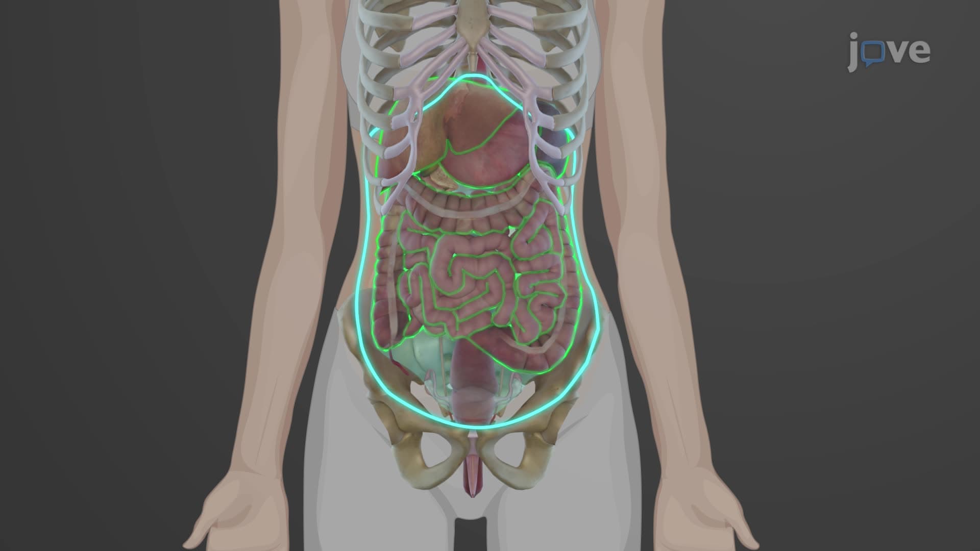
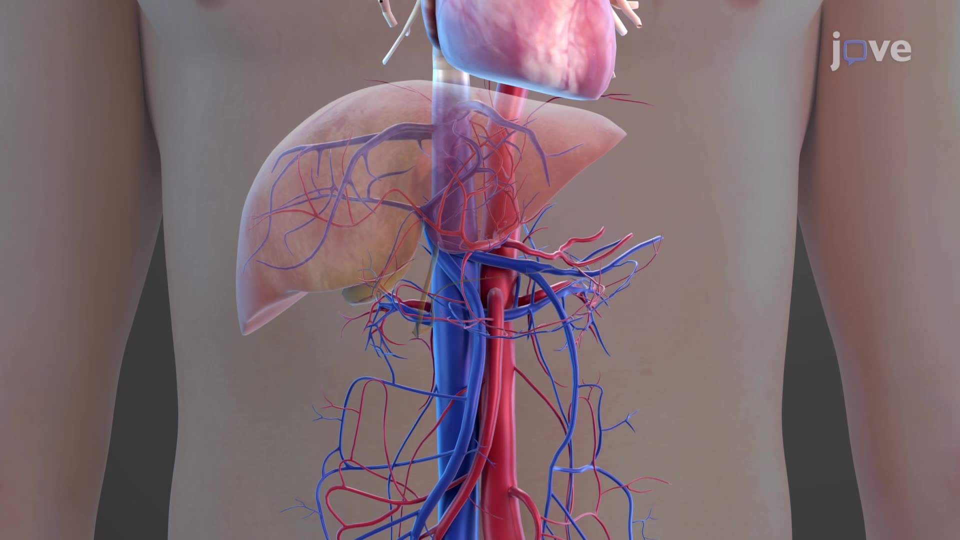
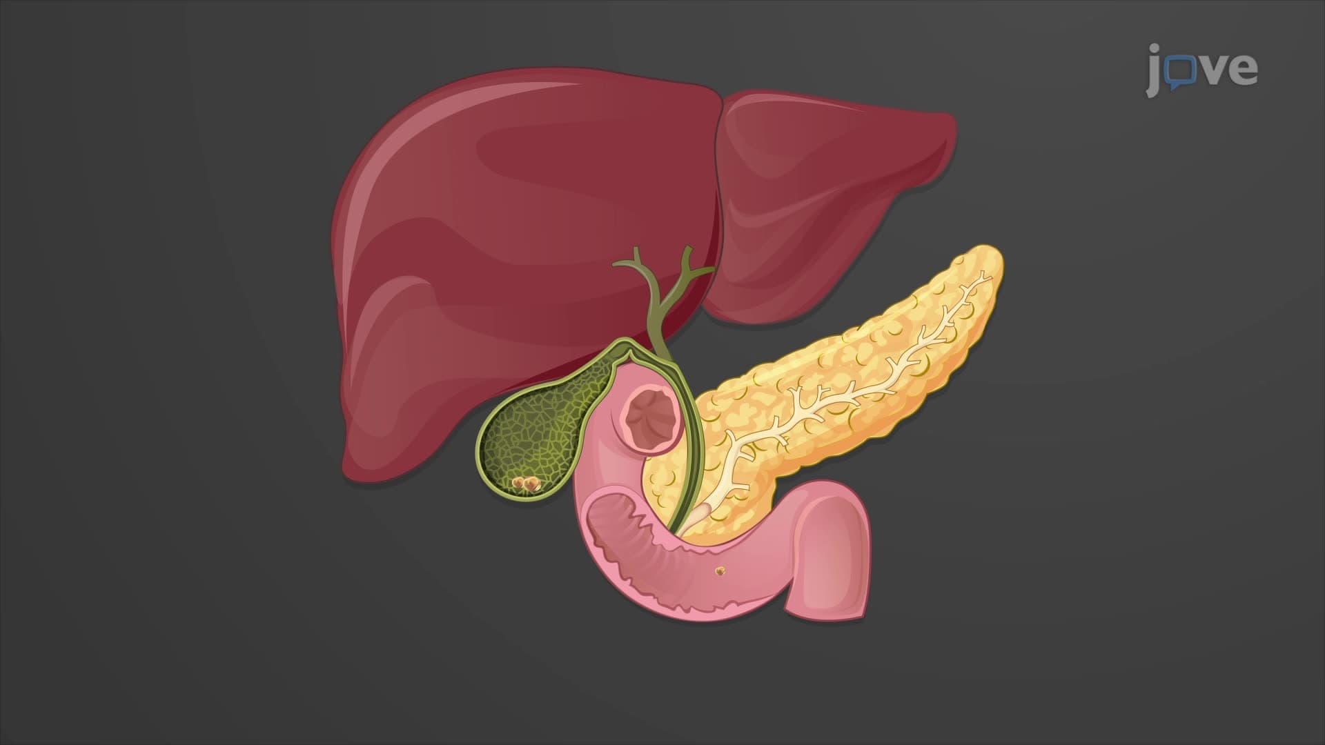
The liver gross anatomy biology represents one of medicine's most intricate architectural marvels. Unlike other organs with simple geometries, the liver's wedge-shaped design maximizes surface area while fitting perfectly beneath the protective dome of the diaphragm. This positioning isn't accidental-the liver's location in the right hypochondriac and epigastric regions provides optimal access to incoming nutrients from the digestive system while maintaining protection from external trauma.
The liver anatomy overview explained through its four-lobe system reveals surgical and diagnostic importance. The right lobe, significantly larger than its left counterpart, handles the majority of hepatic functions and commonly becomes the focus during liver resection procedures at major US medical centers like Mayo Clinic. The caudate and quadrate lobes, though smaller, possess independent blood supplies-a fact crucial for transplant surgeons when determining viable tissue sections.
The hepatic ligament falciform serves dual purposes: structural support and anatomical landmark. This mesenteric fold not only suspends the liver from the diaphragm but also carries the round ligament-a fibrous reminder of fetal development when the umbilical vein delivered oxygenated blood directly to the developing liver. Students preparing for the MCAT or AP Biology exams frequently encounter questions about this developmental transition from fetal to adult circulation patterns.
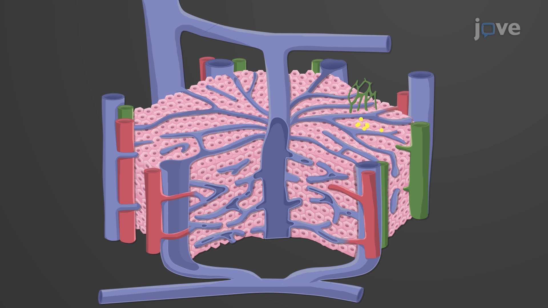
The porta hepatis liver region functions as the organ's central hub, where major vessels converge through the lesser omentum's connective tissue framework. This area becomes critically important during hepatobiliary surgery, as surgeons must navigate the hepatic artery, portal vein, and bile ducts-collectively known as the portal triad. Understanding this anatomy proves essential for students pursuing healthcare careers, particularly those targeting USMLE Step 1 success.
Related Micro-courses