Loading subject page…


89,753 views

Video Summary: What Is Histology Gastrointestinal Gi Tract
Did you know your digestive system processes over 2 gallons of fluid daily through four precisely organized tissue layers? GI tract histology biology reveals how the stomach, small intestine, and colon maintain this complex function through specialized cellular architecture. From the mucus-secreting cells protecting your stomach lining during a spicy meal at Chipotle to the absorption mechanisms in your small intestine, understanding what is histology gastrointestinal GI tract structure explains digestive health and disease. Watch the full video on JoVE Coach to master this concept with expert-led visuals and step-by-step explanations.
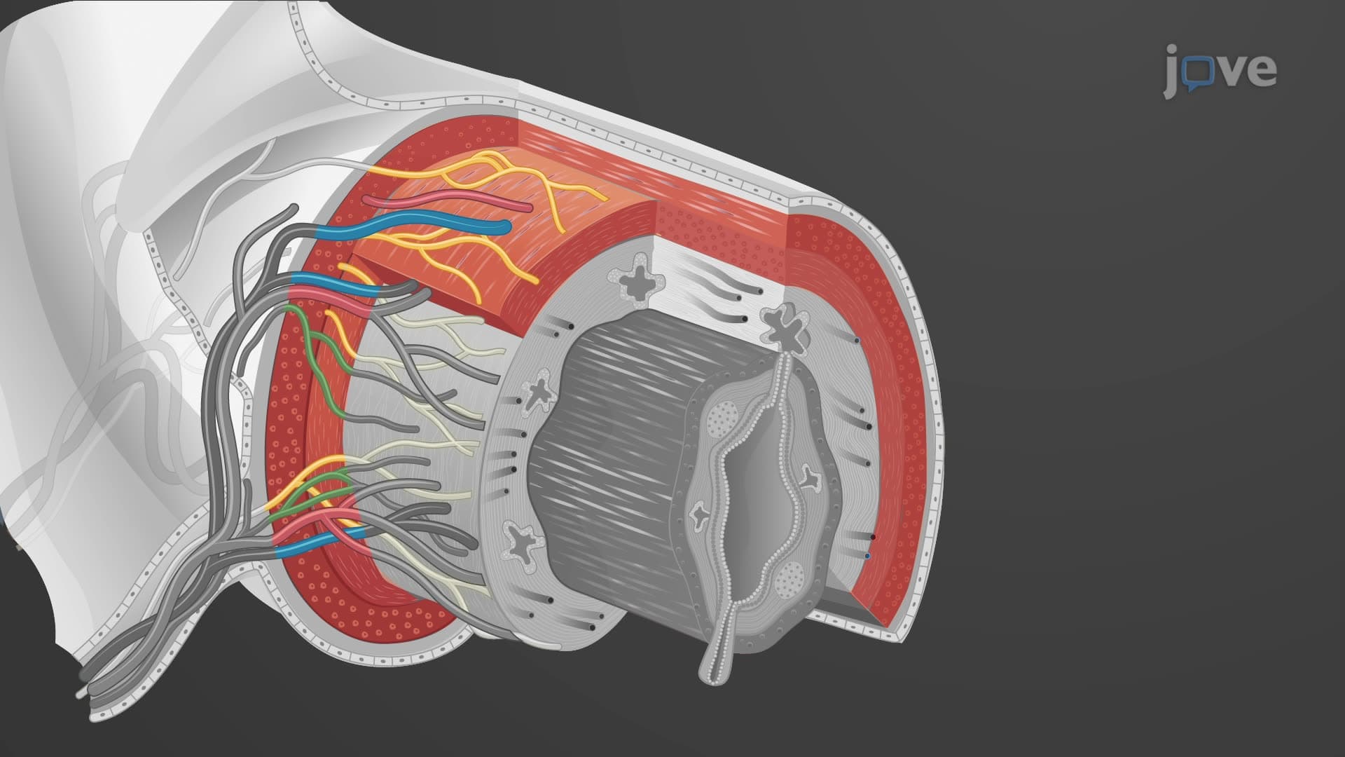
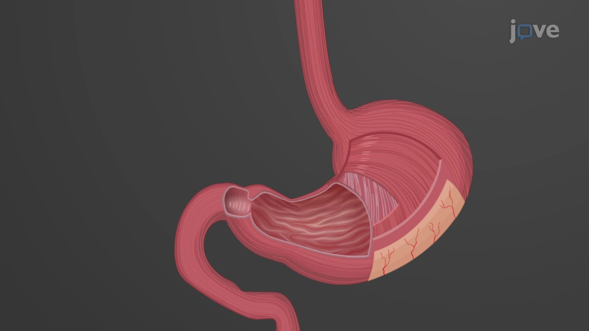
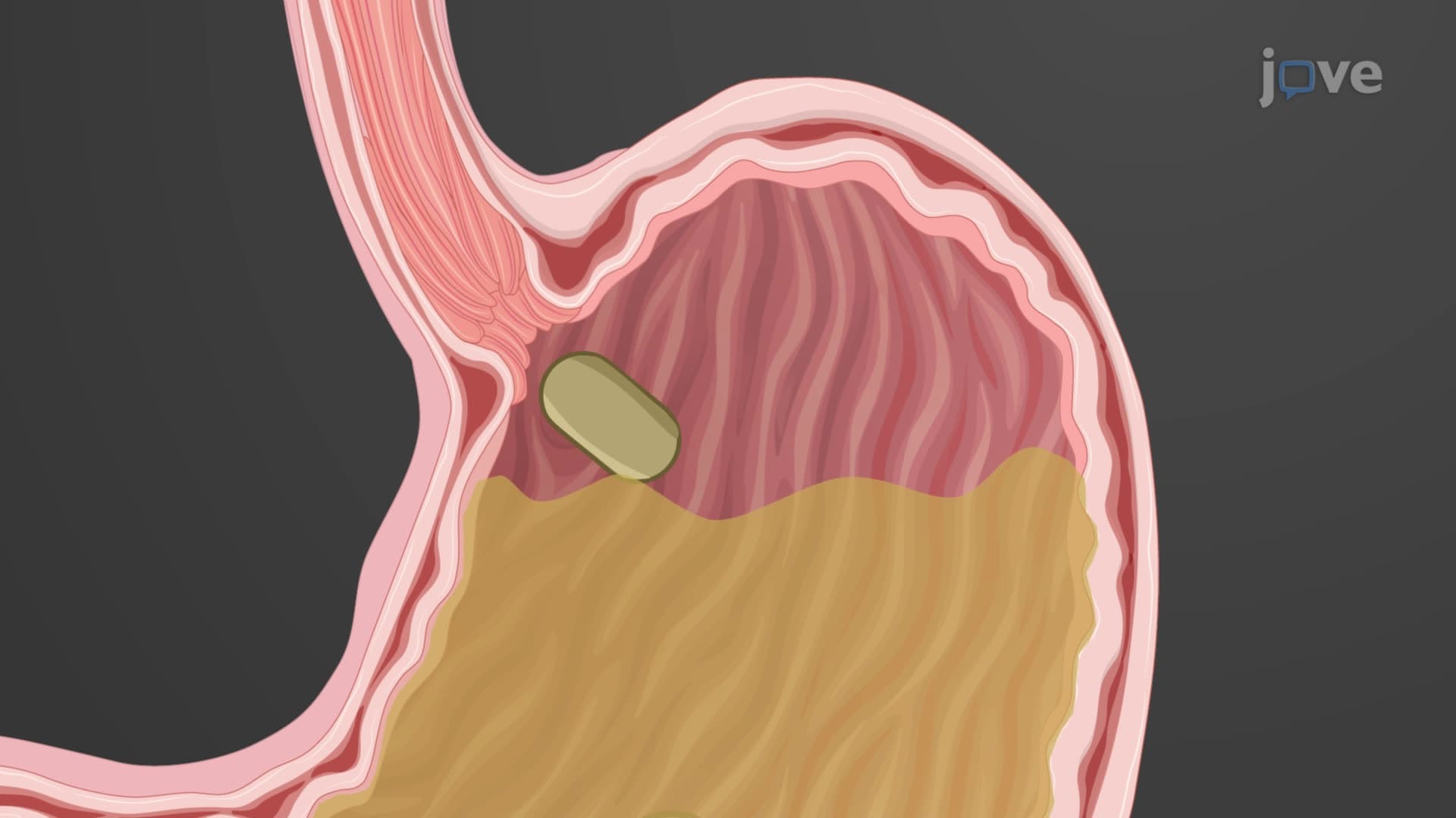
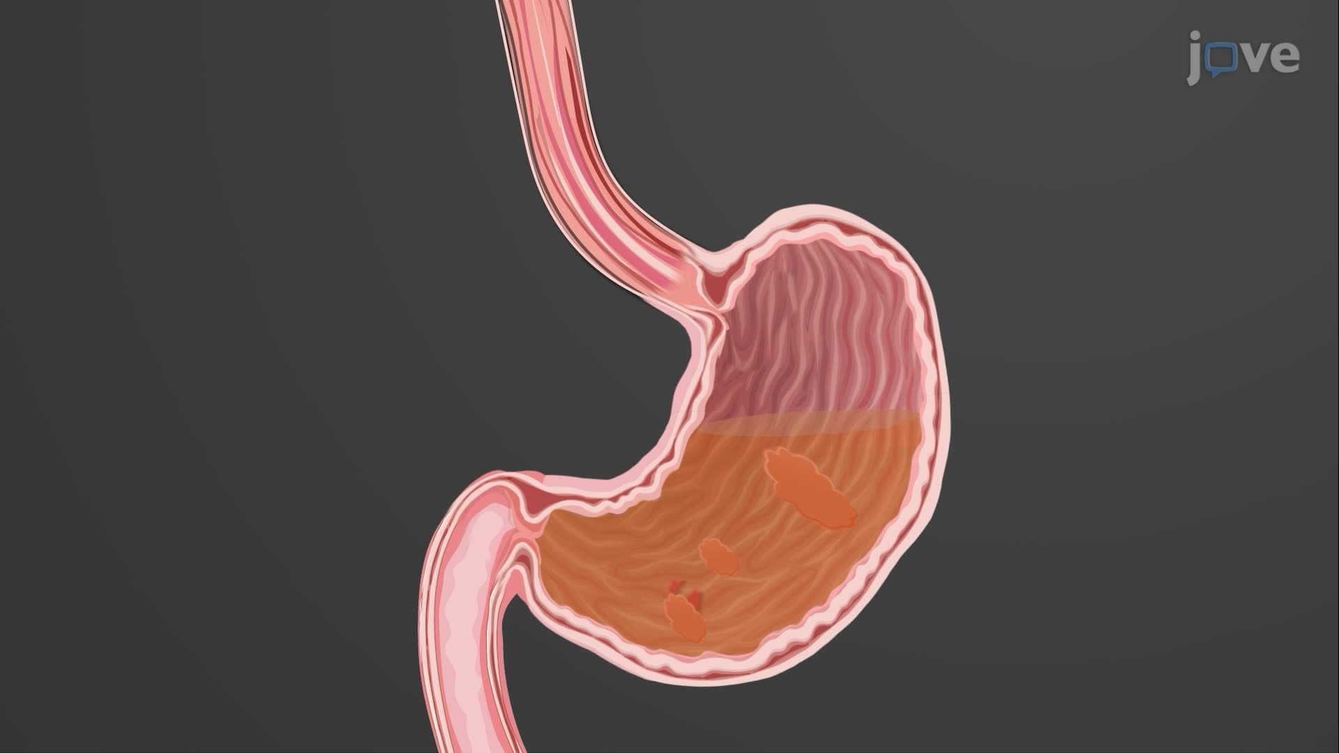
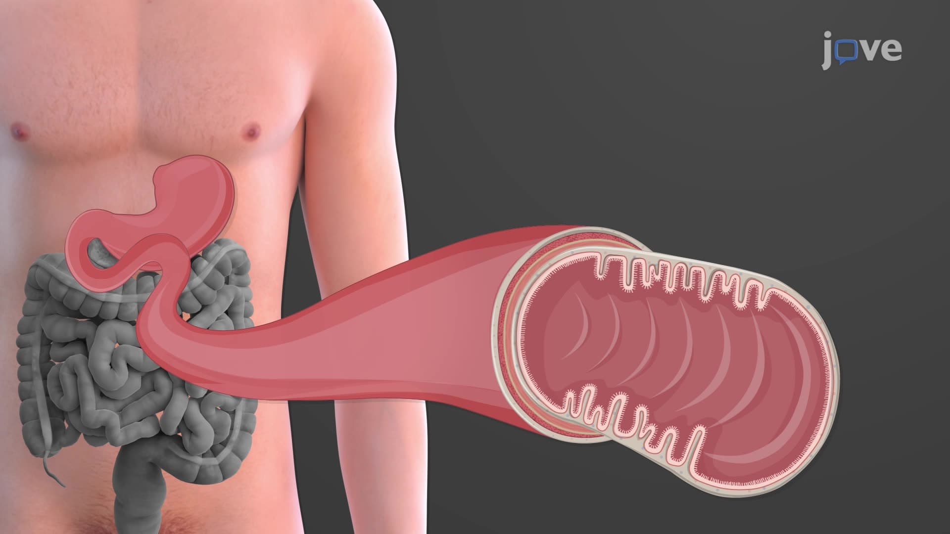
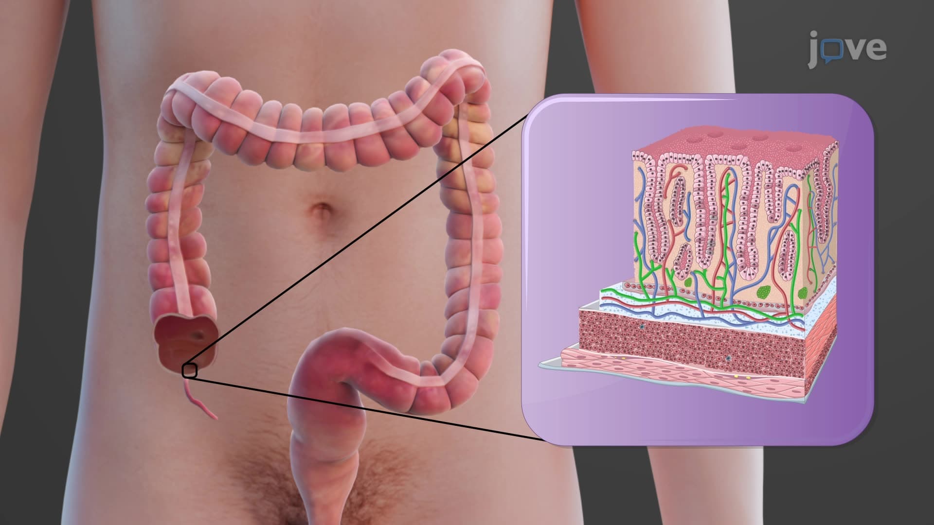
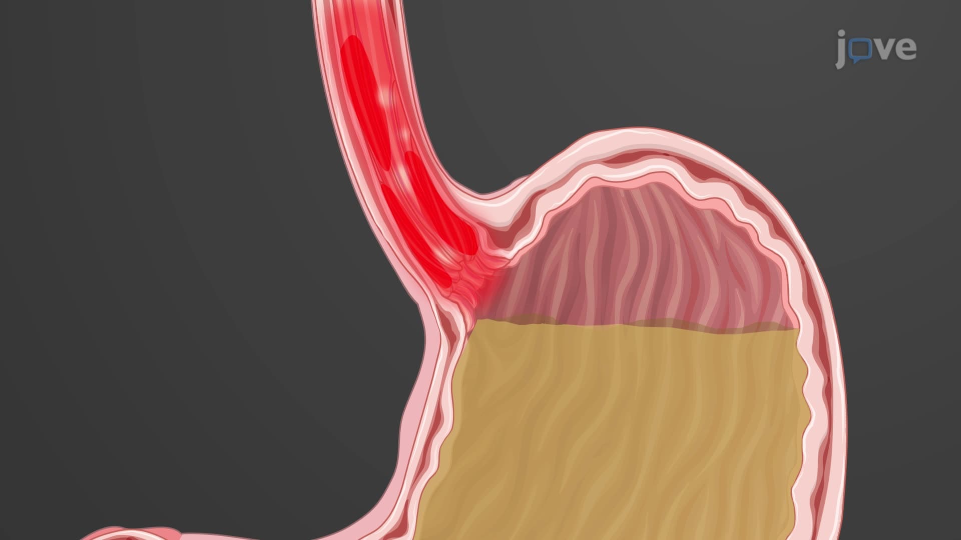
GI tract histology biology examines how microscopic tissue organization enables macroscopic digestive functions. The gastrointestinal wall maintains remarkable consistency from esophagus to rectum, with four layers working synergistically. This architectural precision allows a single bite of food to travel 30 feet through your digestive system while undergoing chemical breakdown, absorption, and waste elimination.
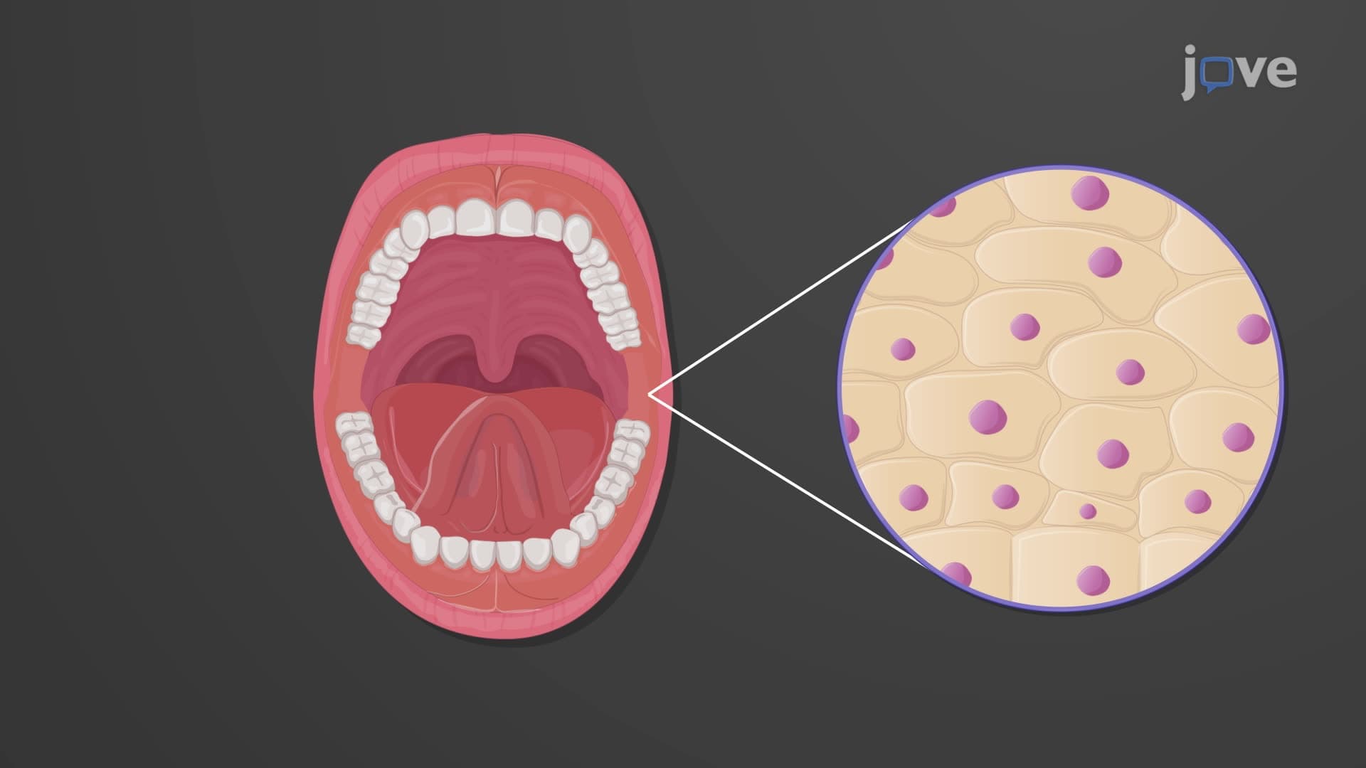
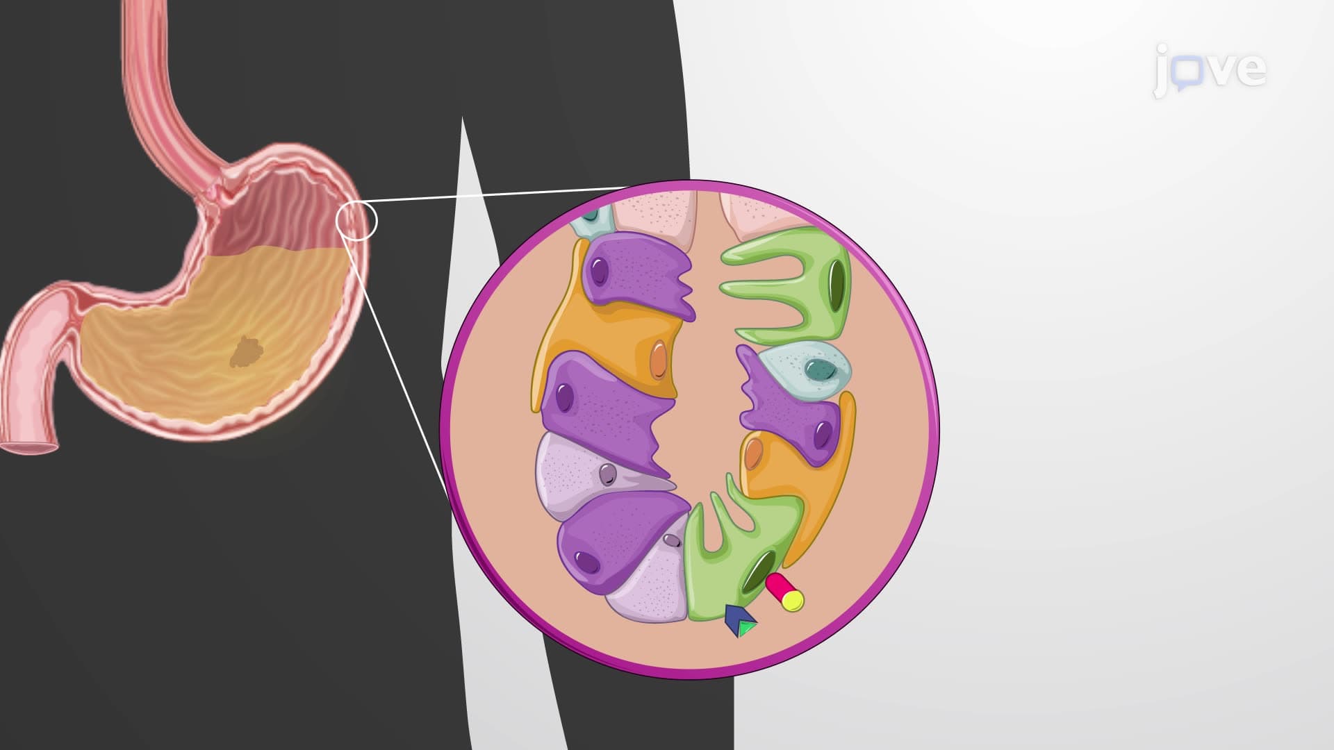
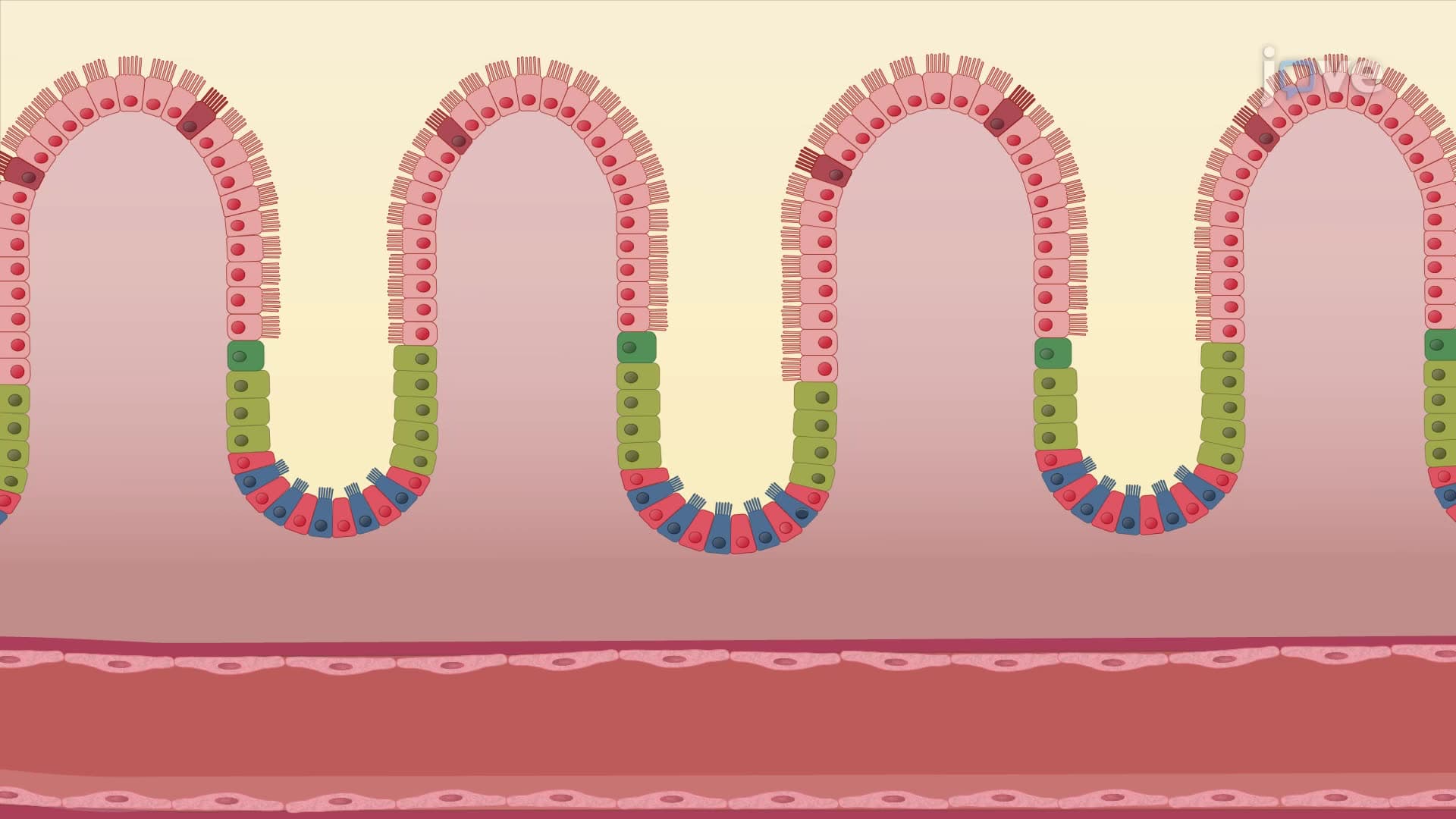
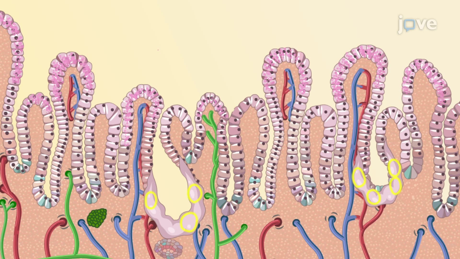
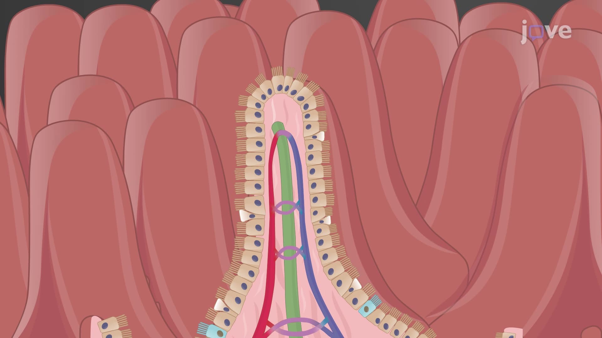
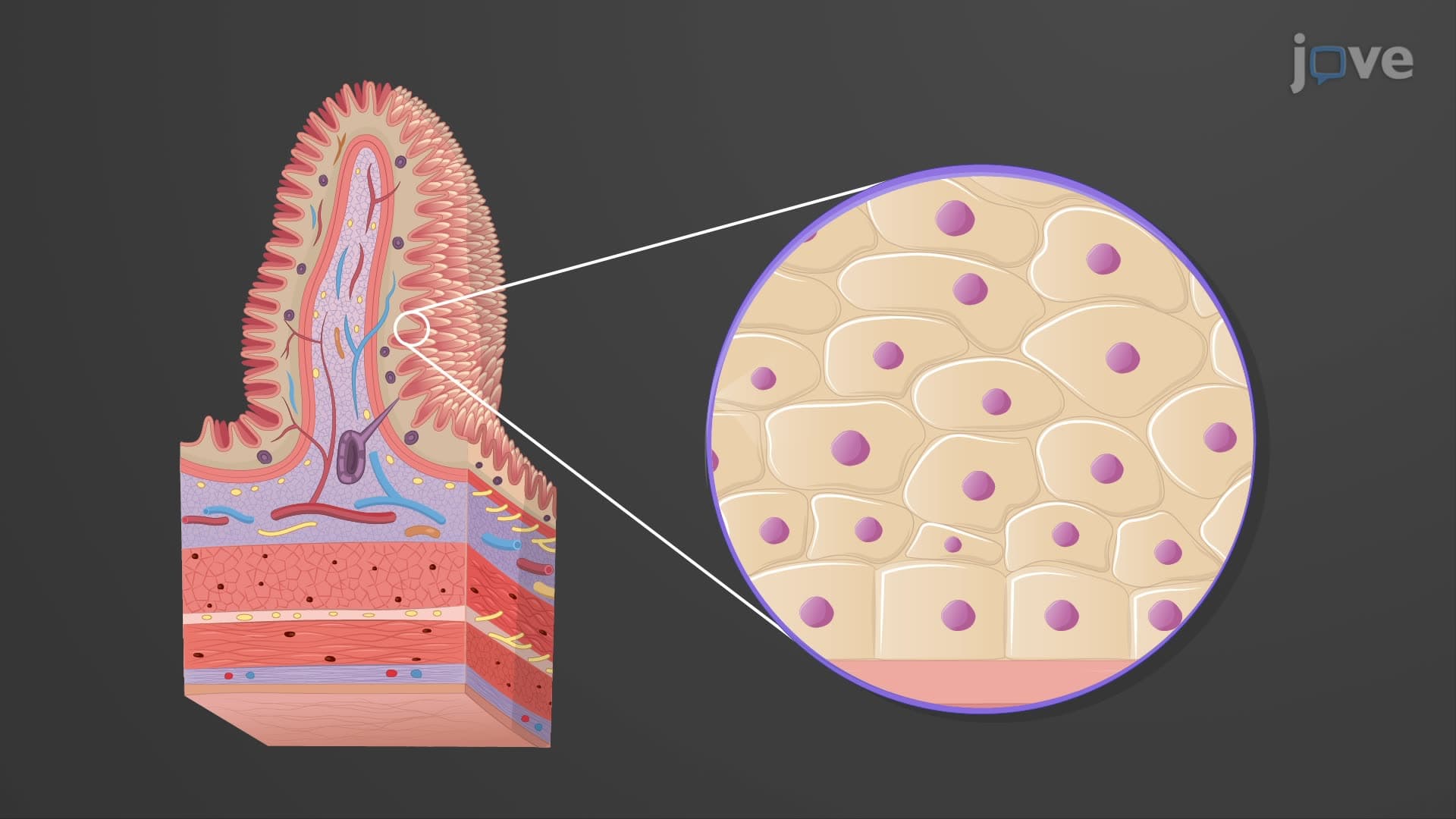
The innermost mucosa layer directly contacts ingested materials, requiring specialized cellular defenses. The epithelial component varies strategically-nonkeratinized stratified squamous epithelium in the esophagus resists mechanical damage from swallowing, while simple columnar epithelium in the intestines maximizes absorption surface area through microvilli.
Enteroendocrine cells scattered throughout this epithelium release hormones like gastrin and secretin, coordinating digestive processes. The underlying lamina propria houses extensive MALT populations, providing immune surveillance against pathogens in food. Students preparing for the MCAT should understand how inflammatory bowel diseases like Crohn's disease primarily affect this mucosal immune system.
The submucosa contains Meissner's plexus (submucosal plexus), part of the enteric nervous system controlling glandular secretions and local blood flow. This autonomic control explains why stress affects digestion-neuronal signals alter mucus production and vascular perfusion.
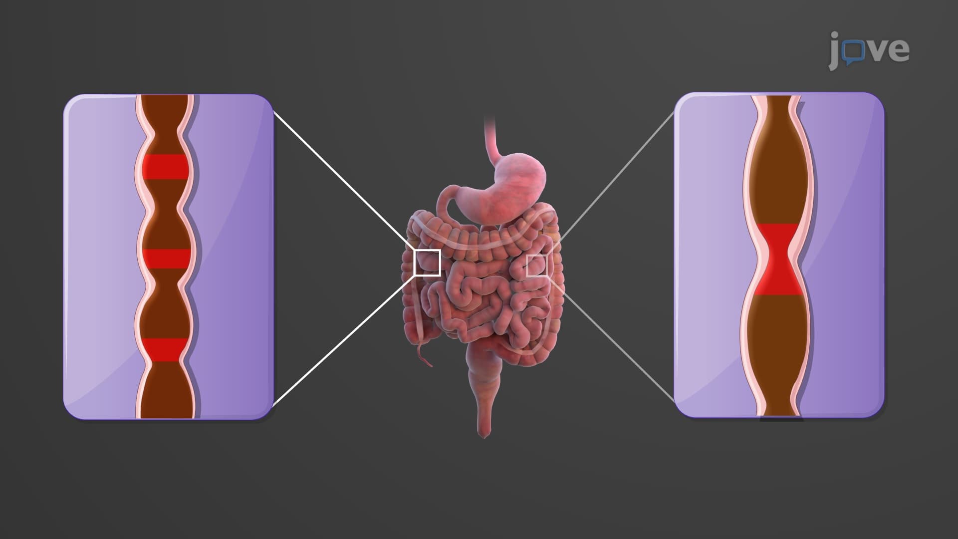
The muscularis layer typically features inner circular and outer longitudinal smooth muscle arrangements. Coordinated contractions create peristaltic waves, propelling contents aborally. Auerbach's plexus (myenteric plexus) between these muscle layers orchestrates this movement. AP Biology students should recognize how parasympathetic stimulation enhances peristalsis while sympathetic activation inhibits it.
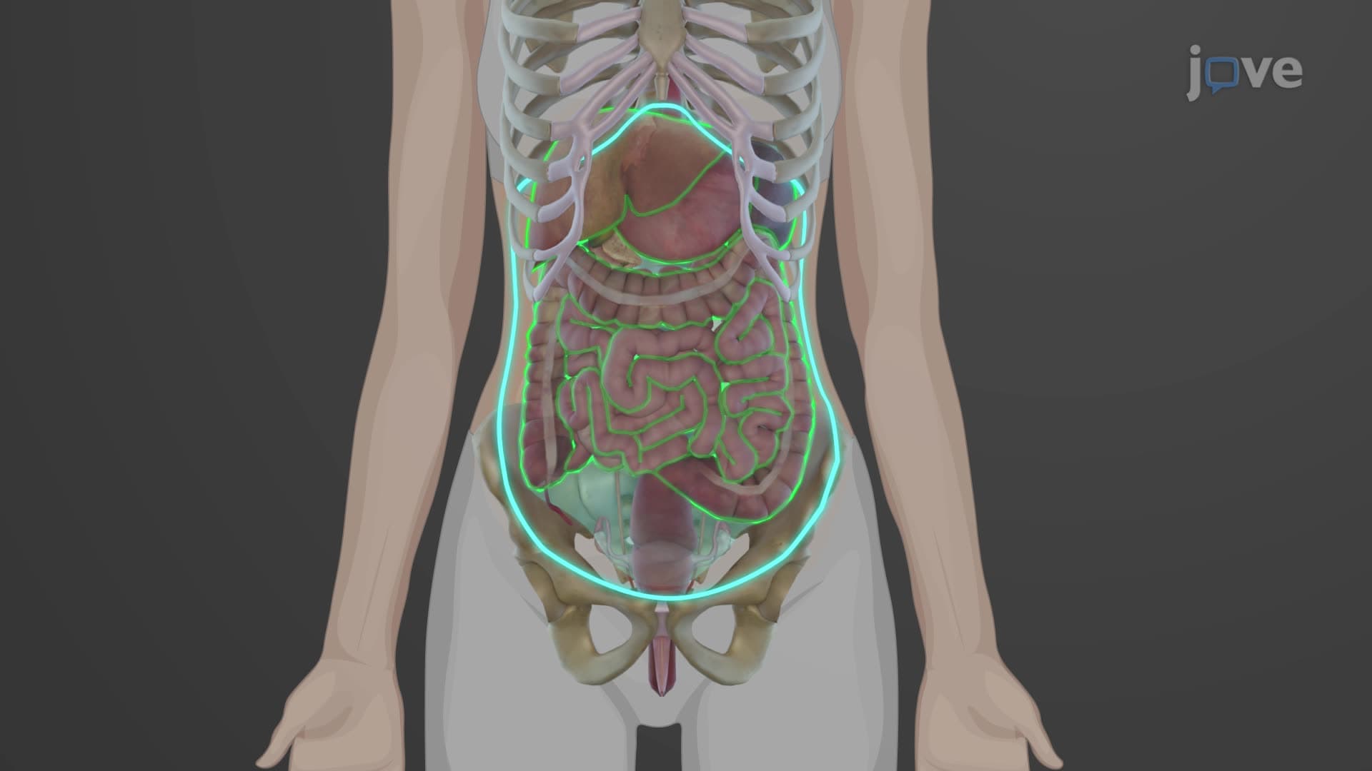
The outermost layer designation depends on anatomical location. Intraperitoneal organs (stomach, jejunum, ileum, transverse colon) have serosa-a smooth, glistening peritoneal covering enabling frictionless movement during digestion. Retroperitoneal structures (duodenum, ascending colon, descending colon) have adventitia-fibrous connective tissue anchoring organs to posterior abdominal wall.
Related Micro-courses