Loading subject page…


89,753 views

Video Summary: What Is Histology Large Intestine
Did you know that your colon's distinctive puckered appearance comes from specialized muscle bands that contract like drawstrings? Large intestine histology biology reveals four distinct wall layers working together to complete digestion and form waste. Unlike the small intestine, the colonic mucosa lacks villi but contains abundant goblet cells producing protective mucus-crucial for patients with inflammatory bowel disease treated at institutions like Johns Hopkins. Watch the full video on JoVE Coach to master this concept with expert-led visuals and step-by-step explanations.
The histology of the large intestine represents a masterpiece of functional anatomy, where each microscopic structure serves the organ's primary roles: water absorption, electrolyte balance, and waste formation. This anatomical knowledge forms the foundation for understanding diseases affecting millions of Americans, from irritable bowel syndrome to colorectal cancer-the third most common cancer in the United States.
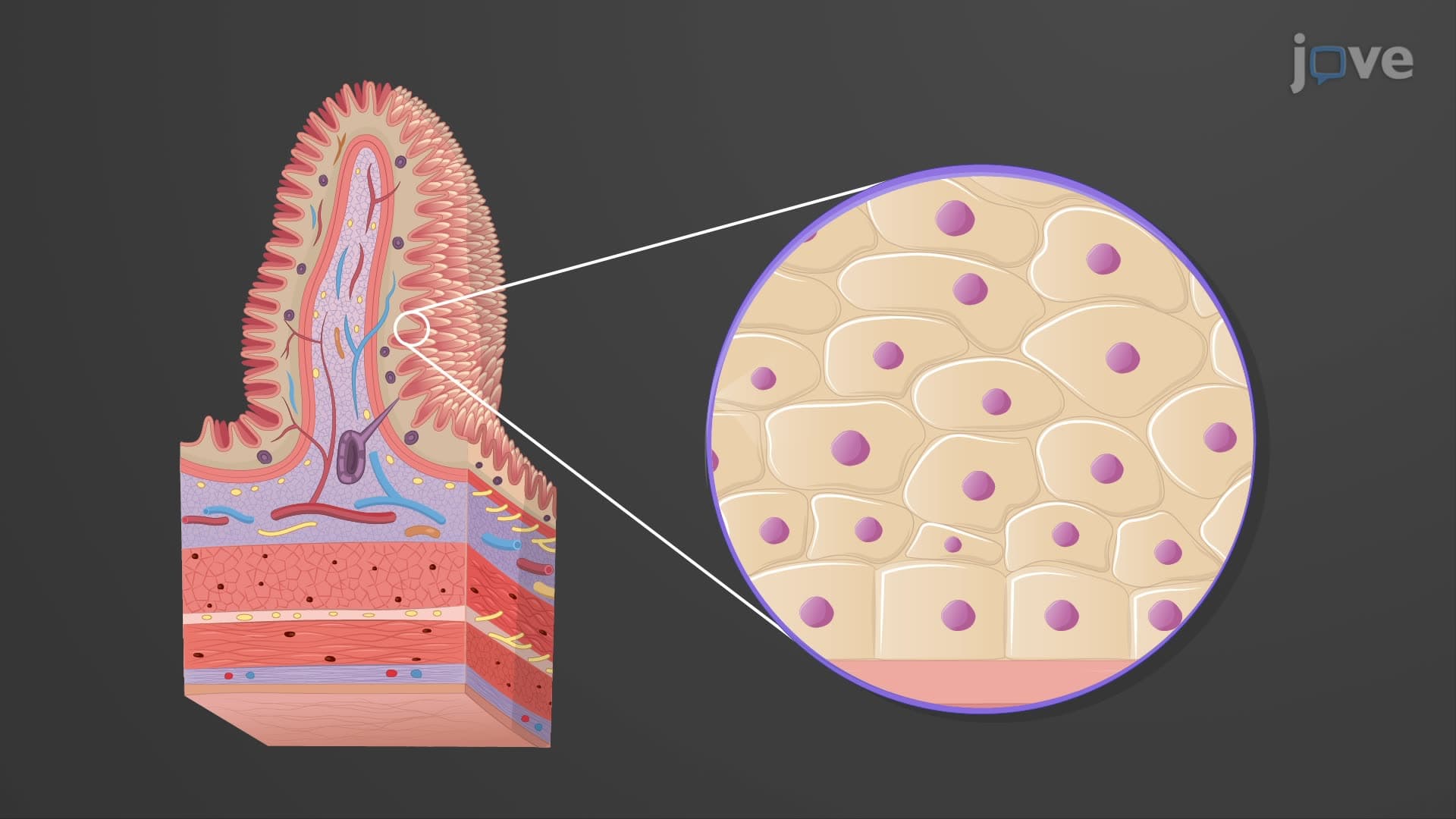
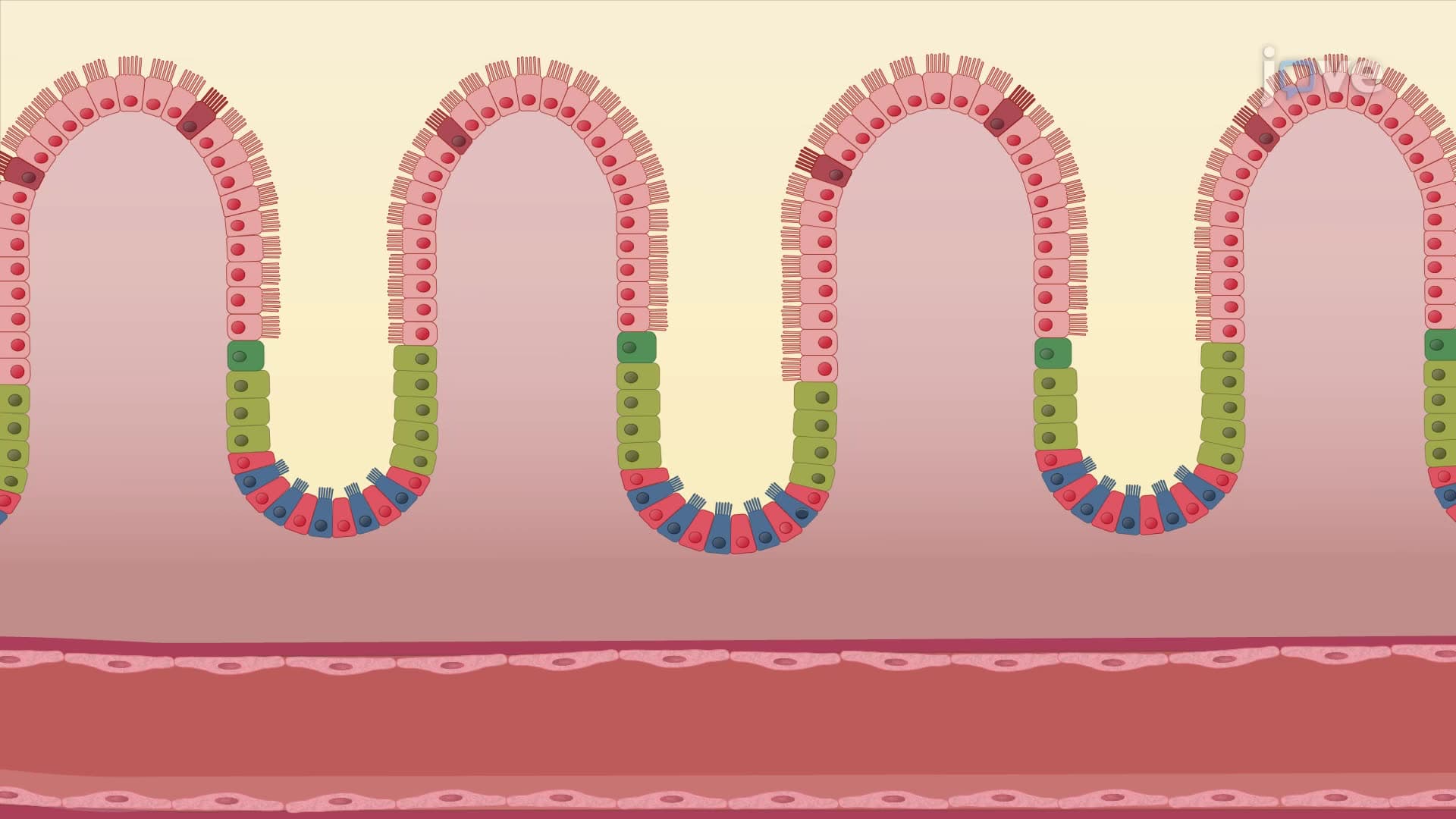
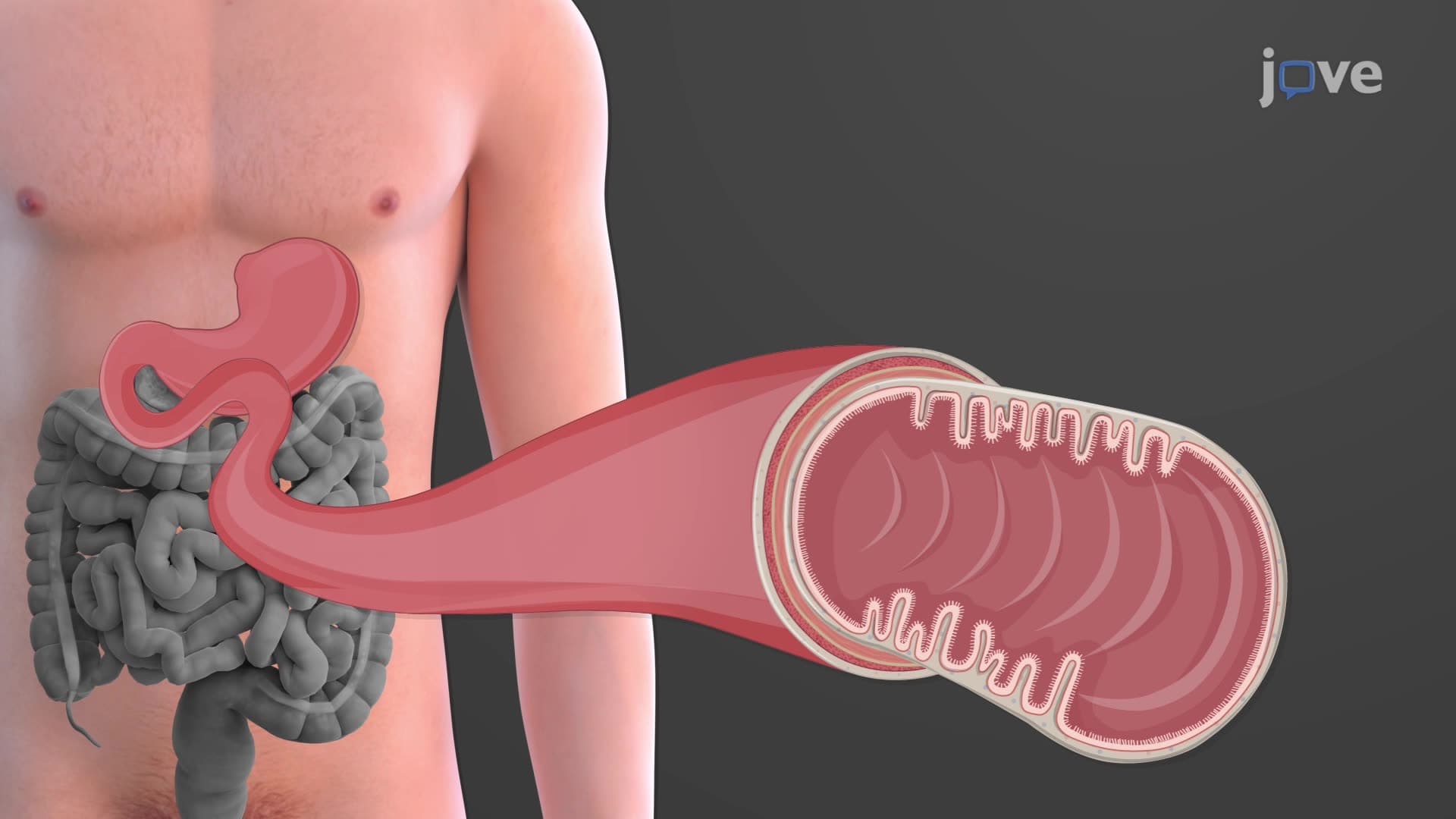
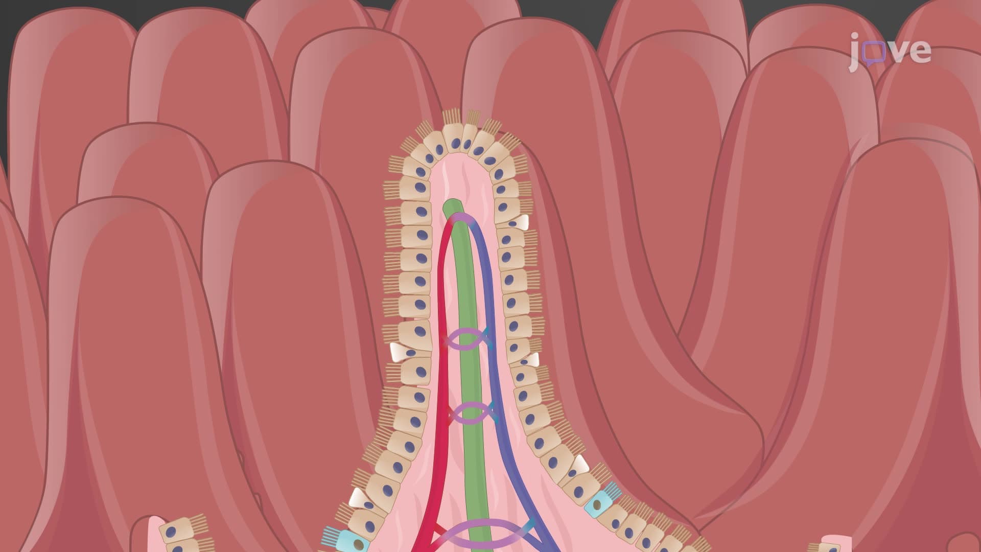
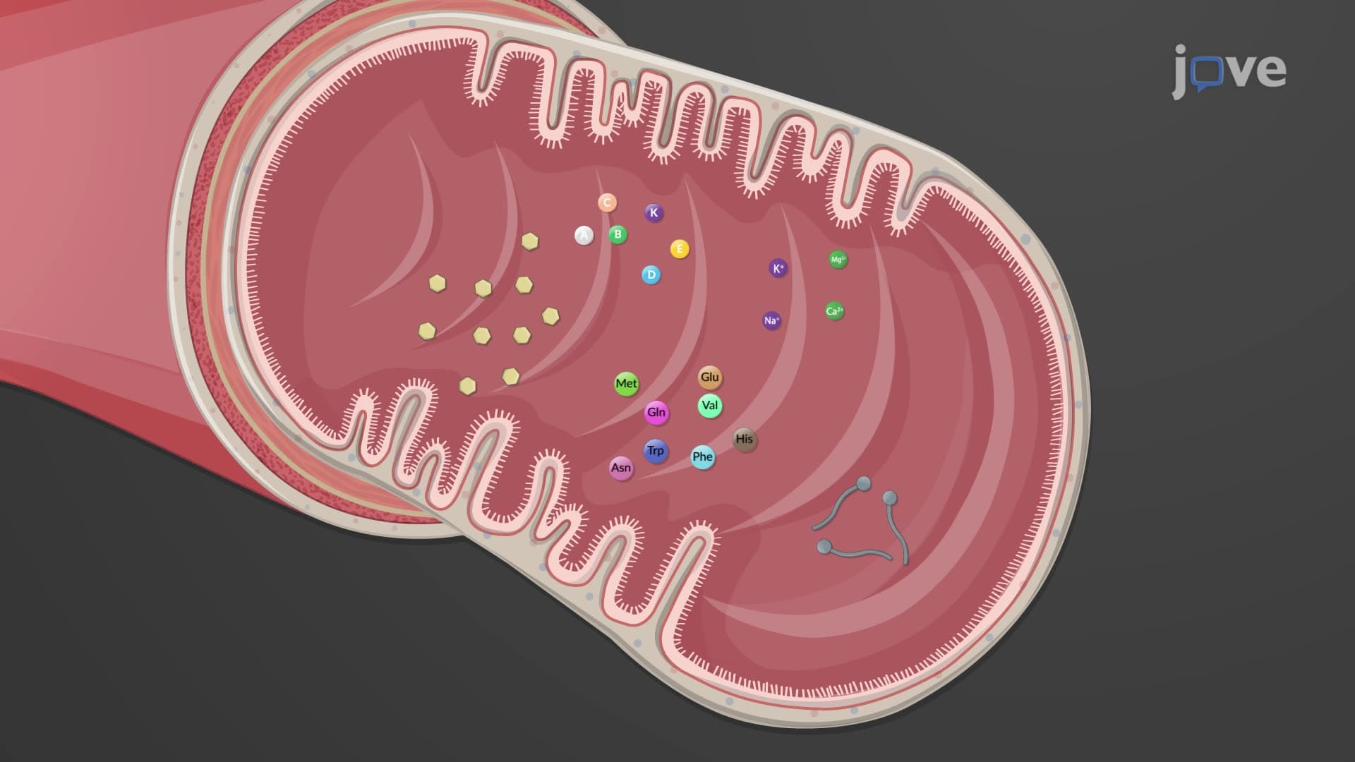
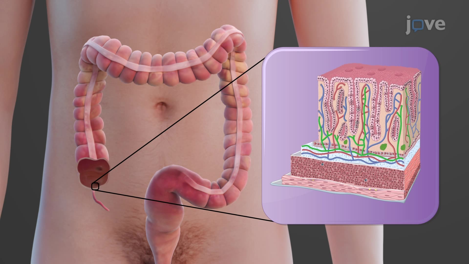
Unlike the small intestine's villous projections, the colonic mucosa features a smooth surface punctuated by deep intestinal crypts. This colon histology explained reveals simple columnar epithelium dominated by two cell types: absorptive cells and goblet cells. Absorptive cells lack villi but maintain microvilli on their apical surface, creating the brush border essential for water and sodium absorption. The absence of villi reflects the colon's specialized function-unlike the small intestine's nutrient absorption requiring maximum surface area, the colon prioritizes controlled water recovery.
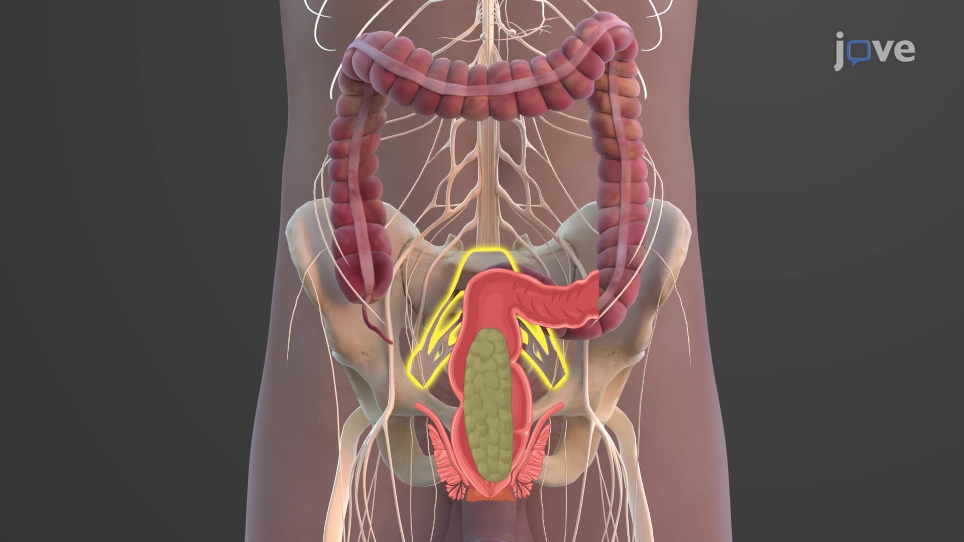
Goblet cell colon histology shows these mucus-secreting cells increase in density from ascending to descending colon, reaching peak concentration in the rectum. This distribution pattern protects against the increasingly solid fecal matter, explaining why conditions like ulcerative colitis often begin in the rectum where goblet cell density is highest.
The crypts of Lieberkühn extend deep into the lamina propria, housing stem cells that completely replace the epithelial lining every 3-5 days-one of the body's fastest tissue renewal rates. This rapid turnover makes the colon particularly susceptible to chemotherapy side effects, knowledge crucial for oncology nurses and physicians treating patients at cancer centers like MD Anderson or Memorial Sloan Kettering.
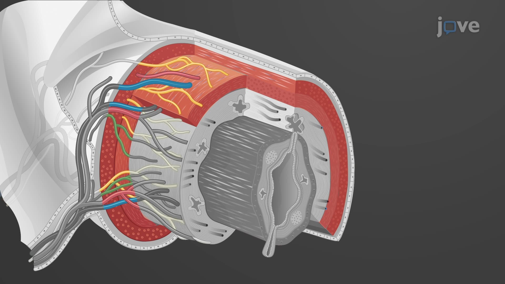
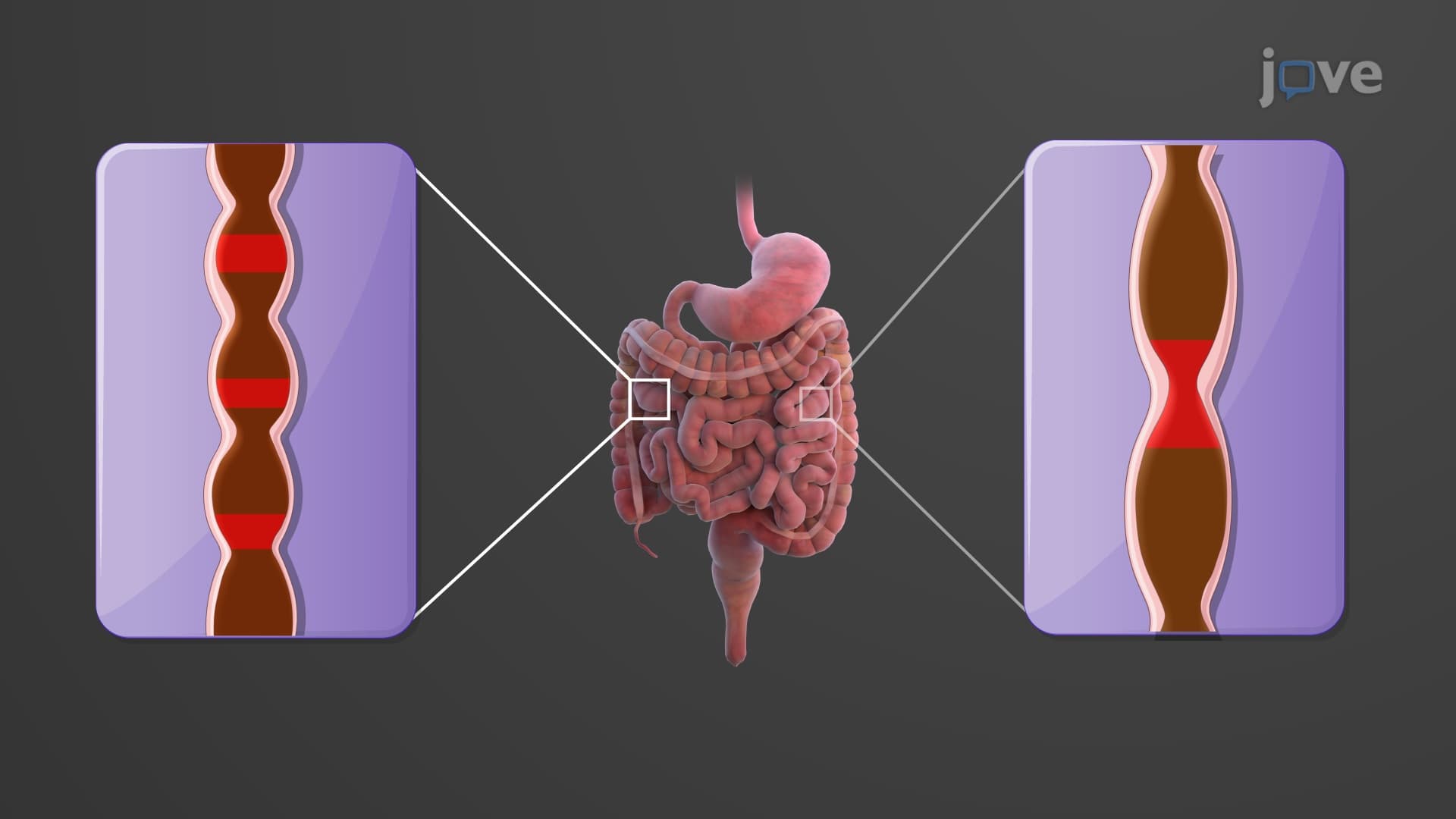
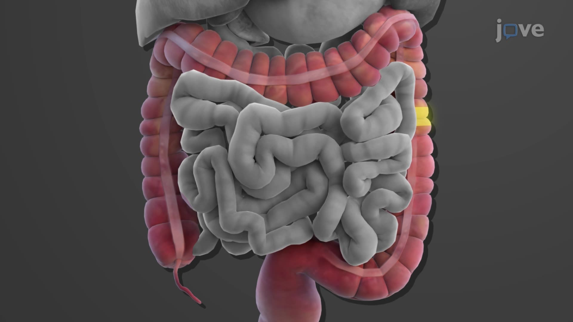
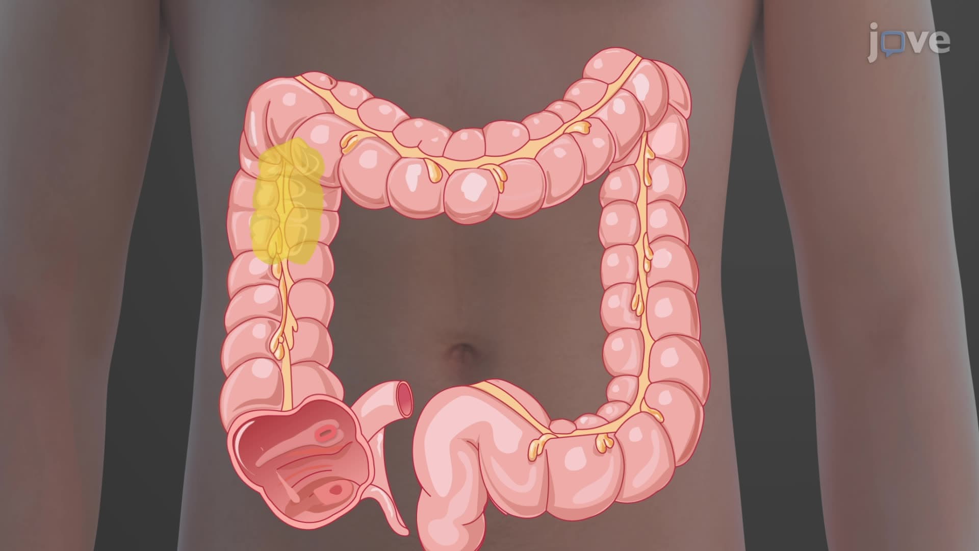
The muscularis teniae coli represents a unique muscular arrangement where longitudinal smooth muscle concentrates into three distinct bands rather than forming a continuous layer. These bands contract to create haustra-the colon's characteristic pouched appearance visible on CT scans and colonoscopies. Understanding this large intestine wall histology proves essential for AP Biology students studying digestive system adaptations and pre-med students preparing for the MCAT's biological systems section.
Related Micro-courses