Loading subject page…


89,753 views

Video Summary: What Is Histology Small Intestine
Did you know that your small intestine contains millions of microscopic fingerlike projections that increase its surface area to roughly the size of a tennis court? Small intestine histology biology reveals how specialized cellular structures enable nutrient absorption in conditions like Crohn's disease, commonly studied at Mayo Clinic. Understanding what is histology of the small intestine shows how villi, crypts, and specialized cells work together to process the food you eat daily. Watch the full video on JoVE Coach to master this concept with expert-led visuals and step-by-step explanations.
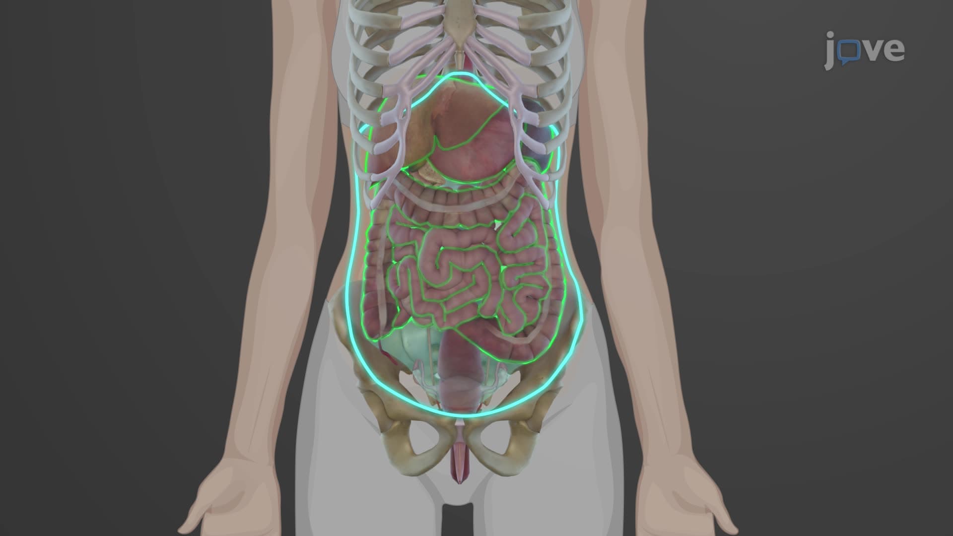
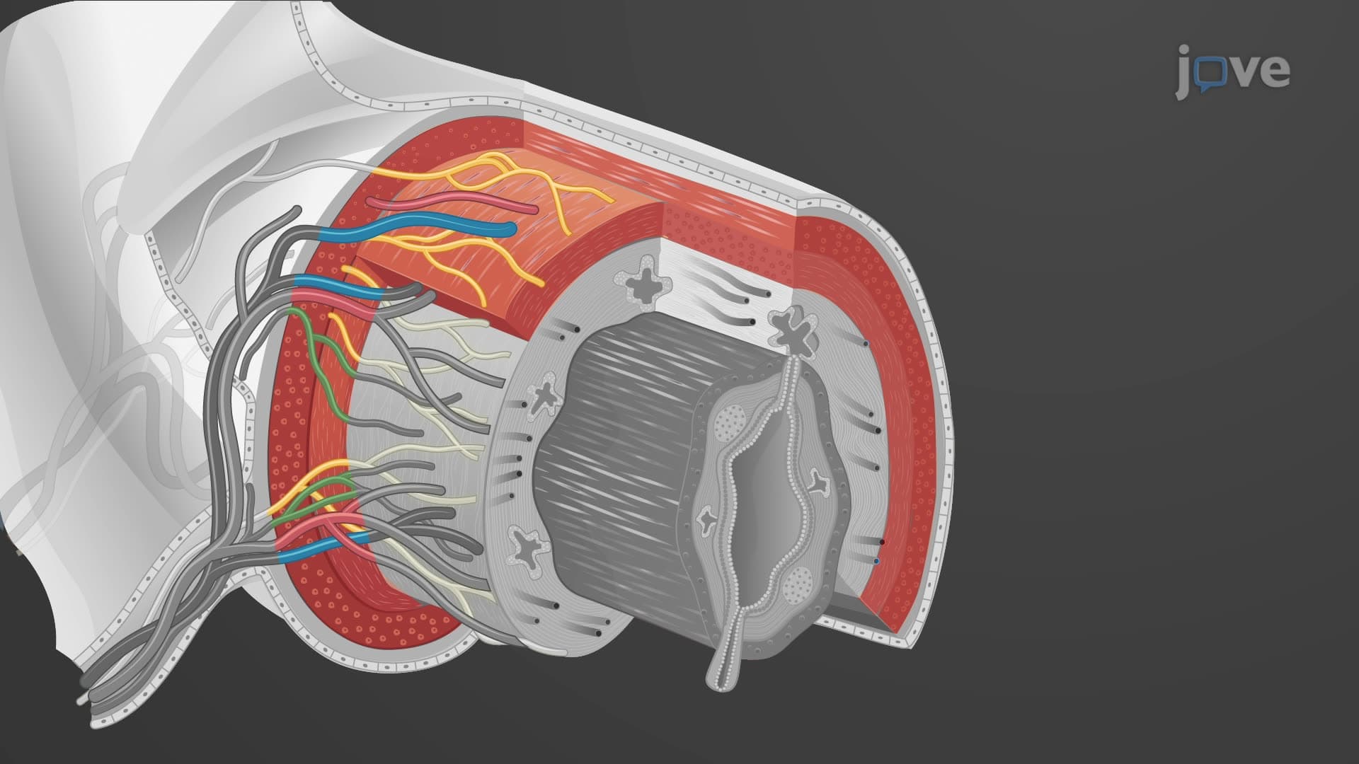
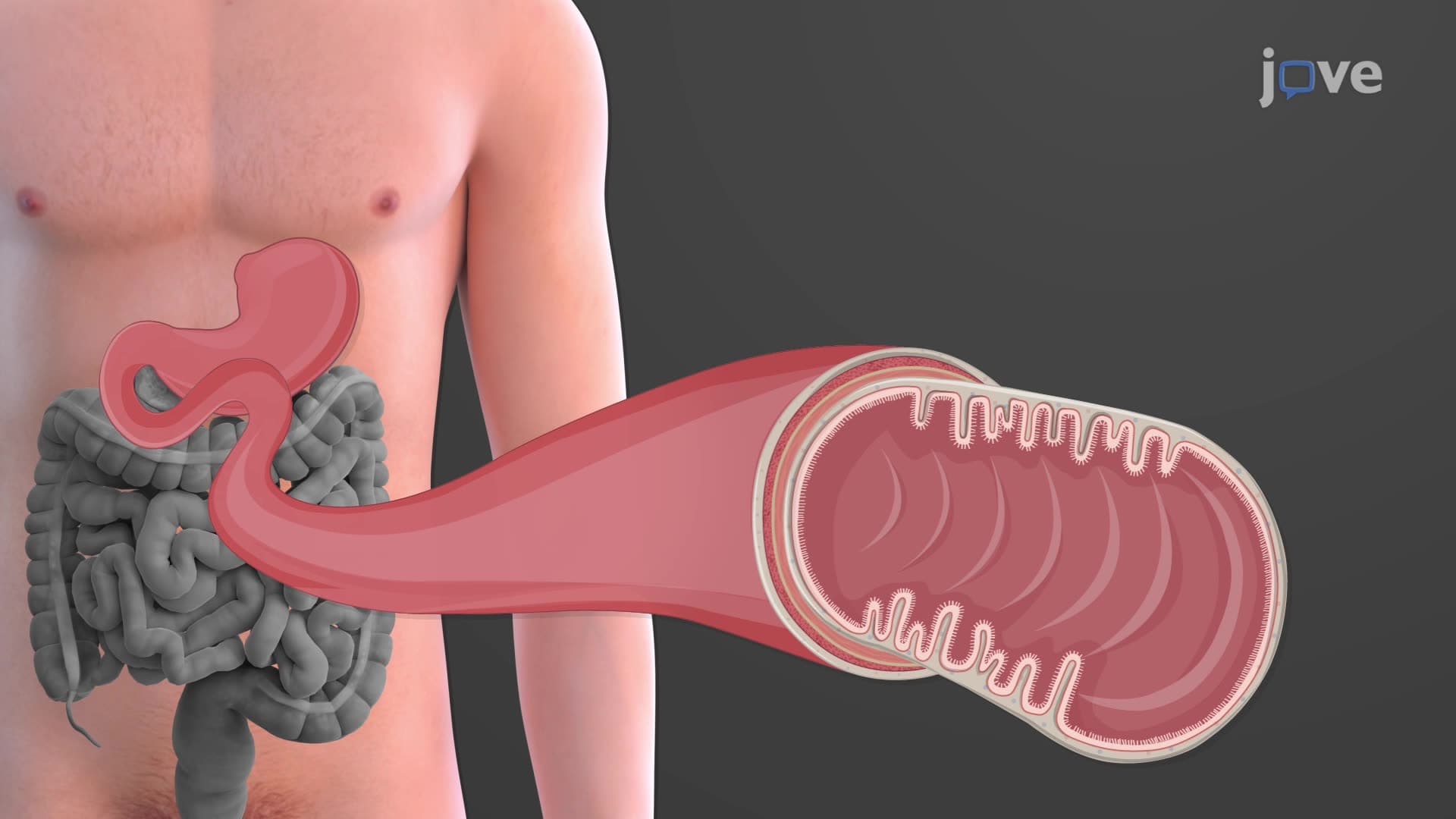
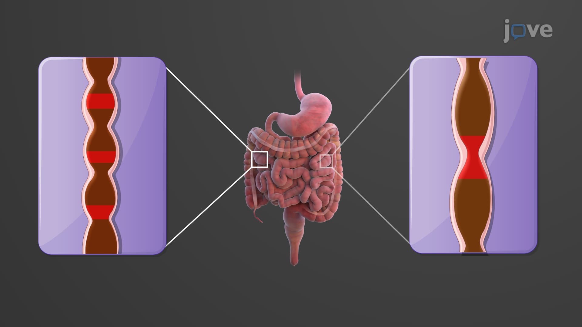
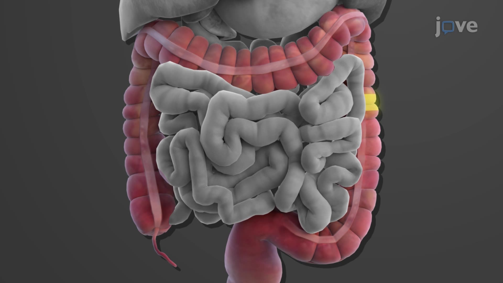
Small intestine histology biology demonstrates one of nature's most elegant solutions to maximizing absorption efficiency. The small intestine's 20-foot length contains three anatomically distinct regions, each optimized for specific digestive functions. The duodenum features Brunner gland duodenal structures that secrete alkaline mucus to neutralize acidic chyme from the stomach. The jejunum displays the most prominent circular folds and longest villi, making it the primary site for nutrient absorption. The ileum, while having shorter villi, contains specialized Peyer's patches-organized lymphoid tissue crucial for immune surveillance.
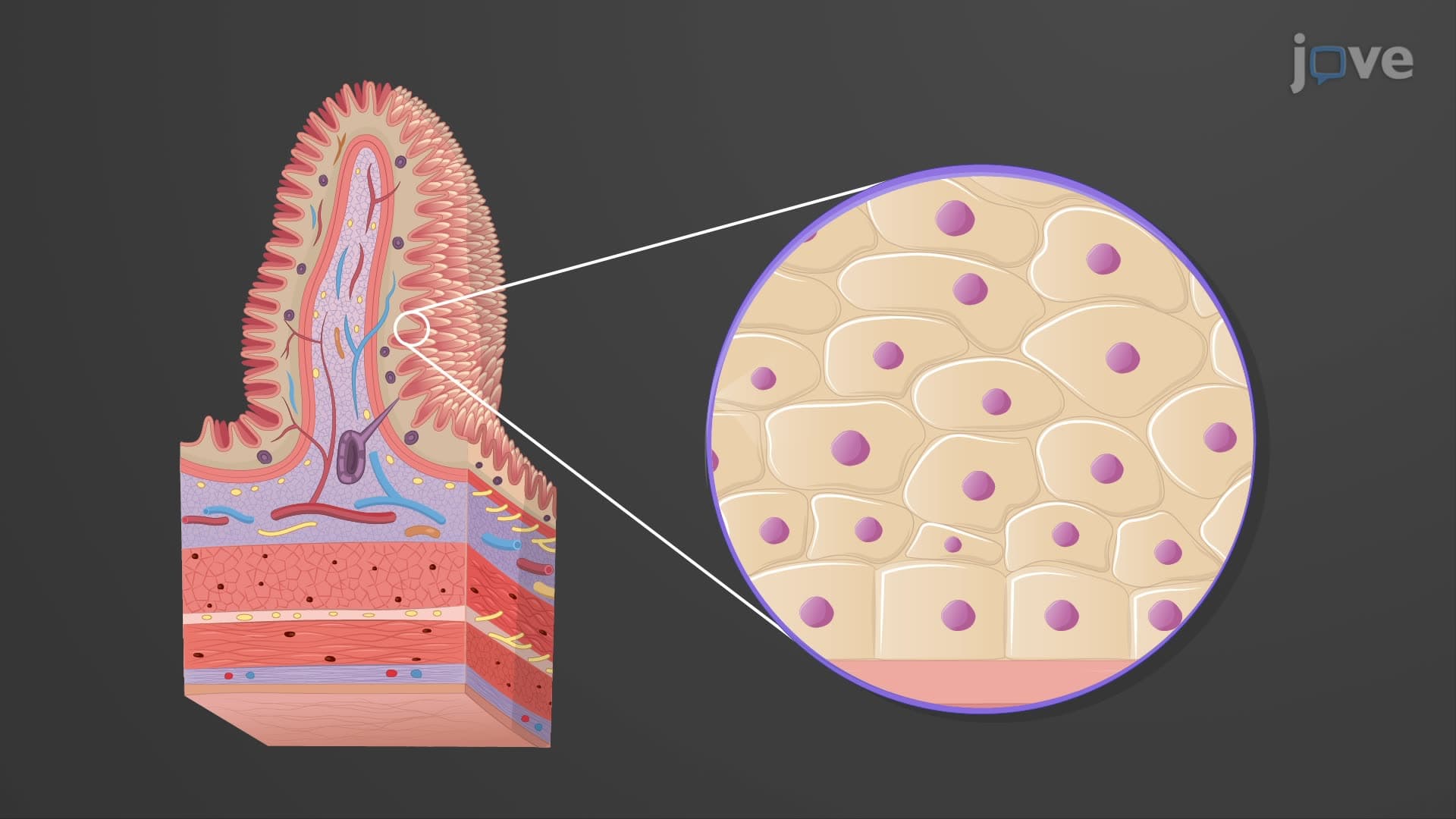
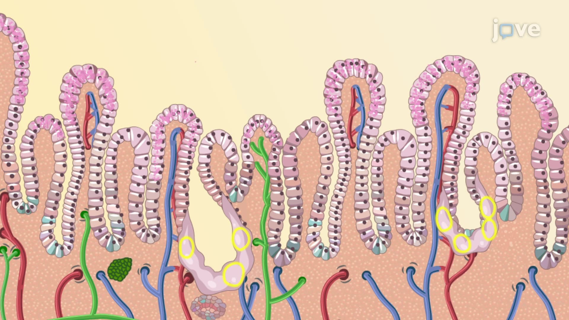
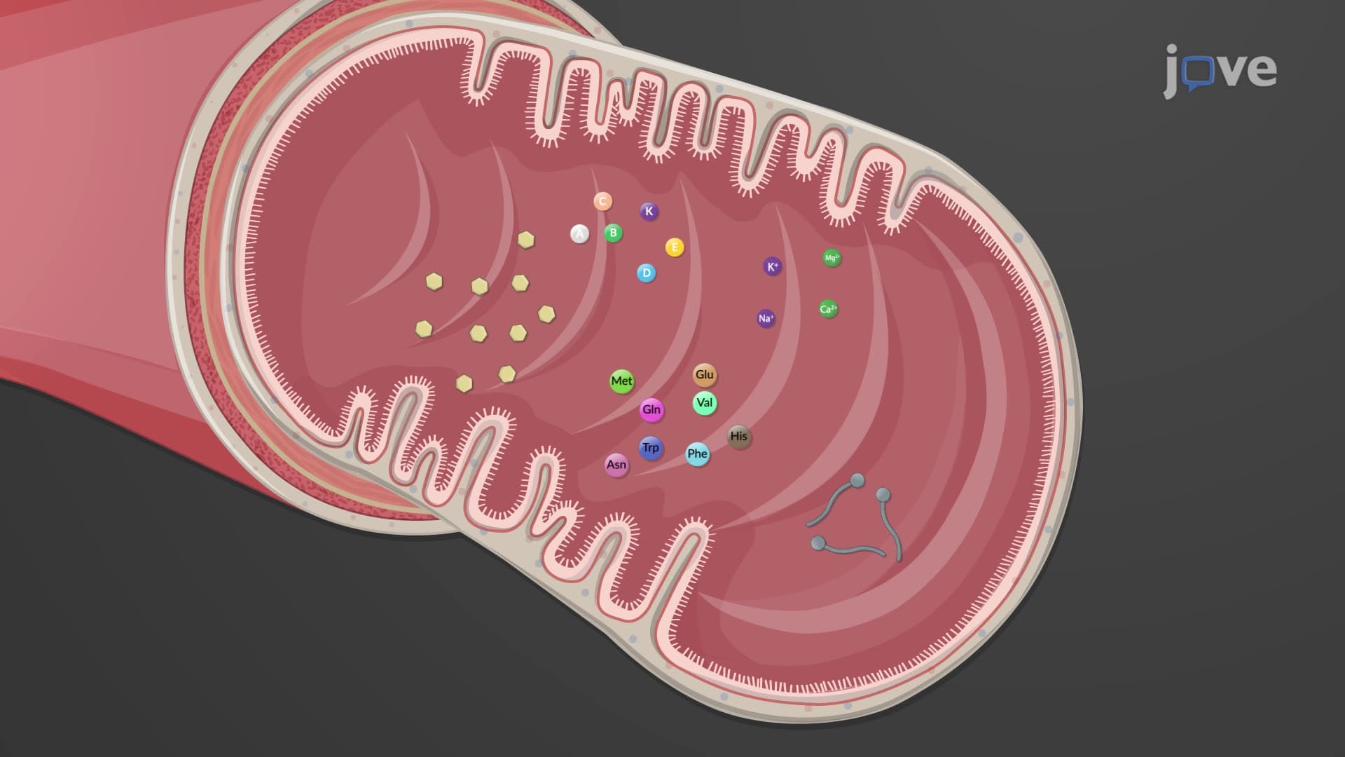
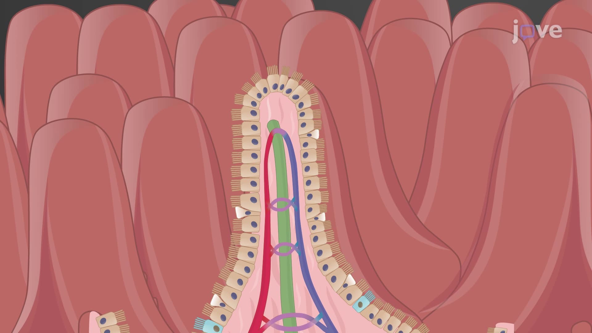
The hierarchical organization of intestinal surface amplification involves three levels. Circular folds (plicae circulares) increase surface area threefold, while intestinal villi provide an additional 10-fold increase. Finally, intestinal villi microvilli on enterocyte surfaces create a 20-fold amplification, resulting in a total 600-fold surface area increase. This concept frequently appears on AP Biology exams and MCAT questions testing structure-function relationships.
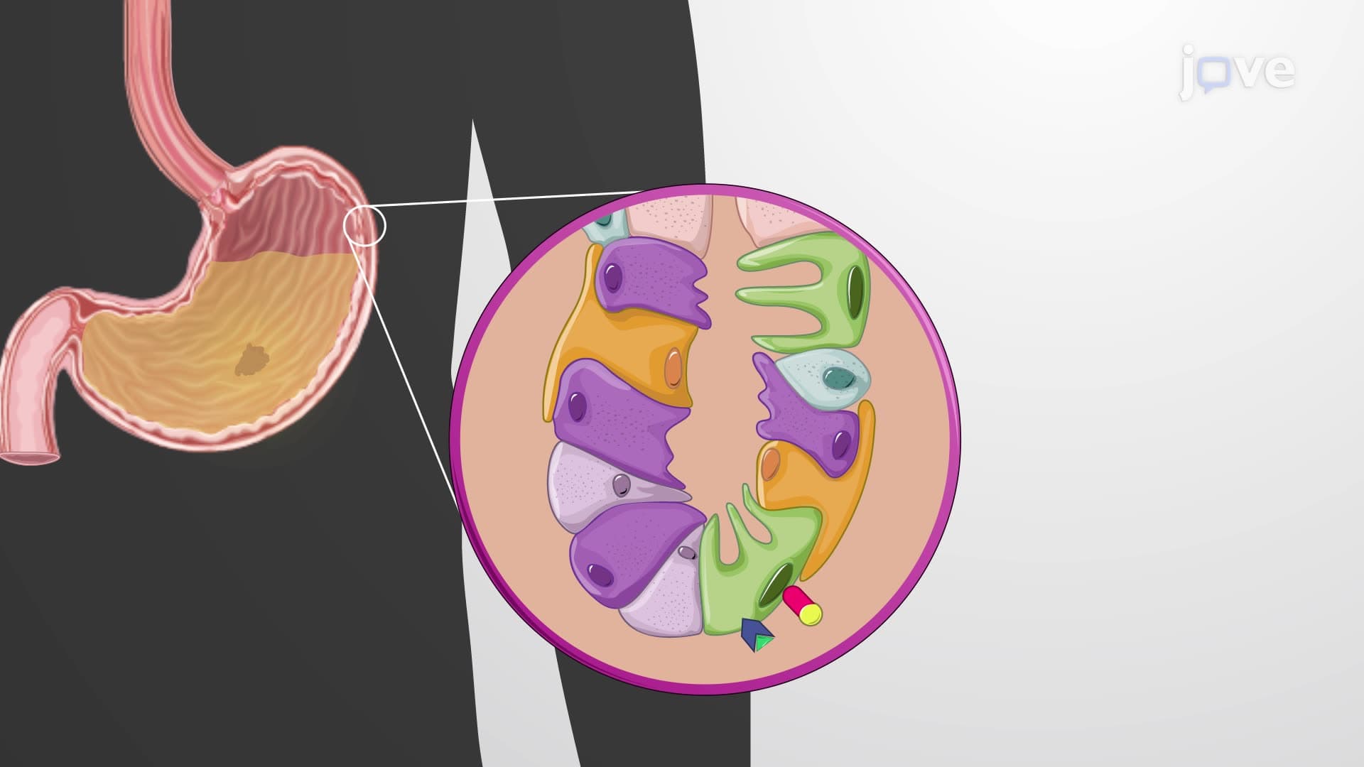
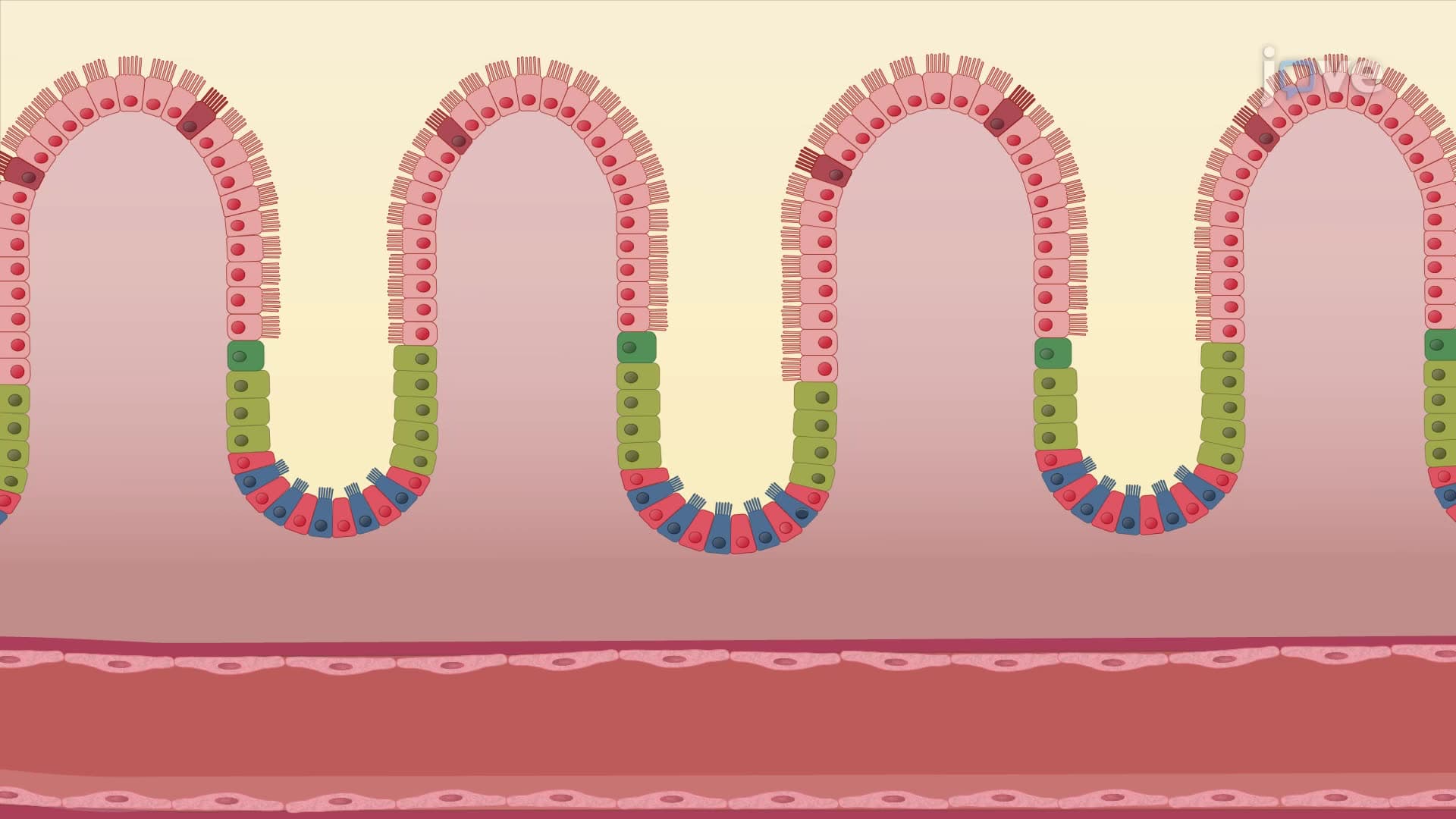
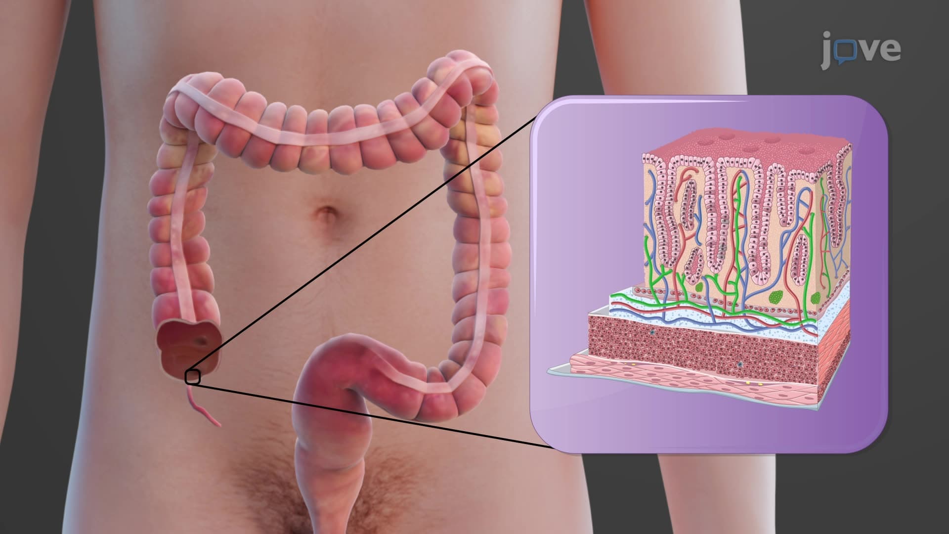
Goblet cell intestinal histology reveals mucus-secreting cells interspersed among absorptive enterocytes. These goblet cells increase in number from duodenum to colon, reflecting increased need for protective mucus. The intestinal crypt Lieberkühn houses the intestinal stem cell niche, where rapid cell division replaces the entire intestinal epithelium every 3-5 days. Medical students studying for USMLE Step 1 must understand how Paneth cells within these crypts secrete antimicrobial peptides like lysozyme and defensins.
Understanding normal small intestine histology layers becomes crucial when studying diseases like celiac disease, where gluten triggers autoimmune villous atrophy. Crohn's disease, commonly seen at institutions like Johns Hopkins, demonstrates how chronic inflammation disrupts normal histological architecture. Nursing students preparing for NCLEX examinations study how malabsorption syndromes relate to specific histological damage patterns, particularly the loss of brush border enzymes on microvilli surfaces.
Related Micro-courses