Loading subject page…

- Anatomy and Physiology
- Digestive System
- Large Intestine
Micro-course: Digestive System
89,747 views
Learn key concepts and earn your certificate.






Large Intestine

00:00
01:09
Video Summary: What Is Large Intestine
Did you know your large intestine is only about 5 feet long but plays a crucial role in processing the 2-3 gallons of liquid waste your body produces daily? Understanding large intestine anatomy biology reveals how this vital organ transforms liquid chyme into solid waste through water absorption and bacterial fermentation. From the cecum's pouch-like structure to the rectum's final storage chamber, each segment serves specific digestive functions that healthcare professionals at Johns Hopkins and Mayo Clinic study extensively. What is Large Intestine becomes clear when examining its four distinct colon regions and specialized sphincter controls. Watch the full video on JoVE Coach to master this concept with expert-led visuals and step-by-step explanations.
- Understand What is Large Intestine by identifying its anatomical boundaries from the ileocecal valve to the anus and its relationship to surrounding digestive organs
- Identify the five main anatomical regions of the large intestine: cecum, ascending colon, transverse colon, descending colon, and sigmoid colon
- Analyze the structural transitions between small and large intestines, focusing on the ileocecal valve's regulatory function and the appendix's anatomical position
- Compare the four distinct colon segments and their spatial orientations within the abdominal cavity, including hepatic and splenic flexures
- Evaluate the functional significance of the rectum and anal canal in waste storage and elimination through sphincter control mechanisms
- Apply knowledge of large intestine positioning to understand common clinical conditions like appendicitis, diverticulitis, and colorectal disorders
- Recognize the anatomical landmarks that distinguish ascending, transverse, descending, and sigmoid colon regions in medical imaging
- Explore the relationship between large intestine structure and its primary functions of water absorption, electrolyte balance, and waste formation
What is Large Intestine: Anatomical Overview and Positioning
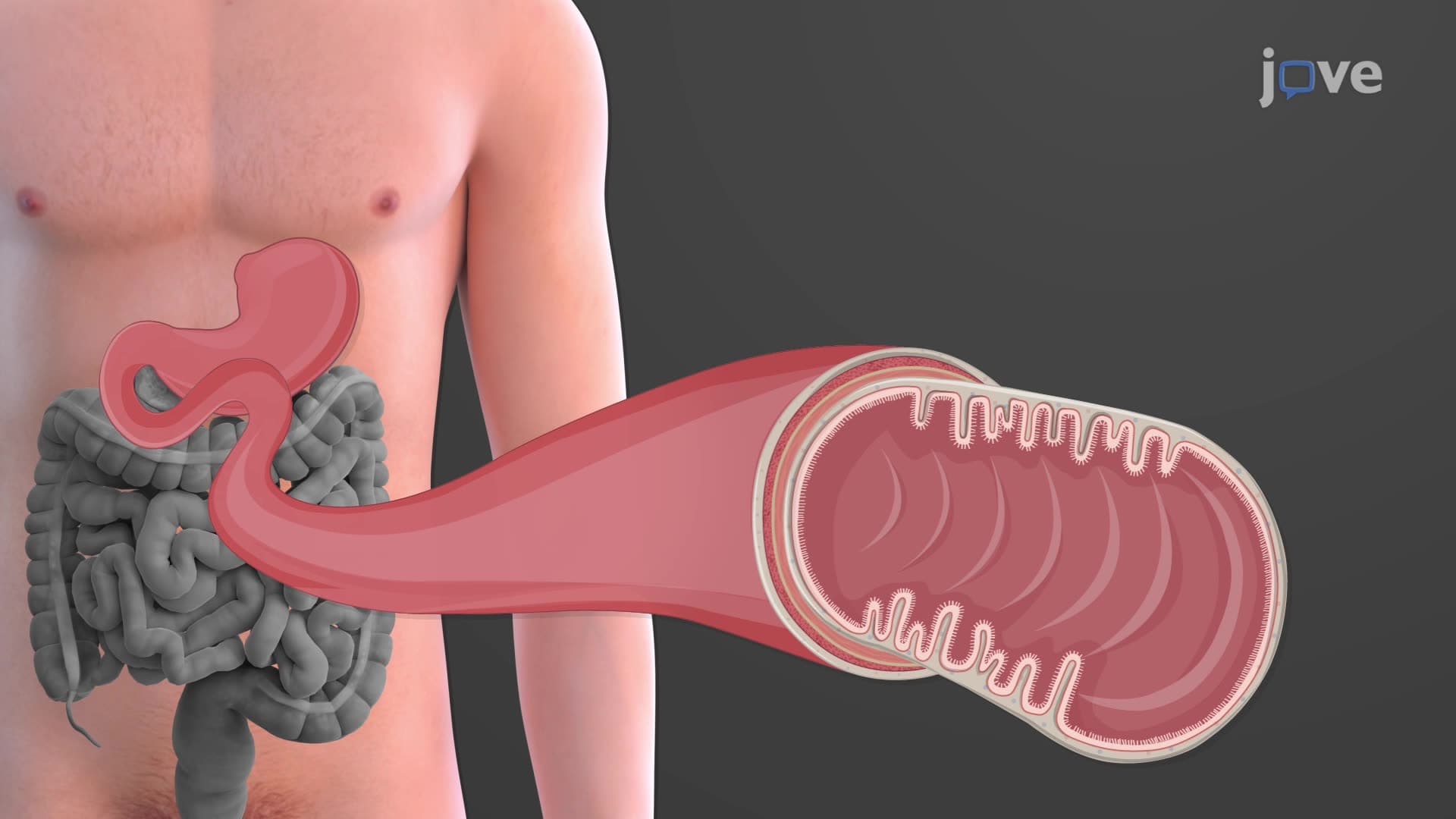
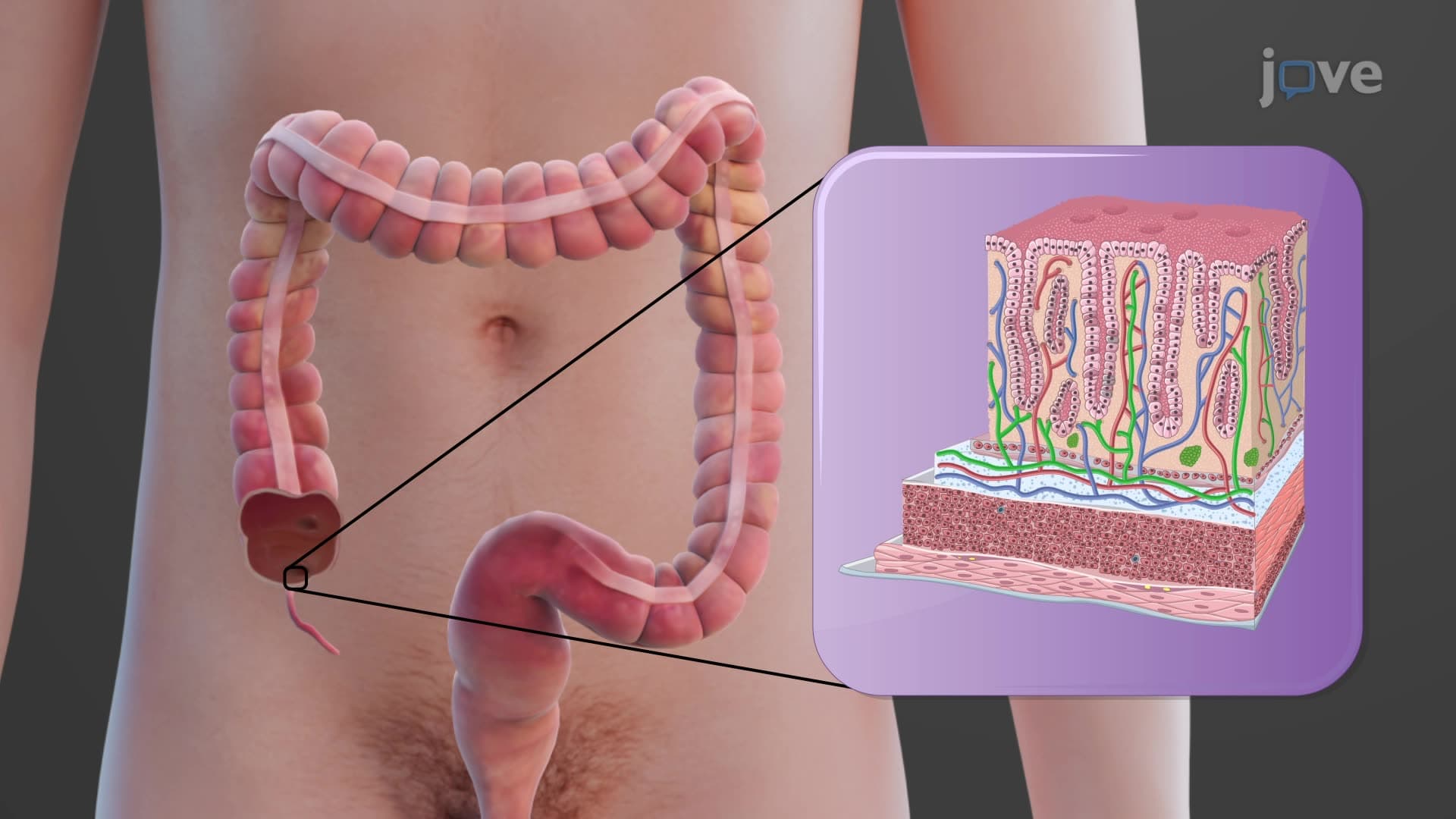
The large intestine anatomy biology encompasses a complex tubular structure that forms the final segment of the human digestive system. Unlike the small intestine's narrow diameter and extensive length, the large intestine measures approximately 5 feet long with a significantly wider diameter of about 2.5 inches. This anatomical arrangement allows for efficient water absorption and waste compaction, processing roughly 1-2 liters of liquid chyme daily into semi-solid fecal matter.
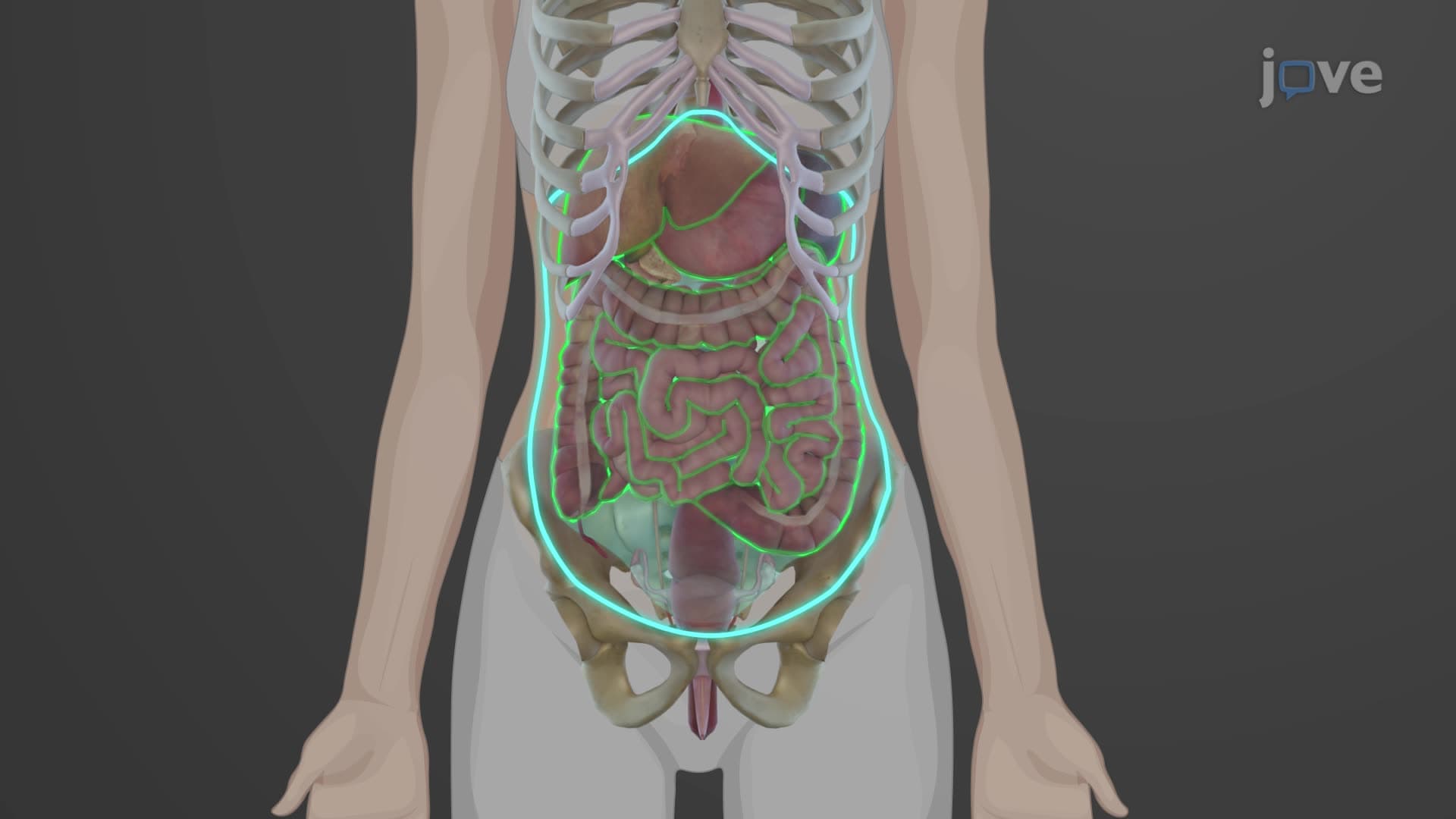
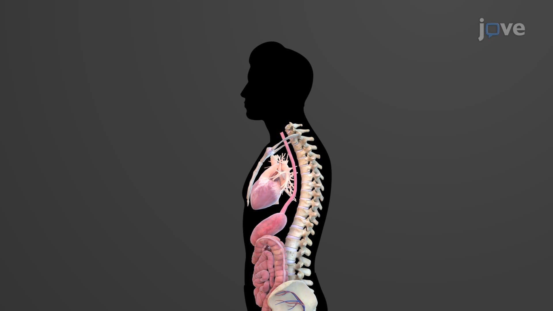
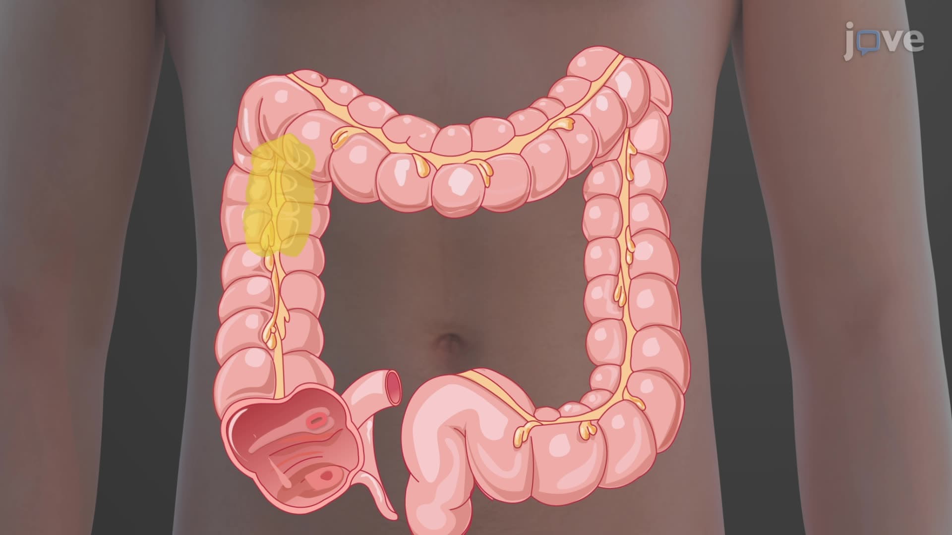
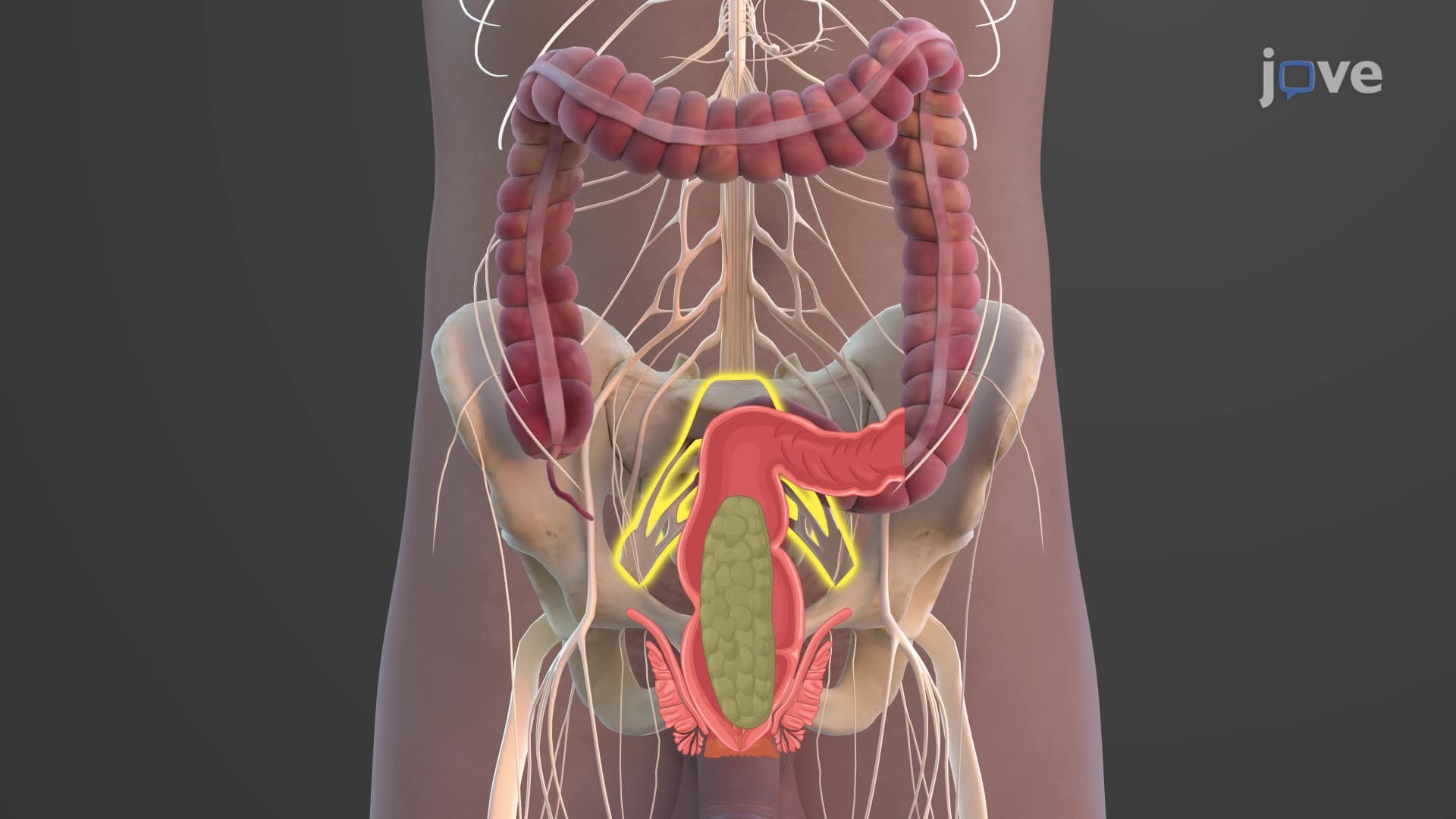
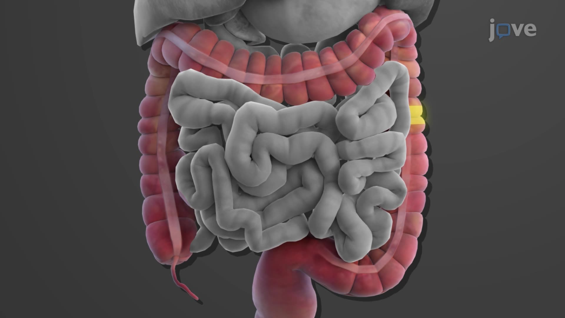
Medical students studying for the MCAT and USMLE Step 1 must understand that the large intestine's positioning creates a distinctive frame around the small intestine. Beginning at the ileocecal valve in the right iliac fossa, this organ ascends along the right side of the abdomen, crosses horizontally below the liver and stomach, then descends on the left side before forming an S-shaped curve toward the pelvis.
Colon Structure Function Explained Through Regional Anatomy
Colon structure function explained requires examining each anatomical segment's unique characteristics. The cecum serves as the initial expanded chamber, typically measuring 6-9 centimeters in diameter. The vermiform appendix, a small tubular structure averaging 3-4 inches long, extends from the cecum's posteromedial wall. While often considered vestigial, recent research from Harvard Medical School suggests the appendix may serve as a bacterial reservoir for gut microbiome recovery.
The ascending colon travels vertically for approximately 15 centimeters before reaching the hepatic flexure near the liver's right lobe. This anatomical landmark proves crucial during colonoscopy procedures performed at facilities like Cleveland Clinic. The transverse colon spans horizontally across the upper abdomen, measuring roughly 45 centimeters and forming the splenic flexure near the spleen's lower pole.
The descending colon continues vertically down the left abdomen for about 25 centimeters, while the sigmoid colon creates characteristic S-shaped curves measuring 15-20 centimeters. This region's mobility and curvature make it particularly susceptible to conditions like diverticulosis, especially prevalent in American adults over 60.
What is the Large Intestine in Digestive Anatomy: Functional Integration
Understanding what is the large intestine in digestive anatomy requires recognizing its integration with other body systems. The rectum and anal canal represent the terminal structures, with the rectum serving as a temporary storage reservoir measuring 12-15 centimeters. The anal canal's 2-4 centimeter length contains both internal and external sphincters, providing voluntary and involuntary control mechanisms essential for continence.
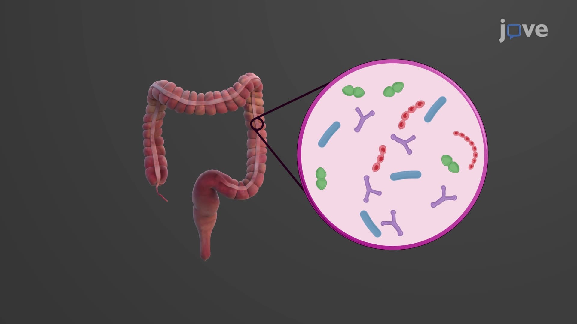
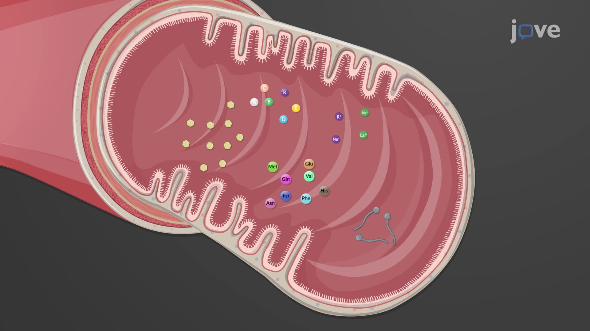
Pre-med students preparing for AP Biology exams should note that large intestine function extends beyond waste elimination. This organ houses over 700 bacterial species comprising the gut microbiome, producing essential vitamins like vitamin K and certain B vitamins. Water and electrolyte absorption occurs primarily in the ascending and transverse regions, concentrating liquid waste from 85% water content to approximately 75% in formed stool.
Clinical Significance and Pathological Considerations
The large intestine's anatomical structure directly impacts common pathological conditions studied in American medical curricula. Appendicitis typically occurs when the narrow appendiceal lumen becomes obstructed, leading to inflammation and potential rupture. Colorectal cancer, the third most common cancer in the United States according to the American Cancer Society, frequently develops in the sigmoid colon due to prolonged fecal contact time.
Inflammatory bowel diseases like ulcerative colitis specifically target the large intestine's mucosal lining, unlike Crohn's disease which can affect any GI tract region. Understanding normal anatomical landmarks helps healthcare providers at institutions like Mayo Clinic and Johns Hopkins identify pathological changes during diagnostic procedures and surgical interventions.
Frequently Asked Questions
The large intestine is the final section of the digestive tract, extending from the ileocecal valve to the anus, measuring about 5 feet long with a wider diameter than the small intestine. Unlike the small intestine which focuses on nutrient absorption, the large intestine primarily absorbs water and electrolytes while forming and storing waste. Its distinctive anatomical features include the cecum, four colon regions, rectum, and specialized sphincter controls for waste elimination.
For MCAT success, focus on understanding the large intestine's role in water absorption and waste formation, particularly in the ascending and transverse colon regions. Test questions often emphasize the ileocecal valve's function, the appendix's anatomical position, and the rectum's storage capacity. Practice identifying anatomical landmarks like hepatic and splenic flexures, as these frequently appear in passage-based questions about digestive physiology and pathological conditions.
AP Biology exams typically test large intestine concepts through questions about structure-function relationships and evolutionary adaptations. Students should understand how the colon's increased diameter facilitates water absorption, why the sigmoid colon's S-shape affects transit time, and how the appendix represents vestigial organ evidence. Focus on connecting anatomical features to physiological processes like bacterial fermentation and vitamin production for comprehensive exam preparation.
In clinical settings like those at Cleveland Clinic, large intestine function directly impacts patient recovery through fluid balance, medication absorption, and infection prevention. Post-surgical patients require careful monitoring of bowel function, as prolonged immobility can lead to complications like ileus or constipation. The large intestine's bacterial populations also influence antibiotic effectiveness and C. difficile infection risk, making gut health crucial for overall patient outcomes.
No advanced chemistry background is required to master large intestine anatomy concepts. Basic understanding of water absorption, pH changes, and bacterial fermentation processes provides sufficient foundation for high school and introductory college courses. Focus on anatomical relationships, structural organization, and physiological functions rather than complex biochemical pathways. Most educational programs introduce these concepts gradually, building from simple anatomical identification to more complex physiological processes.
Use the mnemonic "Can't Always Tell Digestive System" for Cecum, Ascending, Transverse, Descending, Sigmoid regions, then practice with anatomical diagrams and virtual models. Create visual associations linking anatomical positions to familiar landmarks - ascending colon "climbs toward the liver," transverse colon "crosses like a bridge." Regular practice with labeled diagrams and self-testing using flashcards helps reinforce spatial relationships crucial for practical exams and clinical applications.
Large intestine anatomy provides an excellent foundation for understanding gastrointestinal physiology, including water balance, electrolyte absorption, and gut microbiome interactions. This knowledge directly connects to topics like bacterial symbiosis, vitamin synthesis, and immune system development through gut-associated lymphoid tissue. Advanced students can explore connections to endocrinology through gut hormone production and neuroscience through the enteric nervous system's complex regulatory mechanisms.
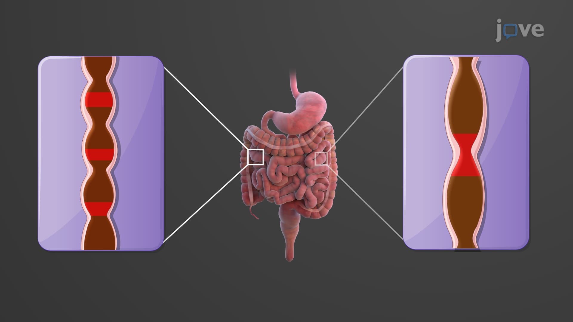
Pre-med students should investigate the enteric nervous system's role in large intestine motility, studying peristaltic patterns and defecation reflexes that appear on MCAT and medical school exams. Explore gut microbiome diversity, antibiotic resistance patterns, and the appendix's potential immunological functions based on recent research from institutions like Harvard and Stanford. Understanding inflammatory bowel disease pathophysiology and colorectal cancer development provides valuable clinical context for future medical studies.
Related Micro-courses
25 Concepts

25 Concepts

20 Concepts