Loading subject page…


89,749 views

Video Summary: What Is Liver Histology
Ever wondered why your liver can regenerate after damage while your heart cannot? Liver histology biology reveals the remarkable cellular architecture that makes this possible. The microscopic structure includes specialized hepatocytes arranged in three-dimensional plates, intricate portal triads, and a unique blood flow system through sinusoids. Students at Johns Hopkins University study these components to understand how liver transplants work and why hepatitis affects specific cell types. Watch the full video on JoVE Coach to master this concept with expert-led visuals and step-by-step explanations.
Liver histology biology forms the cornerstone for understanding how this vital organ performs over 500 different functions. Unlike other organs with simpler architectural patterns, the liver's microscopic structure reflects its complex metabolic roles. Medical students preparing for the MCAT and AP Biology students both encounter this topic as essential knowledge for understanding human physiology.
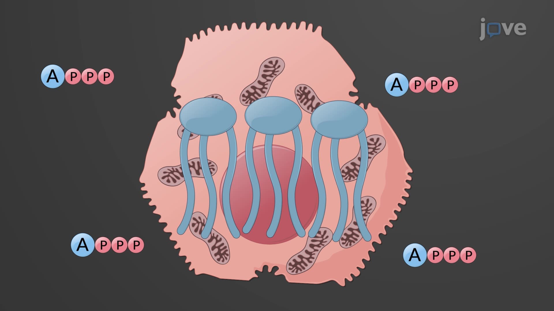
Hepatocytes represent approximately 80% of the liver's cellular mass and demonstrate remarkable versatility. These specialized epithelial cells arrange themselves in hepatic laminae-three-dimensional plates that maximize surface area for metabolic exchange. Each hepatocyte contains abundant mitochondria and endoplasmic reticulum, reflecting their intense metabolic activity.
In clinical practice, hepatocyte damage from conditions like alcoholic liver disease or viral hepatitis directly correlates with elevated liver enzymes (ALT and AST) measured in blood tests at hospitals like Mayo Clinic or Cleveland Clinic. College students studying biochemistry learn how hepatocytes synthesize albumin, clotting factors, and cholesterol while simultaneously detoxifying ammonia to urea.
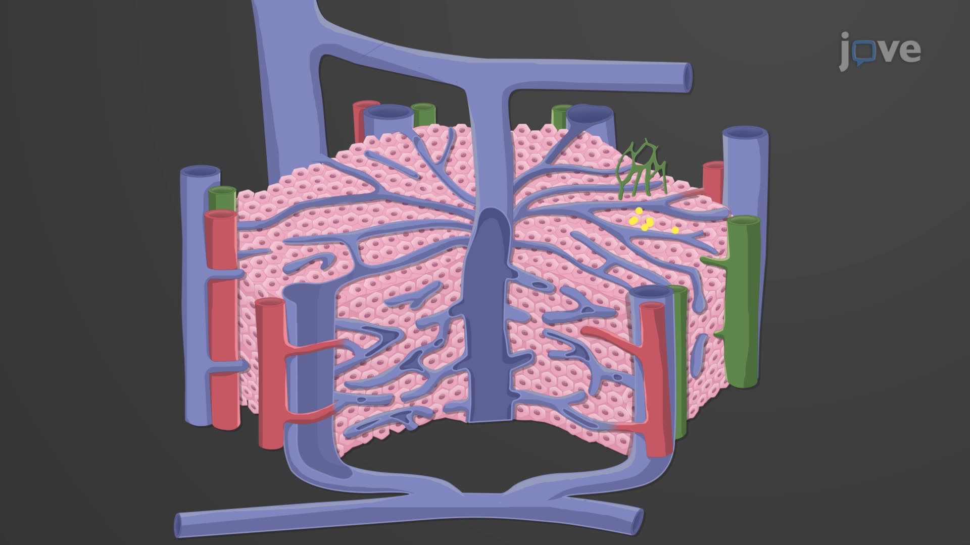
The portal triad liver histology reveals a sophisticated supply system positioned at hepatic plate boundaries. Each triad contains a bile duct, hepatic artery branch, and portal vein branch-creating a functional unit that supports hepatocyte metabolism. This arrangement ensures hepatocytes receive both oxygen-rich arterial blood and nutrient-rich portal blood from the digestive tract.
Students preparing for the USMLE Step 1 must understand how portal hypertension develops when this system becomes compromised, leading to complications like esophageal varices seen in cirrhotic patients at major medical centers.
Liver sinusoid histology demonstrates unique features not found in typical capillaries. These specialized blood vessels contain fenestrations (small pores) that allow direct contact between blood plasma and hepatocytes. Stellate macrophages embedded in sinusoidal walls serve as the liver's primary filtration system, removing bacteria, cellular debris, and aged blood cells.
This knowledge proves essential for nursing students taking the NCLEX-RN, as they must understand how liver dysfunction affects immune function and blood cell counts in hospitalized patients.
Related Micro-courses