Loading subject page…

Micro-course: Digestive System
89,747 views
Learn key concepts and earn your certificate.





Pancreas

00:00
01:19
Video Summary: What Is Pancreas
Did you know that the pancreas produces over a liter of digestive juice daily while simultaneously regulating blood sugar levels? Understanding pancreas anatomy biology reveals how this dual-functioning organ serves as both a digestive powerhouse and hormonal control center. For instance, when diabetes patients at Johns Hopkins University Medical Center receive pancreatic transplants, surgeons must carefully preserve both the exocrine ducts and endocrine islets to restore full pancreatic function. The What is Pancreas concept becomes essential for grasping how one organ manages two critical body systems simultaneously. Watch the full video on JoVE Coach to master this concept with expert-led visuals and step-by-step explanations.
- Understand What is Pancreas by identifying its anatomical location, structural divisions, and dual functional roles in human physiology
- Identify the three main anatomical regions of the pancreas: the broad head within the duodenal loop, the elongated body, and the rounded tail extending toward the spleen
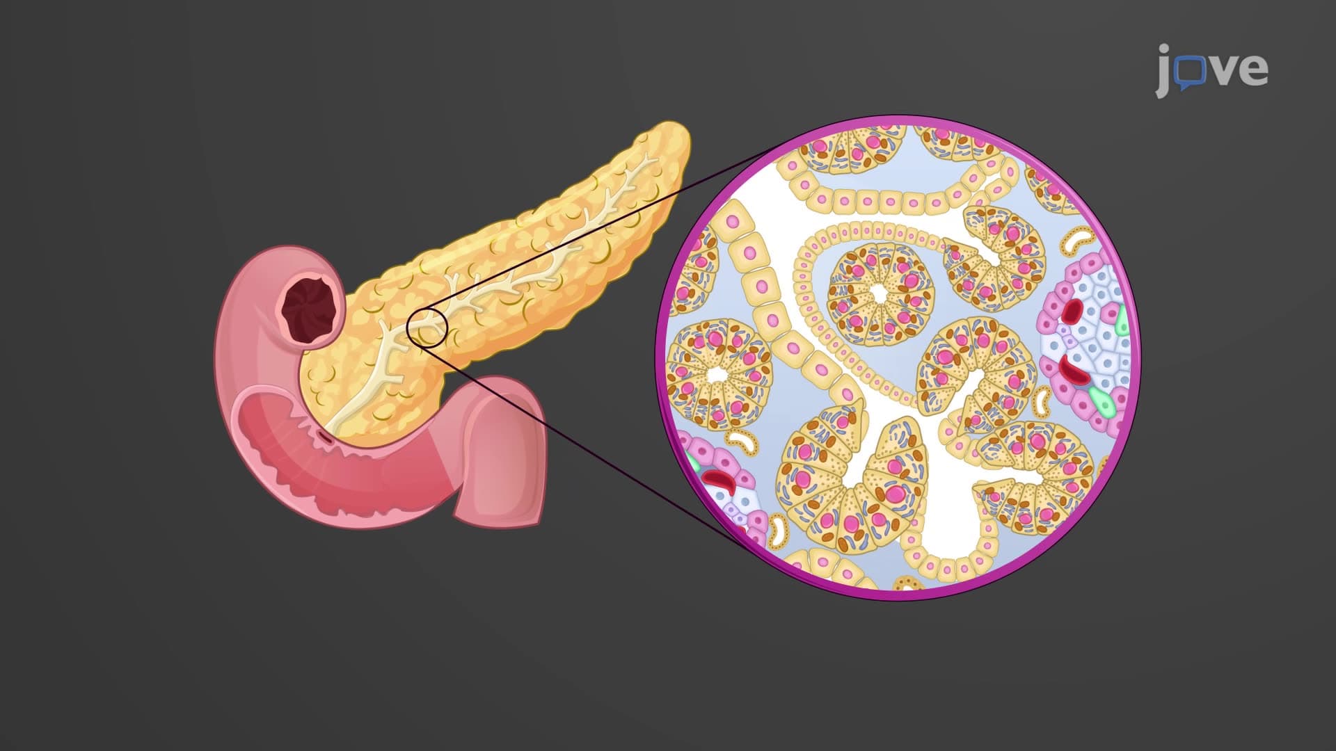
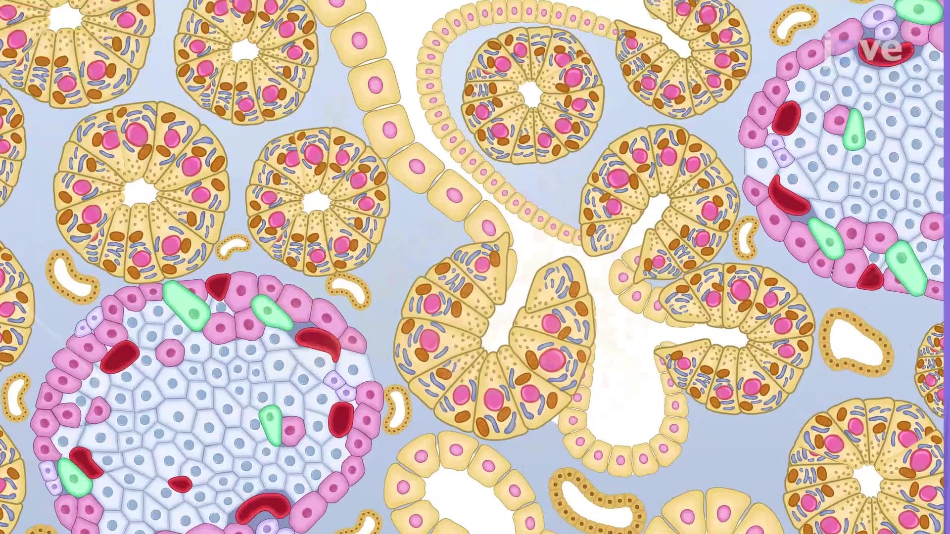
- Analyze the exocrine pancreas structure, including acinar cells, zymogen granules, and the pancreatic duct system that delivers digestive enzymes to the duodenum
- Compare the functions of the main pancreatic duct and accessory duct in enzyme delivery, particularly their relationship with the sphincter of Oddi
- Evaluate the endocrine pancreas components, specifically the islets of Langerhans and their hormone-secreting capabilities for glucose regulation
- Apply knowledge of pancreatic anatomy to understand digestive disorders like pancreatitis and metabolic conditions such as diabetes mellitus
- Recognize the cellular organization of pancreatic acini and their role in producing inactive digestive enzyme precursors (zymogens)
- Explore the integrated relationship between pancreatic exocrine and endocrine functions in maintaining homeostasis
Understanding Pancreas Anatomy and Location
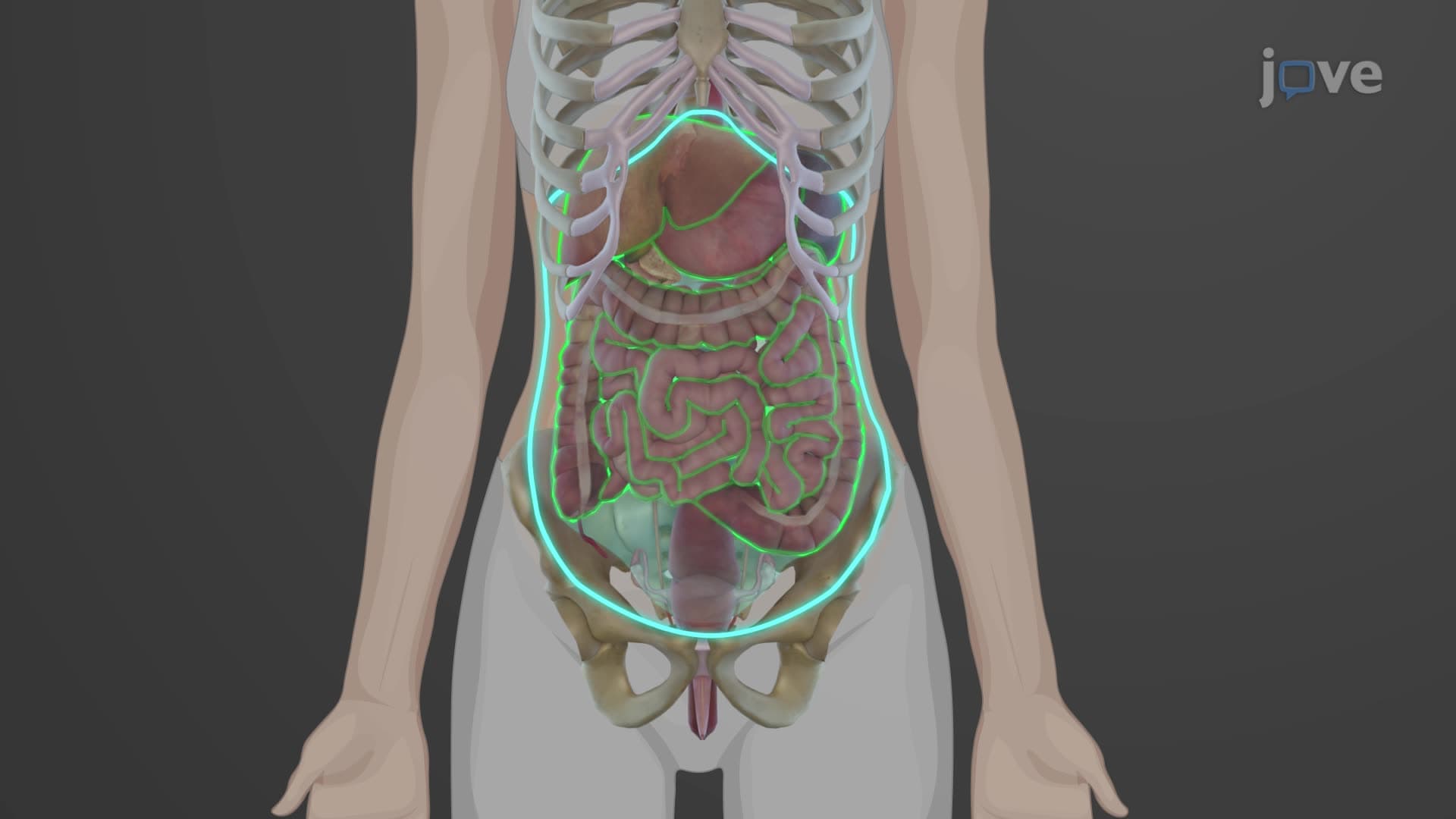
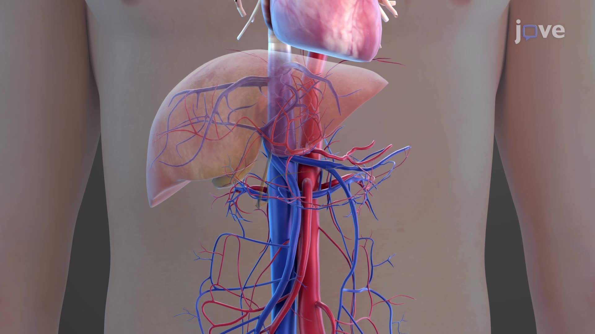
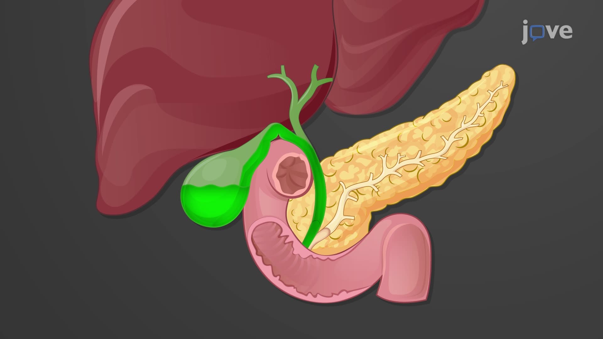
The pancreas anatomy biology reveals a remarkable organ positioned strategically behind the stomach in the retroperitoneal space. This pinkish-gray structure spans approximately 6 inches horizontally, extending from the C-shaped curve of the duodenum to the hilum of the spleen. Medical students at Harvard Medical School often study cadaveric specimens to appreciate how the pancreas lies so deep within the abdominal cavity that it's rarely palpable during physical examination, making pancreatic disorders challenging to diagnose clinically.
Pancreatic Structure and Regional Organization
The pancreas structure function explained becomes clearer when examining its three distinct anatomical regions. The pancreatic head, the broadest portion, nestles within the duodenal loop and contains the uncinate process-a hook-like extension that wraps around major blood vessels. The pancreatic body forms the central portion, while the tail tapers as it approaches the spleen. This anatomical arrangement is crucial for surgical procedures; pancreaticoduodenectomy (Whipple procedure) performed at Mayo Clinic requires precise knowledge of these regional boundaries to preserve surrounding structures.
Exocrine Pancreas: Digestive Enzyme Production
The exocrine component represents about 95% of pancreatic tissue and demonstrates sophisticated cellular organization. Pancreatic acinar cells contain abundant rough endoplasmic reticulum and distinctive zymogen granules storing inactive enzyme precursors like trypsinogen, chymotrypsinogen, and proelastase. This safety mechanism prevents premature enzyme activation that could cause autodigestion. The pancreatic duct anatomy includes the main pancreatic duct (of Wirsung) and accessory duct (of Santorini), which transport enzyme-rich pancreatic juice to the duodenum. Students preparing for the MCAT must understand how the sphincter of Oddi coordinates bile and pancreatic juice release during digestion.
Endocrine Pancreas: Hormonal Regulation
The islet Langerhans pancreas structures function as mini-endocrine organs scattered throughout pancreatic tissue. These islets contain alpha cells producing glucagon, beta cells secreting insulin, delta cells releasing somatostatin, and PP cells generating pancreatic polypeptide. The precise cellular arrangement within islets-with beta cells forming the core and alpha cells at the periphery-optimizes paracrine signaling between different cell types. This organization becomes clinically significant in Type 1 diabetes, where autoimmune destruction specifically targets beta cells while sparing other islet cell populations. AP Biology students frequently encounter questions about insulin's role in glucose homeostasis and how pancreatic dysfunction leads to diabetic complications observed in over 34 million Americans.
Frequently Asked Questions
The pancreas is a mixed exocrine-endocrine gland positioned behind the stomach in the retroperitoneal space. It extends horizontally from the duodenum to the spleen and measures approximately 6 inches in length. This vital organ produces digestive enzymes and hormones essential for metabolism and blood sugar regulation.
MCAT questions frequently test pancreatic enzyme activation, insulin signaling pathways, and anatomical relationships with surrounding organs. AP Biology exams emphasize hormone regulation, particularly insulin-glucagon interactions and cellular organization within pancreatic islets. Students should master both structural components and functional mechanisms for optimal performance.
USMLE Step 1 extensively covers pancreatic pathophysiology, including acute and chronic pancreatitis, pancreatic cancer, and diabetes mellitus. Key topics include zymogen activation mechanisms, islet cell tumors, and anatomical considerations for pancreatic surgery. Understanding embryological development and vascular supply also appears in clinical vignettes.
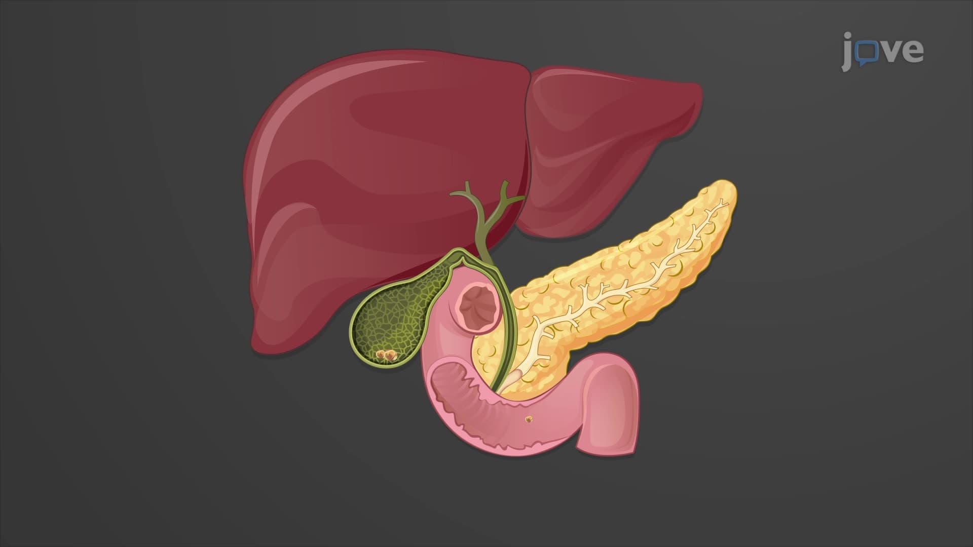
Emergency departments at institutions like Cleveland Clinic frequently treat acute pancreatitis caused by gallstones or alcohol abuse. Patients present with severe epigastric pain radiating to the back, elevated pancreatic enzymes, and potentially life-threatening complications. Understanding pancreatic anatomy helps clinicians interpret CT scans and plan appropriate interventions.
Pancreatic anatomy builds logically on basic digestive system knowledge taught in freshman biology. Students who understand enzyme function and hormone signaling find pancreatic concepts accessible. The dual exocrine-endocrine nature initially seems complex but becomes clearer when studied as two integrated systems working toward common physiological goals.
Create concept maps linking pancreatic structure to function, emphasizing how anatomical features support specific roles. Practice drawing pancreatic anatomy from memory, labeling ducts, islets, and cellular components. Use clinical correlation exercises to connect normal anatomy with pathological conditions frequently tested on standardized exams.
Advanced students should investigate pancreatic development and regeneration, particularly stem cell research conducted at institutions like Stanford University. Explore pancreatic transplantation procedures, artificial pancreas technology for diabetes management, and emerging gene therapies targeting pancreatic disorders currently in clinical trials.
College anatomy exams combine practical identification using cadaveric specimens or anatomical models with written questions about functional relationships. Students must recognize pancreatic structures in cross-sectional anatomy and explain how anatomical organization supports both digestive and endocrine functions through integrated essay responses.
Related Micro-courses
25 Concepts

25 Concepts

20 Concepts