Loading subject page…

Micro-course: Digestive System
89,747 views
Learn key concepts and earn your certificate.



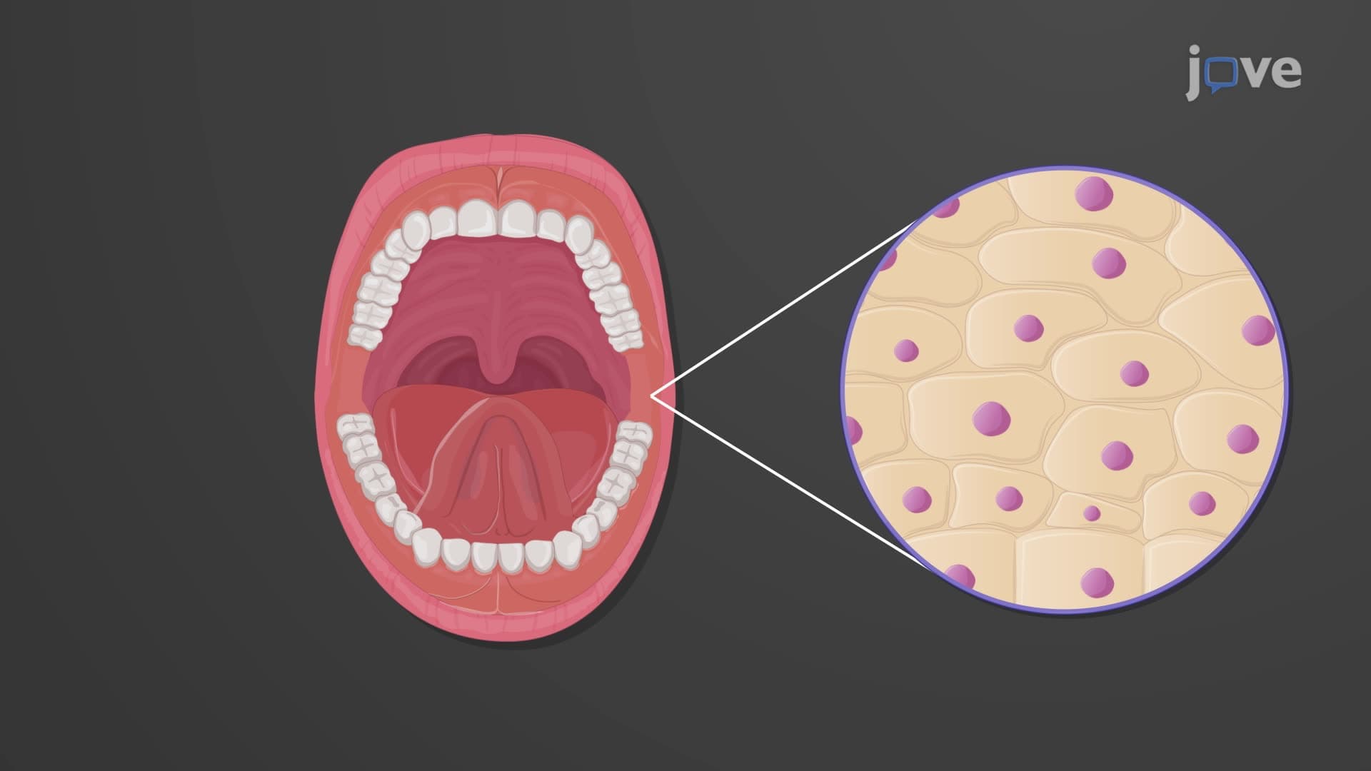

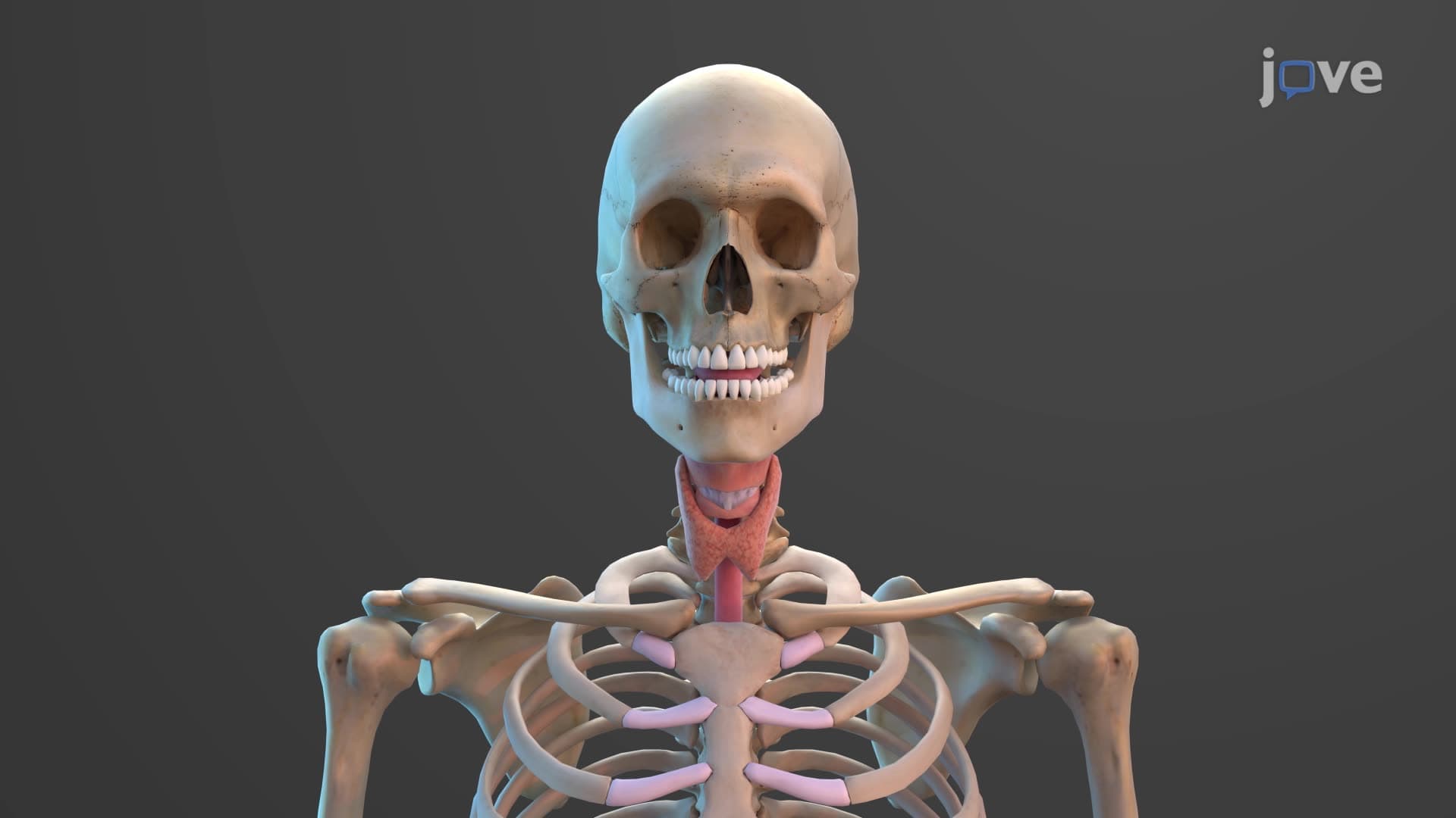
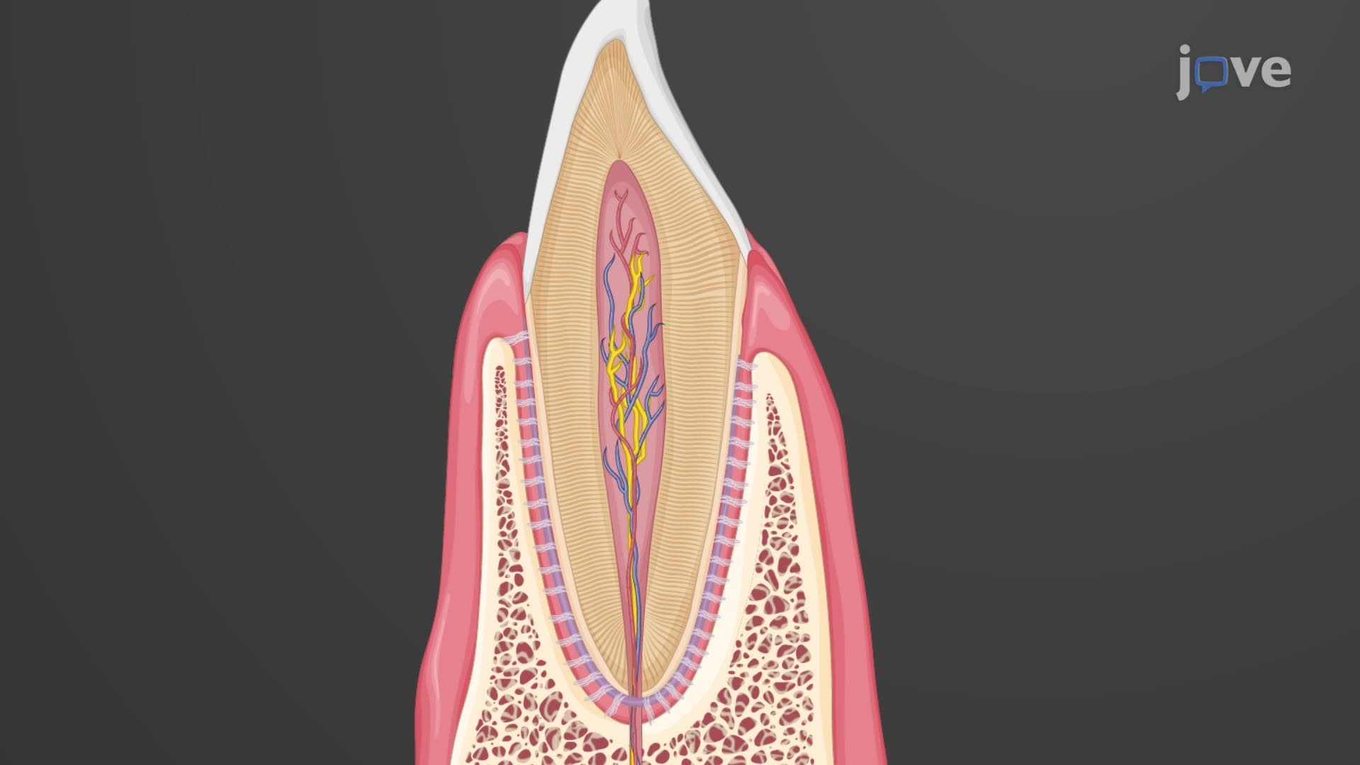
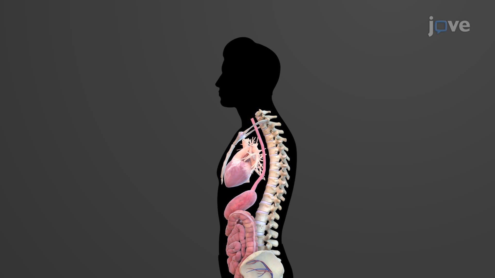
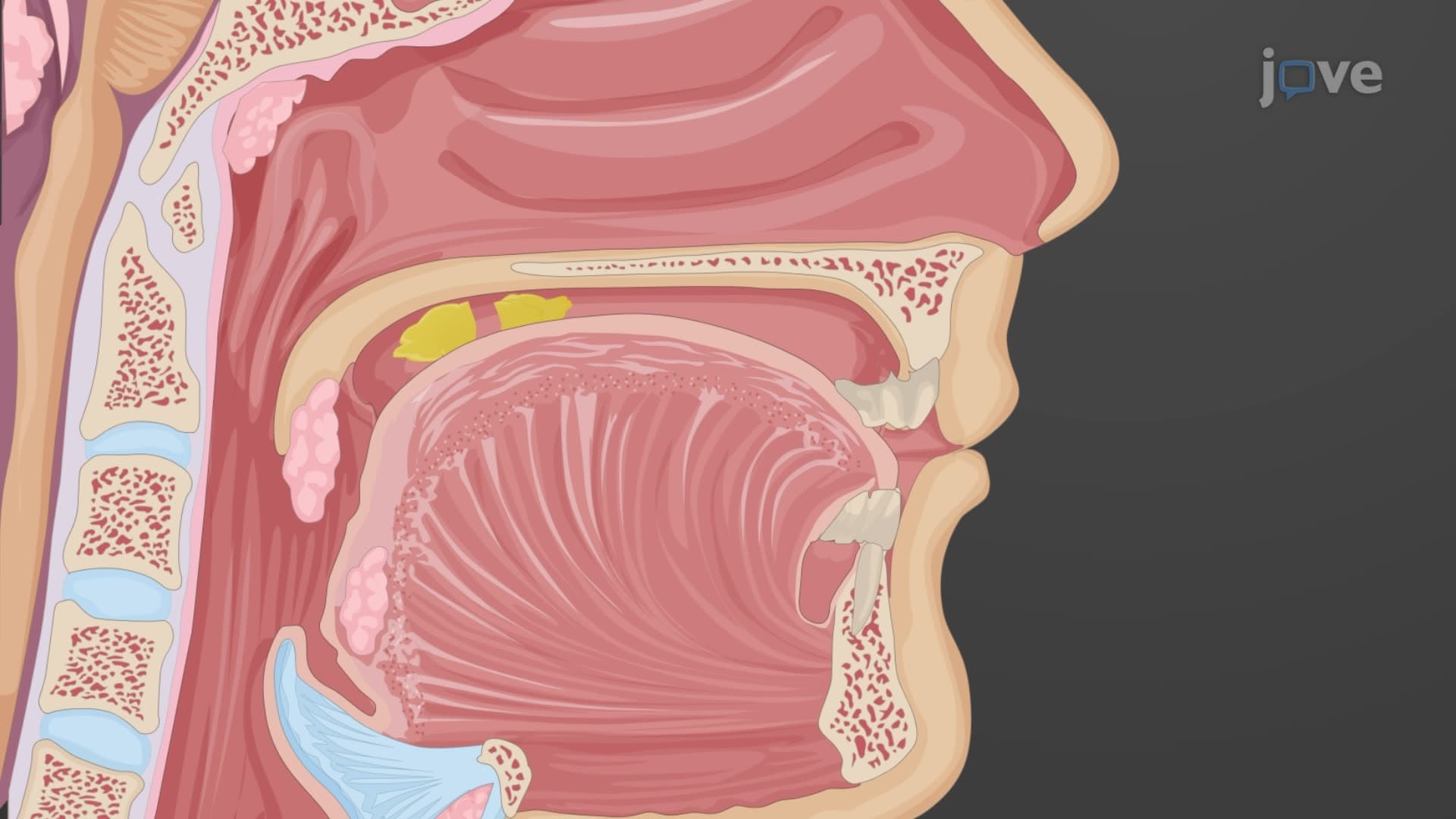
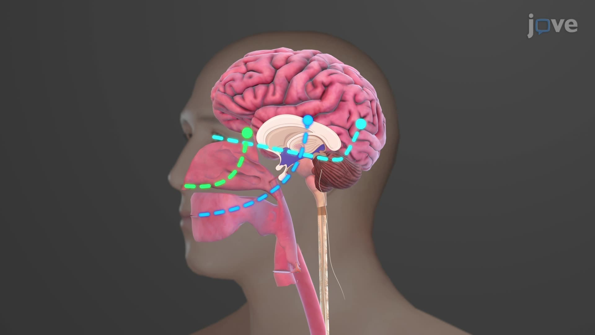
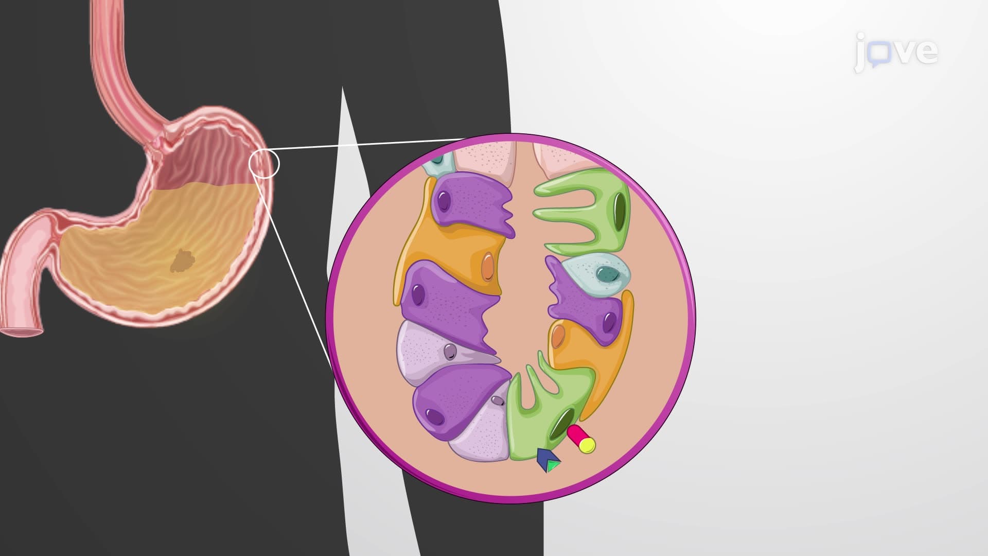
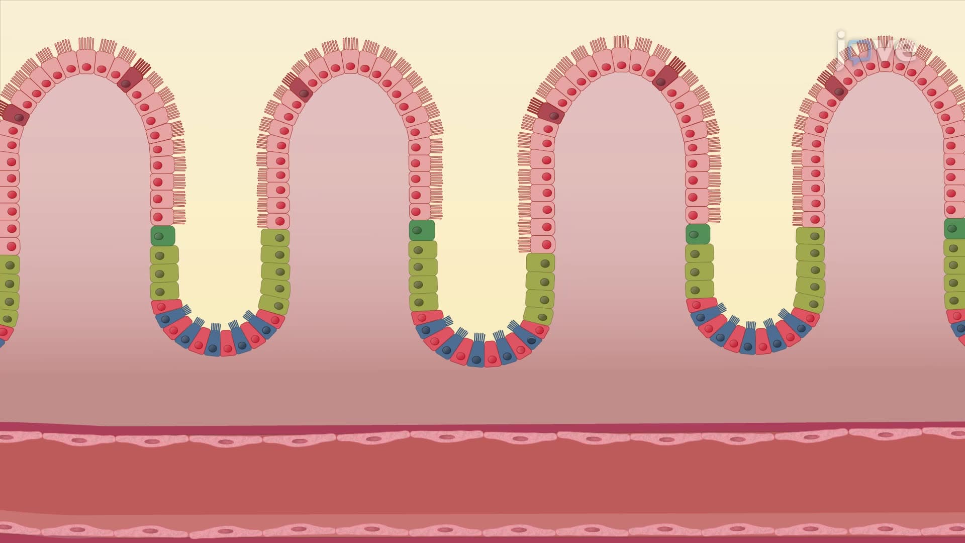
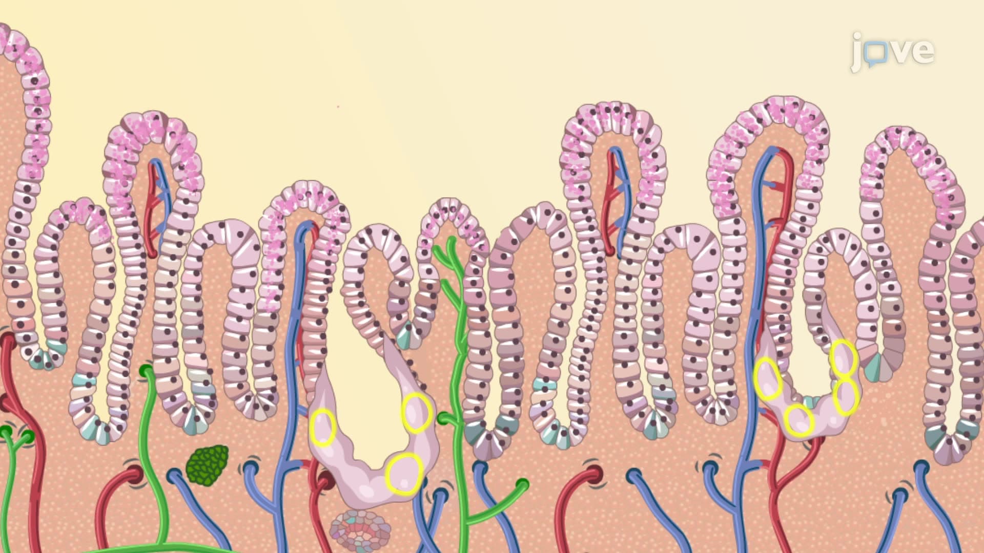

Tongue

00:00
01:01
Video Summary: What Is Tongue
Did you know your tongue contains eight separate muscles working together every time you say "supercalifragilisticexpialidocious"? Understanding tongue anatomy biology reveals how this remarkable organ enables everything from tasting your favorite pizza to delivering a graduation speech. Medical students at Johns Hopkins University study tongue structure extensively because speech pathologists and ENT doctors must understand its intricate design. This muscular organ serves as both an accessory digestive structure and a sophisticated communication tool. Watch the full video on JoVE Coach to master this concept with expert-led visuals and step-by-step explanations.
- Understand what is tongue anatomy and its dual role as both a digestive accessory organ and speech apparatus
- Identify the two main categories of tongue muscles: intrinsic muscles that shape the tongue and extrinsic muscles that position it
- Analyze how tongue structure function explained through its muscular composition enables complex movements for speech and swallowing
- Compare intrinsic and extrinsic muscle functions in tongue positioning and food bolus formation
- Recognize the lingual frenulum's role in anchoring the tongue and preventing excessive backward movement
- Evaluate how the median sulcus creates bilateral symmetry essential for coordinated tongue movements
- Apply knowledge of tongue anatomy to understand speech disorders and swallowing difficulties
- Explore the relationship between tongue positioning and successful food transport from mouth to pharynx
What is Tongue: The Multifunctional Oral Organ
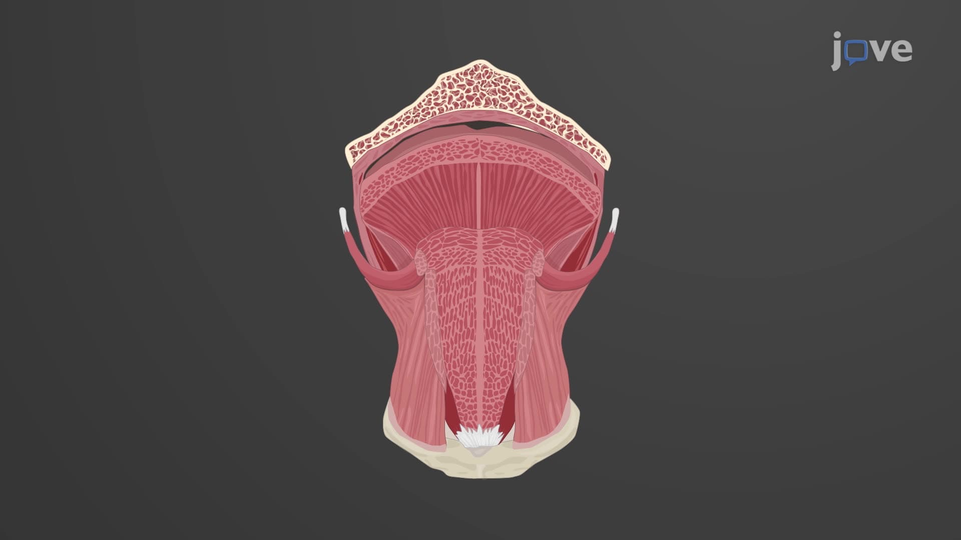
The tongue represents one of the most versatile organs in human anatomy, serving critical roles in digestion, communication, and sensory perception. As an accessory digestive organ, the tongue occupies the oral cavity floor while extending into the oropharynx, creating a bridge between voluntary and involuntary phases of swallowing. This positioning allows the tongue to manipulate food during mastication and initiate the swallowing reflex.
Intrinsic vs. Extrinsic Tongue Muscle Architecture
Understanding intrinsic extrinsic tongue muscle differences is crucial for grasping tongue functionality. Intrinsic muscles, including the superior longitudinal, inferior longitudinal, transverse, and vertical muscles, operate entirely within the tongue without bone attachments. These muscles enable fine motor control necessary for precise speech articulation-think about how your tongue moves differently when pronouncing "th" versus "l" sounds. Students preparing for the MCAT often encounter questions about intrinsic muscle functions in speech pathology contexts.
Extrinsic muscles-the genioglossus, hyoglossus, styloglossus, and palatoglossus-originate from skull bones and the soft palate, providing gross motor control for tongue positioning. The genioglossus, the largest extrinsic muscle, protrudes and depresses the tongue, while the hyoglossus retracts and depresses it. This muscular coordination is essential for creating the tongue movements required in American English phonemes.
Structural Features Supporting Tongue Function
The lingual frenulum tongue attachment prevents excessive posterior movement that could obstruct the airway-a safety mechanism preventing accidental choking. This mucosal fold's clinical significance becomes apparent in conditions like ankyloglossia (tongue-tie), where restricted frenulum length affects speech development and feeding in infants.
The median sulcus creates bilateral symmetry, ensuring coordinated muscle contractions. This groove reflects the tongue's embryological development from paired lateral swellings, explaining why tongue paralysis typically affects one side more than the other following stroke or cranial nerve injuries.
Clinical and Academic Applications
For AP Biology students, tongue anatomy exemplifies how structure determines function. The tongue's muscular composition enables bolus formation-the process of shaping chewed food into a cohesive mass suitable for swallowing. This concept frequently appears on standardized tests when discussing digestive system mechanics.
Pre-health students studying for the MCAT should understand how tongue dysfunction affects both nutrition and communication. Speech-language pathologists at institutions like Northwestern University study tongue biomechanics to treat articulation disorders, while gastroenterologists examine tongue function in dysphagia (swallowing difficulties) common among elderly patients in American healthcare settings.
Frequently Asked Questions
The tongue is a muscular organ composed of intrinsic and extrinsic muscles that enables speech, swallowing, and taste perception. Understanding its anatomy is crucial for healthcare professionals treating speech disorders, swallowing difficulties, and oral health issues. Medical and dental students must master tongue structure to diagnose conditions affecting communication and nutrition.
MCAT questions often test understanding of how muscular coordination enables complex behaviors like speech and swallowing. Students who grasp intrinsic versus extrinsic muscle roles can tackle passages about neurological conditions affecting tongue function. This knowledge also applies to behavioral biology questions about communication mechanisms.
AP Biology exams commonly feature questions about structure-function relationships, using the tongue as an example of how skeletal muscle organization enables specific behaviors. Students should understand how intrinsic muscles create shape changes while extrinsic muscles control positioning, demonstrating evolutionary adaptations for survival functions.
American speech-language pathologists use tongue anatomy knowledge to treat articulation disorders, especially in children with developmental delays or adults recovering from strokes. They design exercises targeting specific muscle groups-intrinsic muscles for sound precision and extrinsic muscles for tongue placement during speech therapy sessions.
Not at all! While the tongue contains multiple muscle types, thinking of it as having "shapers" (intrinsic muscles) and "movers" (extrinsic muscles) simplifies the concept. Students with basic knowledge of skeletal muscle can easily grasp these functional categories without advanced anatomy background.
Create muscle function charts comparing intrinsic versus extrinsic actions, then practice with speech exercises to feel how your tongue moves. Relate muscle actions to familiar words-notice how your tongue positioning differs between "tea" and "key" sounds to understand practical applications.
Students can investigate cranial nerve innervation of tongue muscles, taste bud distribution across papillae types, or embryological tongue development. These topics prepare students for advanced courses in neuroscience, gastroenterology, or speech pathology graduate programs.
The lingual frenulum's length and flexibility affect speech development and feeding ability. Pediatricians and ENT specialists frequently evaluate frenulum restrictions in newborns and children with feeding difficulties or speech delays, making this knowledge essential for healthcare providers working with pediatric populations in American clinical settings.
Related Micro-courses
25 Concepts

25 Concepts

20 Concepts