Loading subject page…

- Anatomy and Physiology
- Digestive System
- Tooth Anatomy
Micro-course: Digestive System
89,747 views
Learn key concepts and earn your certificate.





Tooth Anatomy

00:00
01:21
Video Summary: What Is Tooth Anatomy
Ever wonder why dentists can tell exactly which tooth hurts just by looking at an X-ray? Tooth anatomy biology reveals the intricate structure that makes each tooth uniquely suited for its function. From the visible crown protected by enamel to the hidden root system anchored in your jawbone, understanding what is the anatomy of a tooth helps explain why a root canal procedure at your local US dental office targets specific layers. Each tooth contains multiple specialized tissues working together-enamel, dentin, pulp, and cementum-creating a living structure more complex than it appears. Watch the full video on JoVE Coach to master this concept with expert-led visuals and step-by-step explanations.
- Understand the fundamental components of tooth anatomy and identify the crown, neck, and root regions of any tooth structure
- Recognize the four primary tissues that comprise tooth anatomical structure: enamel, dentin, pulp, and cementum
- Compare root variations among different tooth types, from single-rooted incisors to three-rooted upper molars
- Analyze the relationship between dentin's tubular structure and the odontoblasts that maintain tooth integrity
- Identify the pulp cavity's role in housing nerves and blood vessels that keep teeth alive
- Evaluate how the periodontal ligament and cementum work together to anchor teeth in the jawbone
- Apply knowledge of the apical foramen's location to understand how dental procedures access tooth interiors
- Explore the protective functions of enamel and how its mineral composition makes it the hardest substance in the human body
Understanding the Fundamentals of Tooth Anatomy
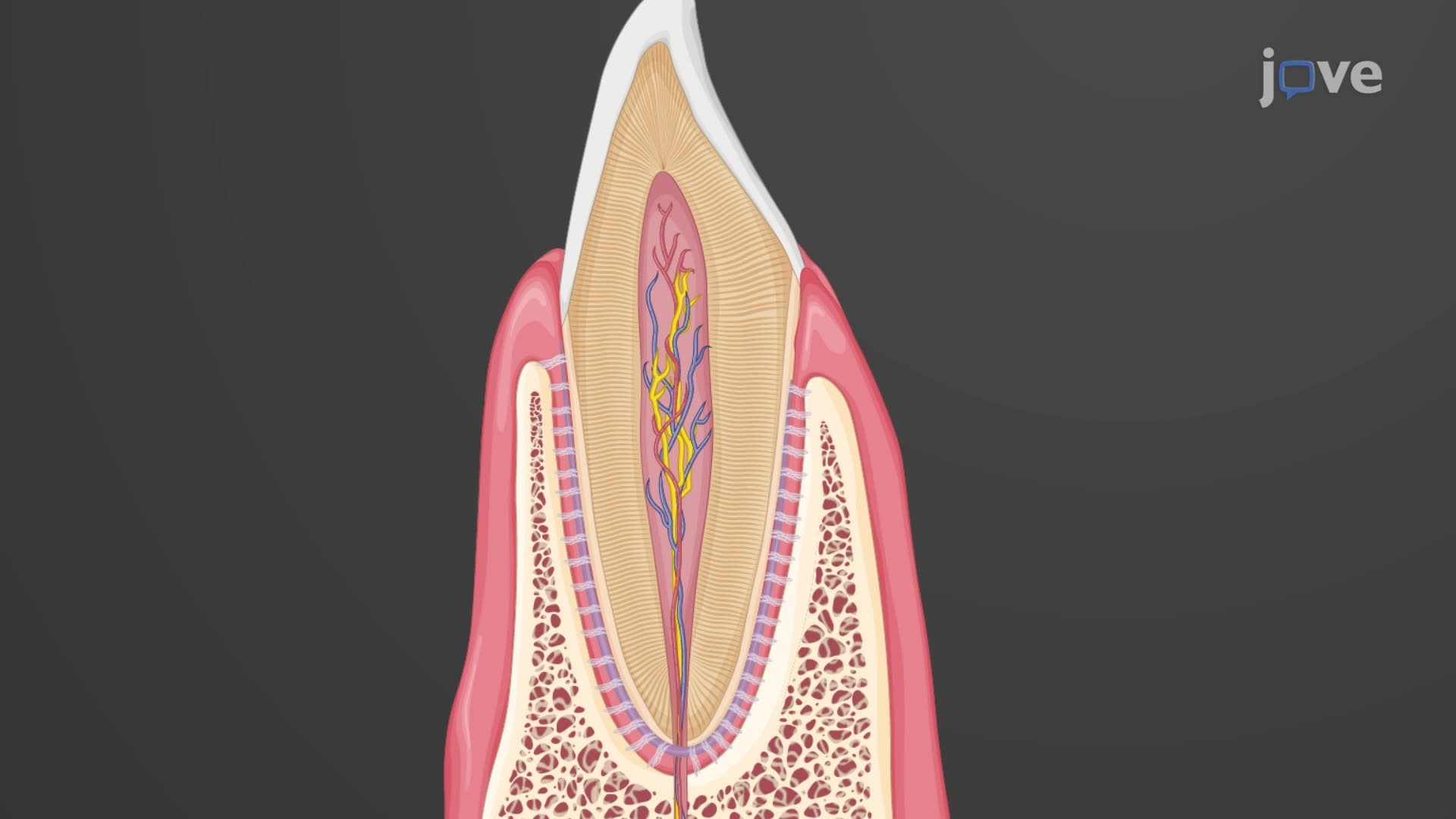
Tooth anatomy biology encompasses the study of dental structures that enable humans to bite, chew, and process food effectively. Each tooth operates as a sophisticated biological unit with distinct regions serving specific functions. The visible portion above the gum line-the crown-handles the mechanical stress of chewing, while the hidden root system provides stable anchorage within the alveolar bone of the jaw.
Crown Structure and Protective Enamel
The crown represents the functional portion of tooth structure explained through its outer protective layer. Enamel, composed of 96% hydroxyapatite crystals, forms the hardest biological material in the human body-harder than steel. This remarkable durability allows enamel to withstand the approximately 200 pounds of pressure generated during normal chewing. US dental schools teach students that enamel's unique prismatic structure, formed by ameloblasts during tooth development, cannot regenerate once damaged, making prevention crucial for long-term dental health.
Root Variations and Anchoring Systems
Root anatomy varies systematically across tooth types, reflecting their specialized functions. Crown root tooth anatomy demonstrates this relationship clearly: incisors and canines possess single roots optimized for cutting and tearing, while molars feature multiple roots to handle grinding forces. Upper premolars typically exhibit two roots, and upper molars contain three distinct roots-a configuration that explains why molar extractions require more complex surgical techniques in US oral surgery practices.
Internal Architecture: Dentin and Pulp Systems
Beneath the enamel lies dentin, a living tissue that comprises the tooth's bulk. Enamel dentin pulp tooth structures work synergistically, with dentin containing thousands of microscopic tubules extending from the pulp cavity to the outer surface. These dentinal tubules house odontoblast processes-cellular extensions that maintain dentin throughout life and respond to stimuli. When students encounter questions about tooth sensitivity on the MCAT or dental school entrance exams, understanding this tubular architecture proves essential.
The pulp cavity tooth anatomy houses the tooth's vital tissues: nerves, blood vessels, and connective tissue. This soft tissue core extends through the root canal system, connecting to the body's circulatory and nervous systems via the apical foramen-a small opening at the root tip. US endodontists rely on this anatomical knowledge when performing root canal treatments, accessing infected pulp through carefully planned entry points.
Cementum and Periodontal Integration
Cementum periodontal tooth connections represent the final component of dental anchorage. This specialized calcified tissue covers the root surface, providing attachment sites for periodontal ligament fibers. These collagen fibers extend from the cementum to the surrounding alveolar bone, creating a dynamic suspension system that allows slight tooth movement during chewing while maintaining stability. Understanding this relationship proves crucial for AP Biology students studying connective tissues and for pre-dental students preparing for the Dental Admission Test (DAT).
Frequently Asked Questions
Tooth anatomy is the study of dental structures including the crown, root, and internal tissues like enamel, dentin, pulp, and cementum. Understanding these components helps explain how teeth function, why dental problems occur, and how treatments work. This knowledge forms the foundation for dental and medical education, appearing frequently on MCAT biology sections and AP Biology exams covering human body systems.
MCAT biology sections often test tooth anatomy through questions about tissue types, embryological development, and physiological functions. Expect questions comparing enamel's mineral composition to bone, analyzing dentin's tubular structure, or explaining how pulp cavity anatomy enables root canal procedures. The DAT dedicates entire sections to dental anatomy, requiring detailed knowledge of root variations and tissue relationships.
AP Biology emphasizes tooth anatomy as an example of structure-function relationships and tissue specialization. Focus on how enamel's crystalline structure provides hardness, dentin's tubular organization enables sensation, and cementum's fiber attachments create periodontal connections. These concepts frequently appear in free-response questions about human body systems and evolutionary adaptations.
US dentists use anatomical knowledge daily when diagnosing problems through X-rays, planning treatments, and performing procedures. Endodontists rely on root canal anatomy to navigate treatment paths, while oral surgeons use root variation knowledge for extraction planning. Orthodontists apply understanding of periodontal ligament properties when moving teeth, and periodontists use cementum-ligament relationships in treating gum disease.
Tooth anatomy builds logically from basic biology concepts, making it accessible to high school students with introductory biology knowledge. The structure follows clear organizational patterns-outer protective layers, inner living tissues, and supportive attachment systems. Visual learning tools and anatomical models help students grasp three-dimensional relationships, while the systematic variation in root numbers creates memorable patterns rather than random facts.
Create visual associations linking structure to function-remember that molars have multiple roots because they handle heavy grinding forces, or that enamel cannot repair itself because it lacks living cells. Use anatomical diagrams to trace the path from pulp cavity through root canals to the apical foramen. Practice with dental school flashcards and online quizzes, focusing on root number patterns and tissue layer relationships that commonly appear on standardized tests.
Advanced topics include tooth development (odontogenesis), comparing human dental anatomy to other mammals, and studying how dental anatomy relates to evolutionary diet changes. Pre-dental students should explore clinical anatomy focusing on radiographic interpretation and procedural approaches. Students interested in research might investigate biomaterial science, studying how artificial dental materials interact with natural tooth structures in US dental applications.
Related Micro-courses
25 Concepts

25 Concepts

20 Concepts