Loading subject page…


628,008 views

Video Summary: The Cochlea Structure and Role in Hearing
Ever wonder how your ears can instantly distinguish between a fire truck's wail and a bird's chirp? The cochlea: structure role hearing mechanism works like a sophisticated frequency analyzer, with its spiral design enabling precise sound discrimination. Similar to how piano strings of different lengths produce distinct notes, the cochlea's basilar membrane varies in width and stiffness to create a topographic map of sound frequencies. This remarkable system allows musicians at Juilliard to detect subtle pitch differences and helps audiologists diagnose hearing loss patterns. Watch the full video on JoVE Coach to master this concept with expert-led visuals and step-by-step explanations.
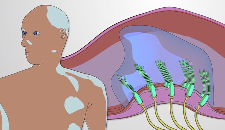
The cochlea represents one of nature's most sophisticated bioengineering solutions for sound processing. This snail-shaped structure in the inner ear transforms mechanical sound waves into the electrical nerve impulses that our brains interpret as music, speech, and environmental sounds. Understanding cochlear function is essential for students pursuing careers in audiology, otolaryngology, biomedical engineering, or neuroscience.
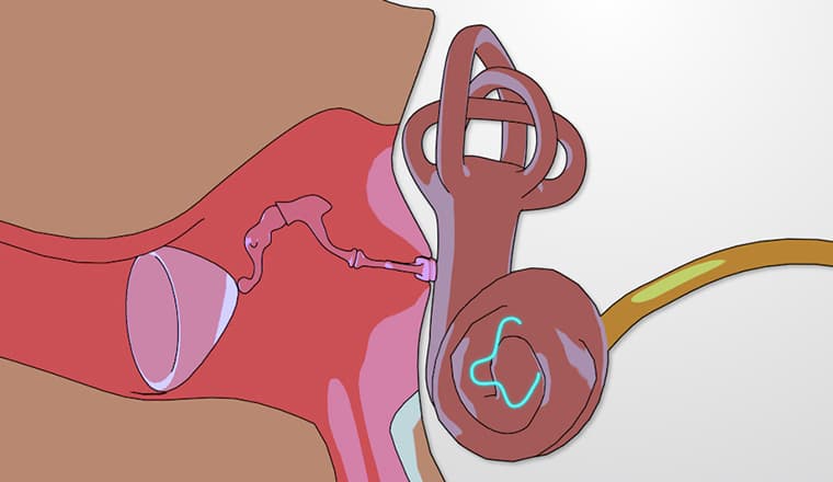
Sound transmission begins when the three tiny bones of the middle ear (malleus, incus, and stapes) amplify vibrations and transfer them to the oval window. This creates pressure waves in the fluid-filled cochlear chambers, setting the basilar membrane into motion. The genius of cochlear design lies in its frequency-selective architecture: the basilar membrane's physical properties change dramatically along its length, creating distinct resonance zones for different frequencies.
This frequency separation, called tonotopy, mirrors how a piano's strings work-shorter, tighter strings vibrate at higher frequencies, while longer, looser strings produce lower tones. In the cochlea, the basal region (near the oval window) responds maximally to high frequencies around 20,000 Hz, while the apical end resonates with low frequencies around 20 Hz. This creates a precise frequency map that remains consistent across individuals, making cochlear implant programming possible for patients with sensorineural hearing loss.
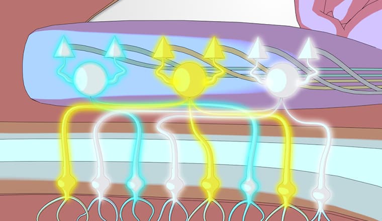
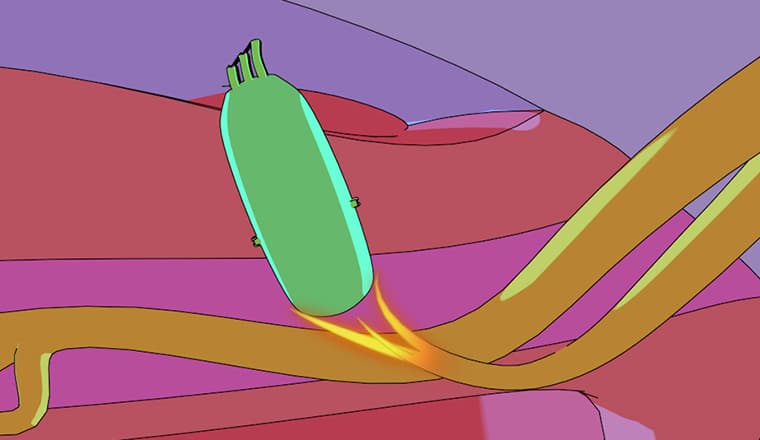
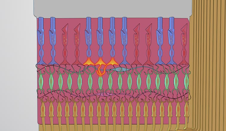
The conversion of mechanical vibration to electrical signals occurs through specialized hair cells sandwiched between the basilar and tectorial membranes. When the basilar membrane vibrates, it creates a shearing motion that bends the hair cell stereocilia, opening mechanosensitive ion channels. This process, called mechanotransduction, generates action potentials in auditory nerve fibers with microsecond precision.
Each cochlear location connects to specific auditory nerve fibers that maintain their frequency selectivity throughout the ascending auditory pathway. This tonotopic organization persists through the cochlear nucleus, inferior colliculus, and auditory cortex, enabling sophisticated sound processing capabilities like speech recognition in noisy environments.
Understanding cochlear structure and function is crucial for MCAT preparation, particularly in the biological and biochemical foundations section. AP Biology students encounter this topic when studying sensory systems and signal transduction. Medical students must master cochlear anatomy for USMLE Step 1, as questions often test the relationship between specific cochlear regions and frequency processing.
Real-world applications include audiometric testing, where clinicians use pure-tone audiometry to map hearing thresholds across frequencies, directly reflecting cochlear function. Cochlear implant technology bypasses damaged hair cells by electrically stimulating auditory nerve fibers at appropriate tonotopic locations, restoring functional hearing to over 324,000 Americans with severe hearing loss.
Related Micro-courses