Loading subject page…

- Biology
- Sensory Systems
- The Retina
Micro-course: Sensory Systems
628,008 views
Learn key concepts and earn your certificate.

The Retina

00:00
01:32
Video Summary: What Is the Retina
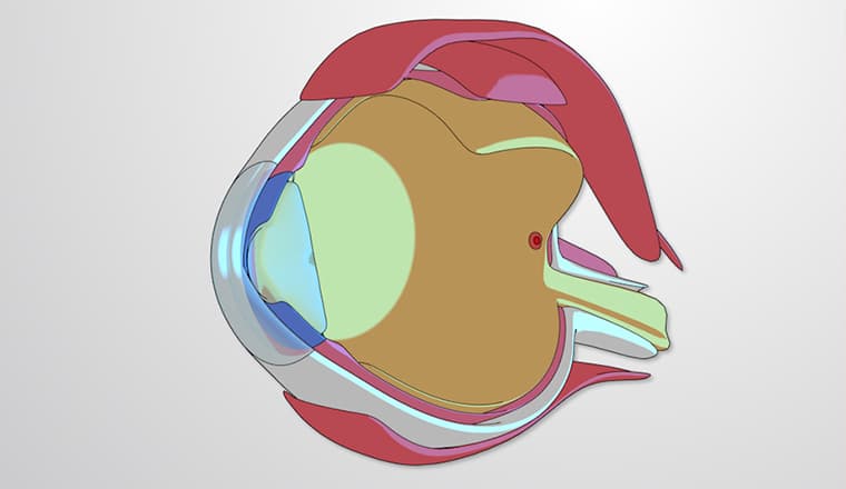
Did you know that the retina contains over 120 million photoreceptor cells working together to create your vision? The retina is a thin layer of tissue at the back of your eye that converts light into electrical signals your brain can interpret. Just like the camera sensor in your smartphone captures images, the retina captures visual information through specialized cells called rods and cones, then processes it through multiple neural layers before sending it to your brain via the optic nerve. Watch the full video on JoVE Coach to master this concept with expert-led visuals and step-by-step explanations.
- Understand what is the retina and its role as the light-sensitive tissue that initiates vision
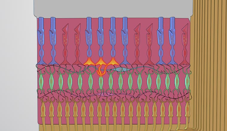
- Identify the two main types of photoreceptors (rods and cones) and their specific functions in different lighting conditions
- Analyze how photopigment molecules in outer segments transduce light energy into electrochemical signals
- Compare the three types of cone cells and explain how their differential activation creates color perception
- Recognize the fovea's importance as the region of highest visual acuity due to dense cone concentration
- Evaluate the sequential signal processing pathway from photoreceptors through bipolar cells to ganglion cells
- Apply knowledge of horizontal and amacrine cell functions in lateral signal processing and visual information refinement
- Explore how retinal ganglion cell axons form the optic nerve to transmit processed visual information to the brain
Understanding What is The Retina: The Eye's Neural Network
The retina represents one of the most sophisticated biological sensors in the human body, functioning as both a light detector and a complex neural processing center. Unlike a simple camera film, the retina actively processes visual information through multiple layers of interconnected neurons before transmitting refined signals to the brain. This remarkable tissue, no thicker than a few sheets of paper, contains approximately 130 million photoreceptors and processes visual information in real-time.
Photoreceptor Types and Visual Function
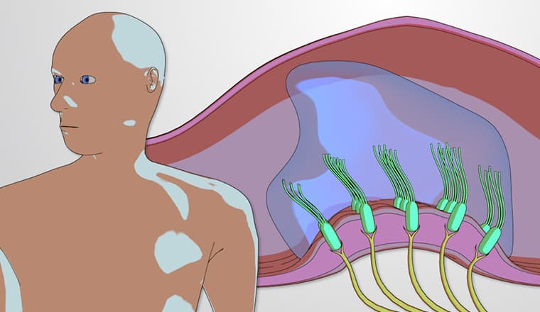
The retina's photoreceptors-rods and cones-serve distinctly different roles in human vision. Rods, numbering about 120 million per eye, excel in low-light conditions and are primarily responsible for night vision and peripheral awareness. These cells contain rhodopsin, a highly sensitive photopigment that can respond to single photons of light. Cones, though fewer at about 6 million per eye, handle daytime vision and color detection through three distinct types: L-cones (sensitive to long wavelengths/red light), M-cones (medium wavelengths/green light), and S-cones (short wavelengths/blue light).
This concept frequently appears on AP Biology exams, particularly in questions about sensory systems and signal transduction. Medical school entrance exams like the MCAT often test understanding of photoreceptor distribution and function, especially regarding color blindness mechanisms and night vision physiology.
The Fovea: Precision Vision Center
The fovea centralis, a small depression measuring only 1.5 millimeters in diameter, contains the highest concentration of cone cells in the retina. This region provides the sharp, detailed vision necessary for activities like reading, driving, and recognizing faces. Unlike the peripheral retina, the fovea contains virtually no rods, explaining why you cannot see faint stars when looking directly at them-you must look slightly to the side to engage your rod-rich peripheral vision.
Neural Processing Layers and Information Flow
Beyond photoreception, the retina performs sophisticated signal processing through its layered neural network. Horizontal cells create lateral connections that enhance contrast and edge detection, while amacrine cells contribute to motion detection and temporal processing. This retinal preprocessing significantly reduces the information load sent to the brain, compressing roughly 130 million photoreceptor inputs into approximately 1 million ganglion cell outputs that form the optic nerve. Understanding this neural architecture helps explain visual phenomena like lateral inhibition and center-surround receptive fields, concepts essential for neuroscience coursework and medical boards like the USMLE Step 1.
Frequently Asked Questions
The retina is the light-sensitive tissue layer at the back of your eye that converts light into electrical signals for your brain to interpret as vision. It contains millions of photoreceptor cells (rods and cones) that capture light and begin processing visual information. The retina functions like a biological camera sensor, but unlike electronic sensors, it also performs complex neural processing before sending refined visual data to your brain through the optic nerve.
MCAT questions frequently focus on photoreceptor types, phototransduction mechanisms, and retinal processing pathways. Expect questions about rod versus cone distribution, color vision mechanisms, and how retinal ganglion cells form the optic nerve. Practice problems often involve experimental scenarios testing night vision, color blindness, or visual field defects to assess your understanding of retinal anatomy and physiology.
The retina appears in AP Biology Unit 7 (Natural Selection) when discussing sensory systems and Unit 3 (Cellular Energetics) regarding signal transduction. Students should understand photoreceptor signal cascades, neurotransmitter release mechanisms, and how retinal processing demonstrates biological information transfer. Free response questions may ask you to trace signal flow from photon absorption to brain processing or explain evolutionary advantages of different photoreceptor types.
Retinal diseases like diabetic retinopathy affect millions of Americans and can cause blindness if untreated. Macular degeneration specifically damages the fovea region, leading to central vision loss while preserving peripheral sight. Retinal detachment, where the retina separates from underlying tissue, requires emergency medical treatment to prevent permanent vision loss. Understanding normal retinal function helps explain why these conditions cause specific visual symptoms and treatment approaches.
The retina concept builds naturally on basic cell biology and nervous system principles you've already learned. Start with the simple idea that the retina converts light to electrical signals, then add layers of complexity about cell types and processing pathways. Visual diagrams and analogies to cameras or electronic sensors help make the concept more accessible, and most students find the real-world connections to vision and eye diseases highly engaging.
Create a flowchart tracing light from photoreceptor activation through all retinal cell layers to the optic nerve. Use active recall by drawing the retinal layers from memory and labeling each cell type's function. Practice explaining why rods work better in dim light while cones handle color vision and detailed sight. Connect structure to function by asking how each retinal feature contributes to overall visual processing.
Explore visual cortex processing to understand how the brain interprets retinal signals, including topics like visual field mapping and cortical columns. Study other sensory systems to compare transduction mechanisms across different stimuli types. Advanced topics include retinal development, stem cell therapy for retinal diseases, and how artificial retinal implants work to restore vision in blind patients.
Related Micro-courses
11 Concepts
6 Concepts
8 Concepts