Loading subject page…


83,181 views
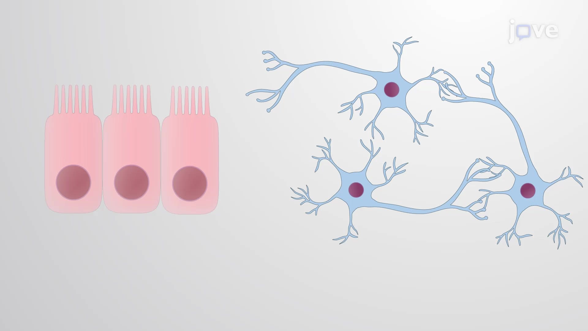

Video Summary: What Is Cadherins in Tissue Organization
Did you know that cancer cells can literally "break free" from tumors by switching off their cellular glue? Cadherins in tissue organization serve as these crucial molecular adhesives that keep our body's tissues properly structured and prevent cells from wandering where they shouldn't. When breast cancer cells lose E-cadherin expression, they can detach from the primary tumor and potentially metastasize throughout the body. Understanding what is cadherins in tissue organization reveals how our bodies maintain cellular order and how diseases disrupt this delicate balance. Watch the full video on JoVE Coach to master this concept with expert-led visuals and step-by-step explanations.
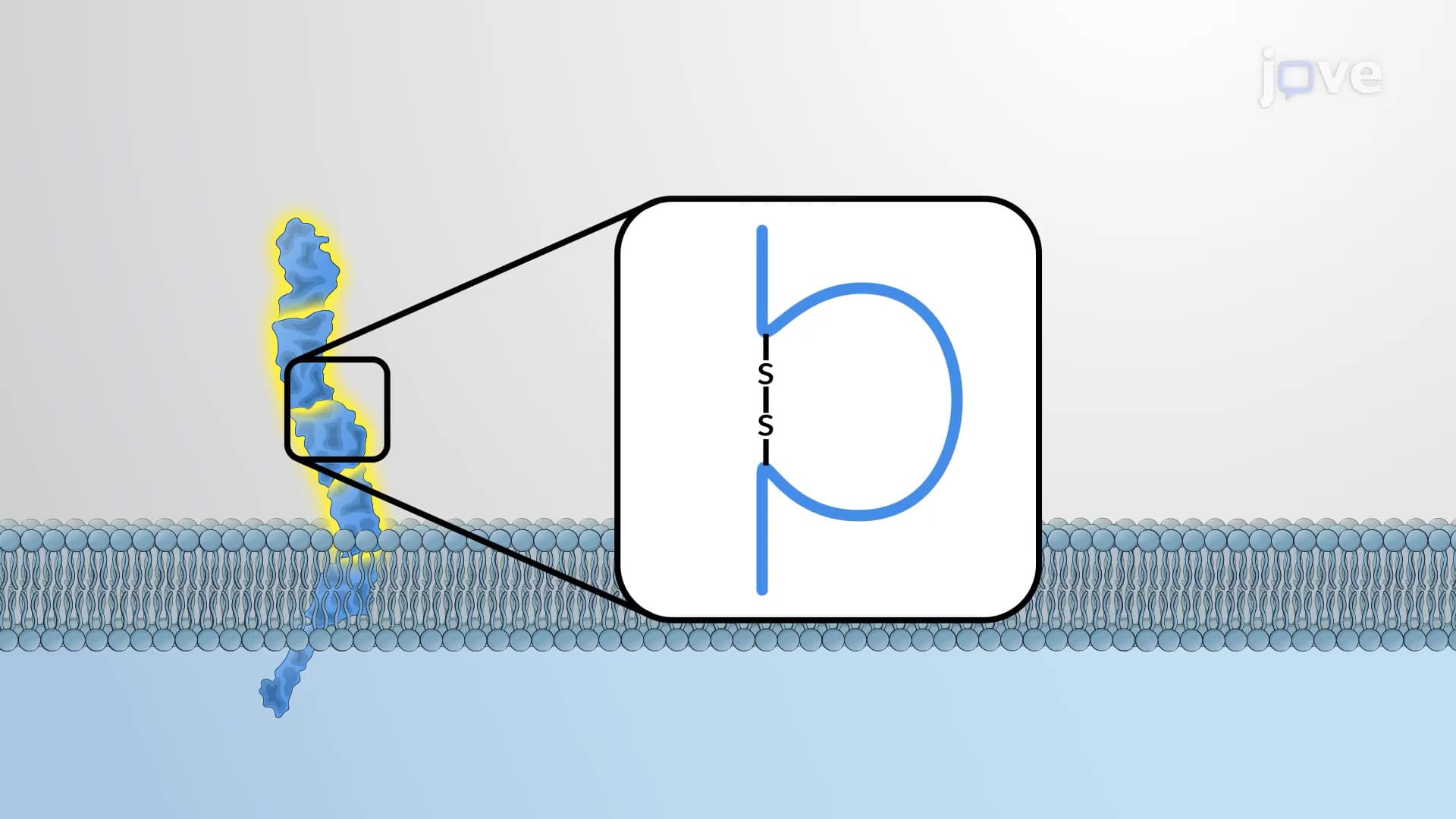
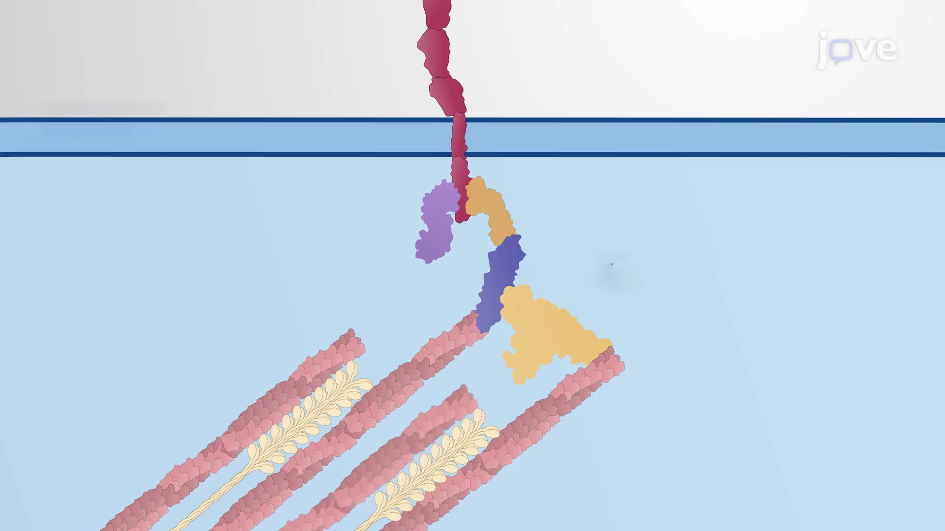
Cadherins in tissue organization represent one of biology's most elegant solutions to a complex architectural challenge: how do billions of cells organize themselves into functional tissues and organs? These transmembrane glycoproteins act as cellular "zip codes," ensuring that cells end up in the right neighborhoods and stay there. Unlike simple adhesive molecules, cadherins provide both specificity and strength, creating dynamic yet stable tissue architectures that can adapt during development while maintaining structural integrity.
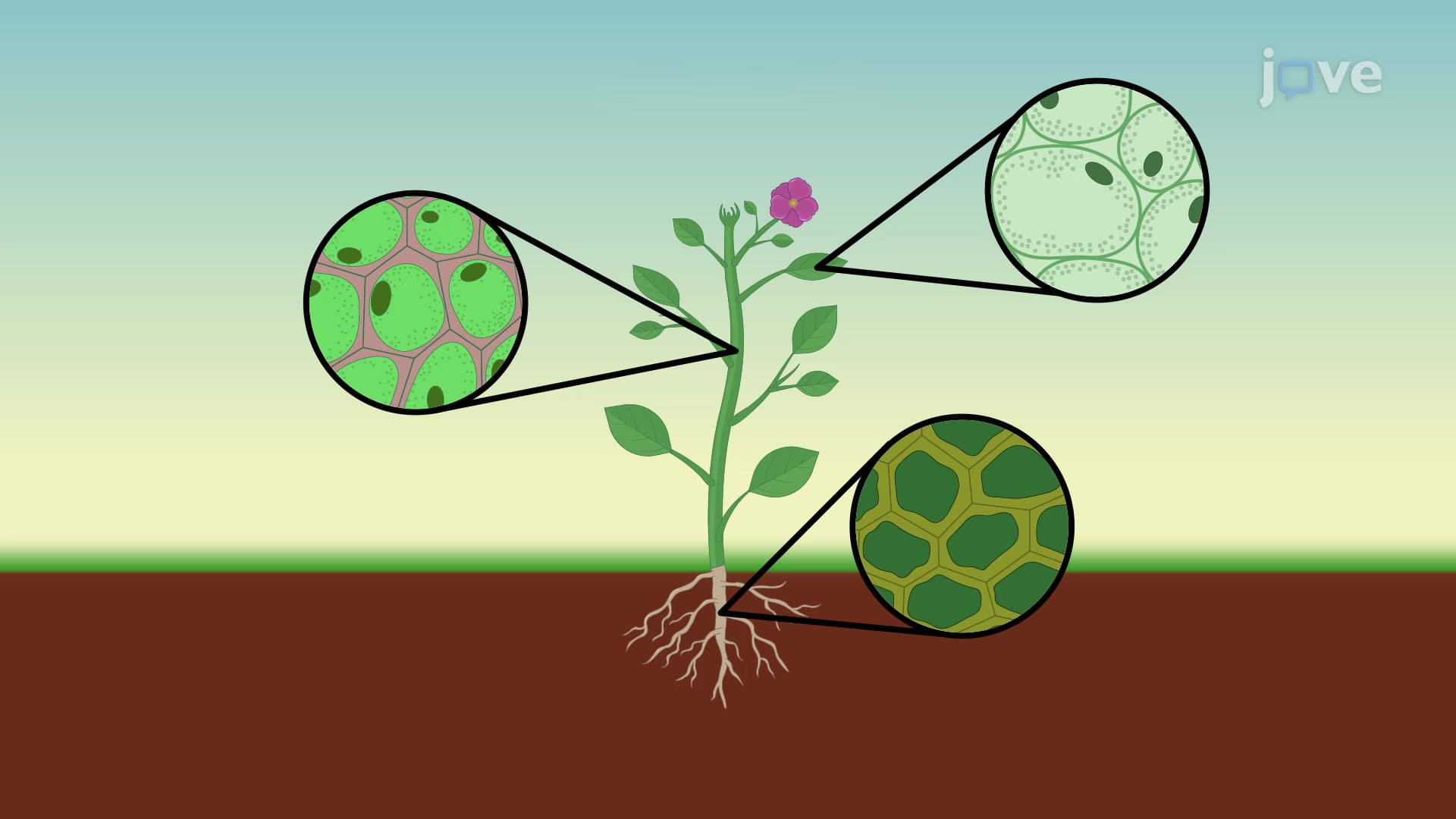
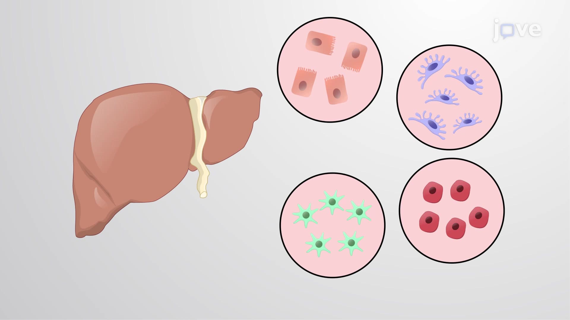
The cadherin family demonstrates remarkable diversity, with over 100 different types identified in humans. E-cadherins (epithelial cadherins) dominate in epithelial tissues like skin, intestinal lining, and glandular structures, while N-cadherins (neural cadherins) concentrate in nervous system tissues and muscle. P-cadherins appear in placental tissues and certain epithelial cells. This tissue-specific distribution isn't random-it reflects evolutionary optimization for particular mechanical and functional demands. For AP Biology students, understanding this specificity helps explain how organ systems maintain distinct identities while coordinating function.
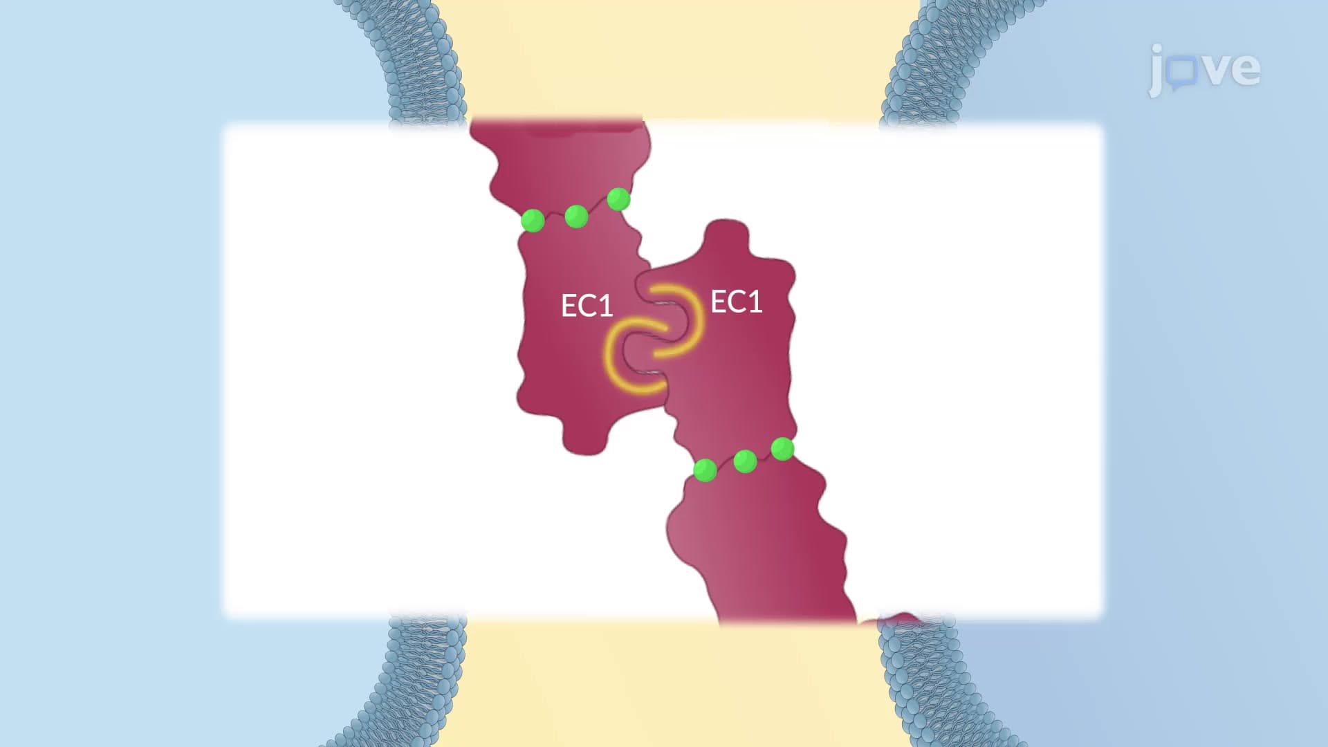
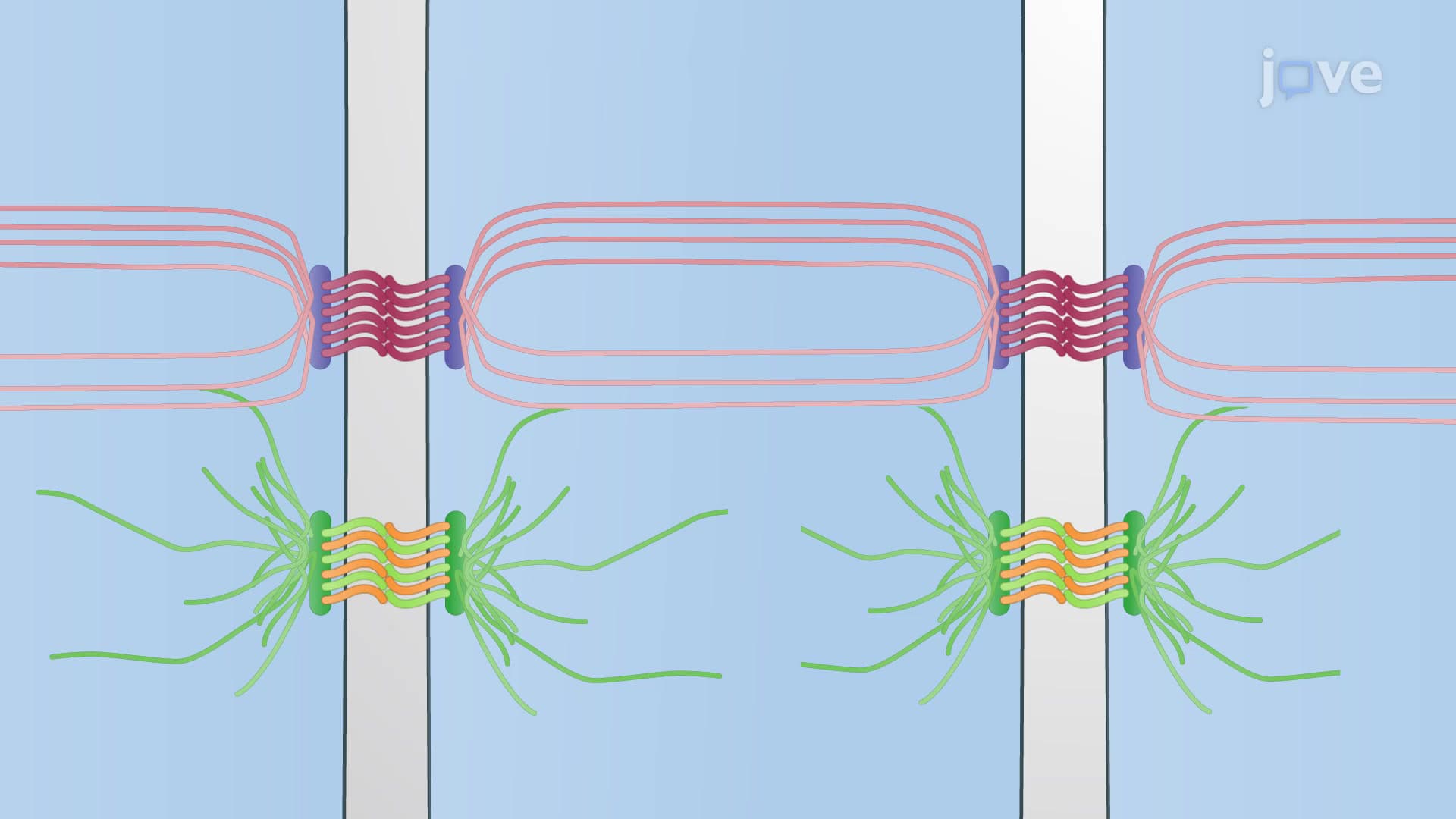
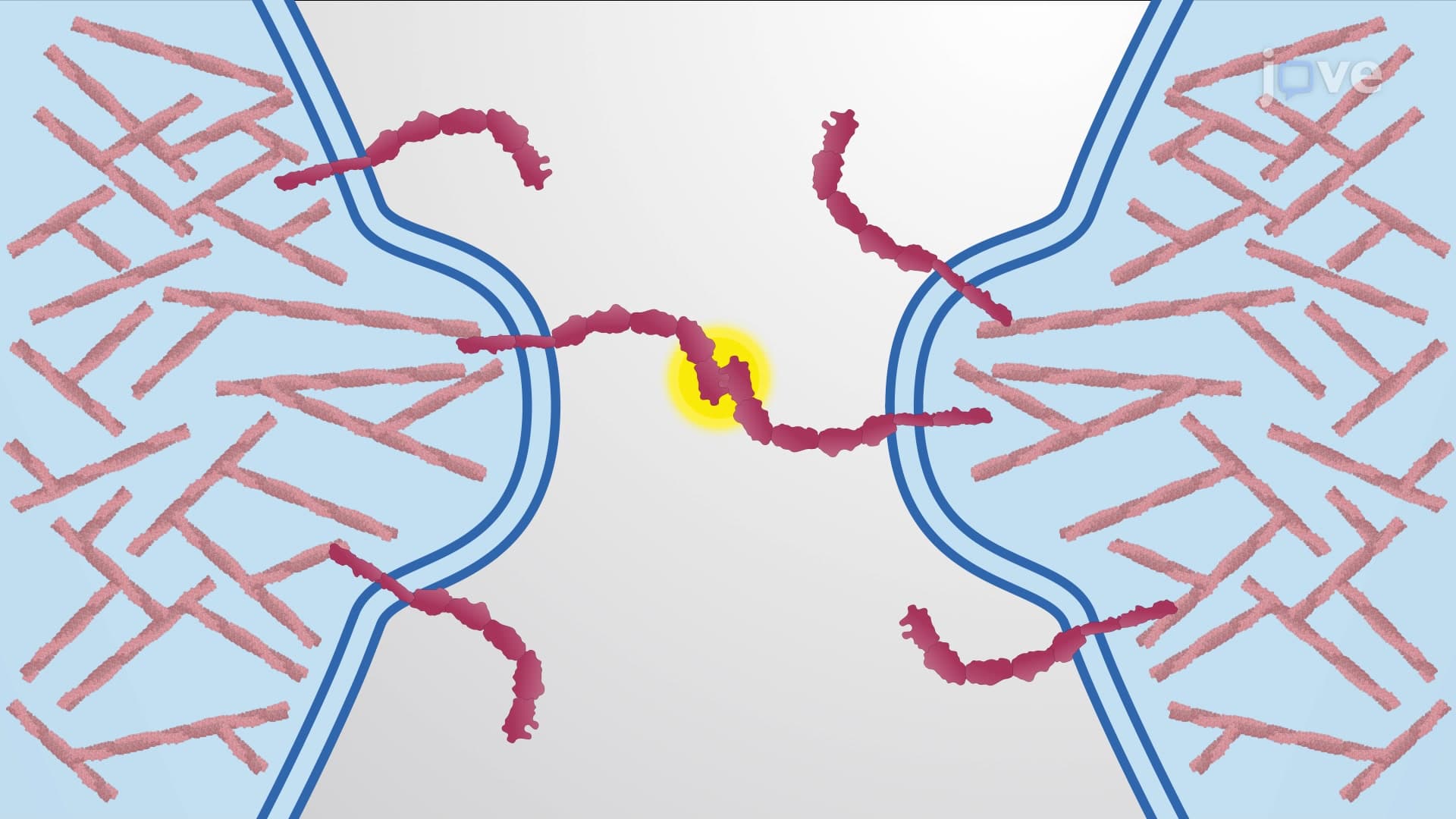
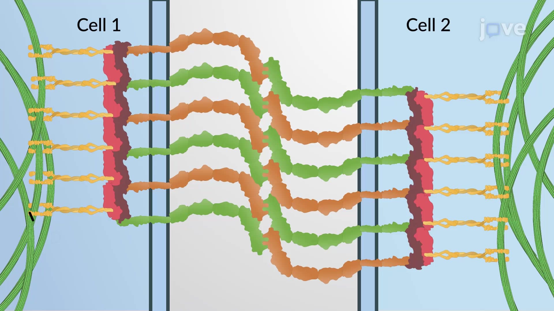
Cadherins primarily exhibit homophilic binding, meaning E-cadherins preferentially bind to other E-cadherins, creating selective adhesion zones. This mechanism drives cellular sorting during embryonic development, similar to how oil and water separate. Cells expressing high cadherin levels form tighter aggregations than those with lower expression, creating tissue boundaries and organizational hierarchies. Research at institutions like Harvard Medical School and Stanford University has shown how quantitative differences in cadherin expression-sometimes just 2-3 fold variations-can dramatically alter tissue organization patterns, a principle frequently tested on MCAT examinations.
The pathological loss of cadherin expression underlies one of cancer's most dangerous characteristics: metastasis. Epithelial-to-mesenchymal transition (EMT) involves coordinated downregulation of E-cadherins, allowing tumor cells to detach from primary sites and invade surrounding tissues. This process, extensively studied at cancer centers like MD Anderson and Memorial Sloan Kettering, represents a critical therapeutic target. Understanding cadherin regulation has led to experimental treatments aimed at preventing or reversing EMT, making this concept increasingly relevant for pre-med students preparing for medical school coursework and USMLE examinations.
Related Micro-courses