Loading video…


 16 Concepts
 424.1K views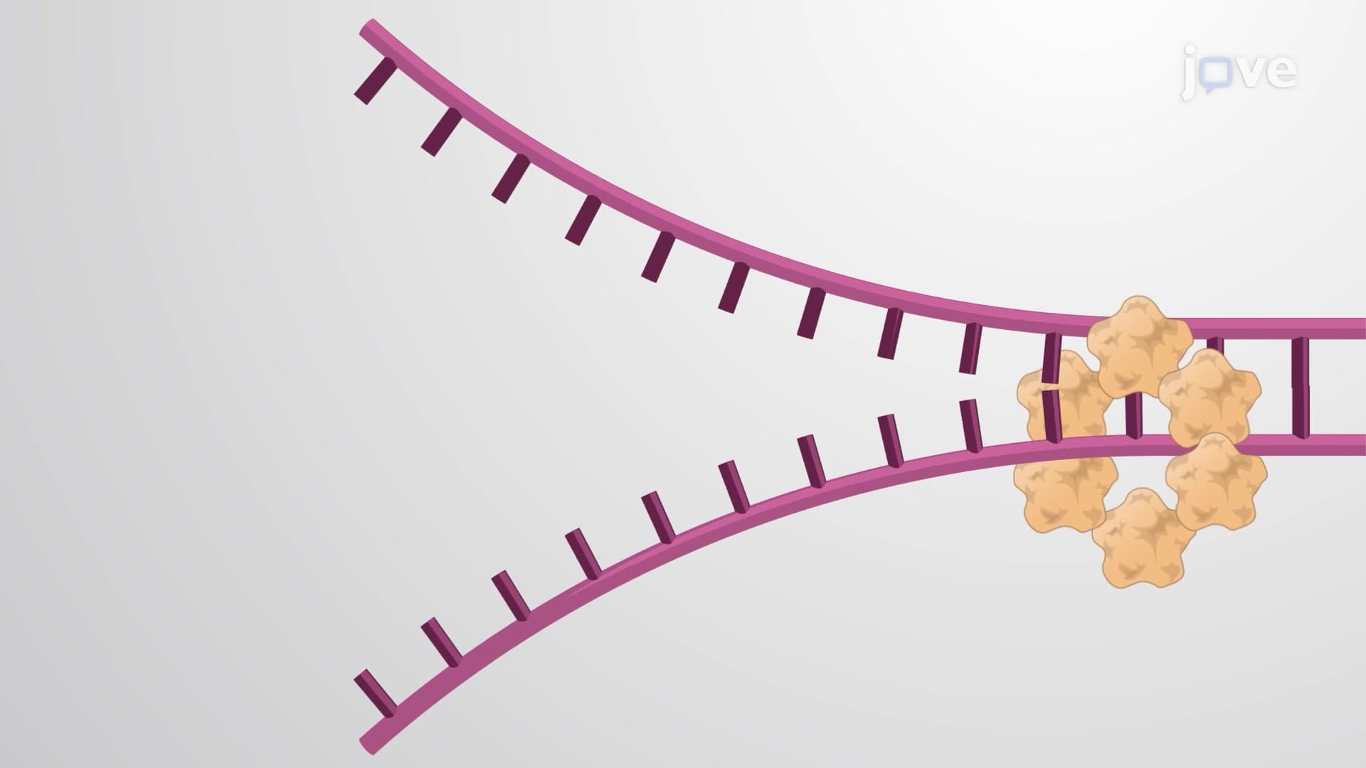

Video Summary: What Is the DNA Replication Fork
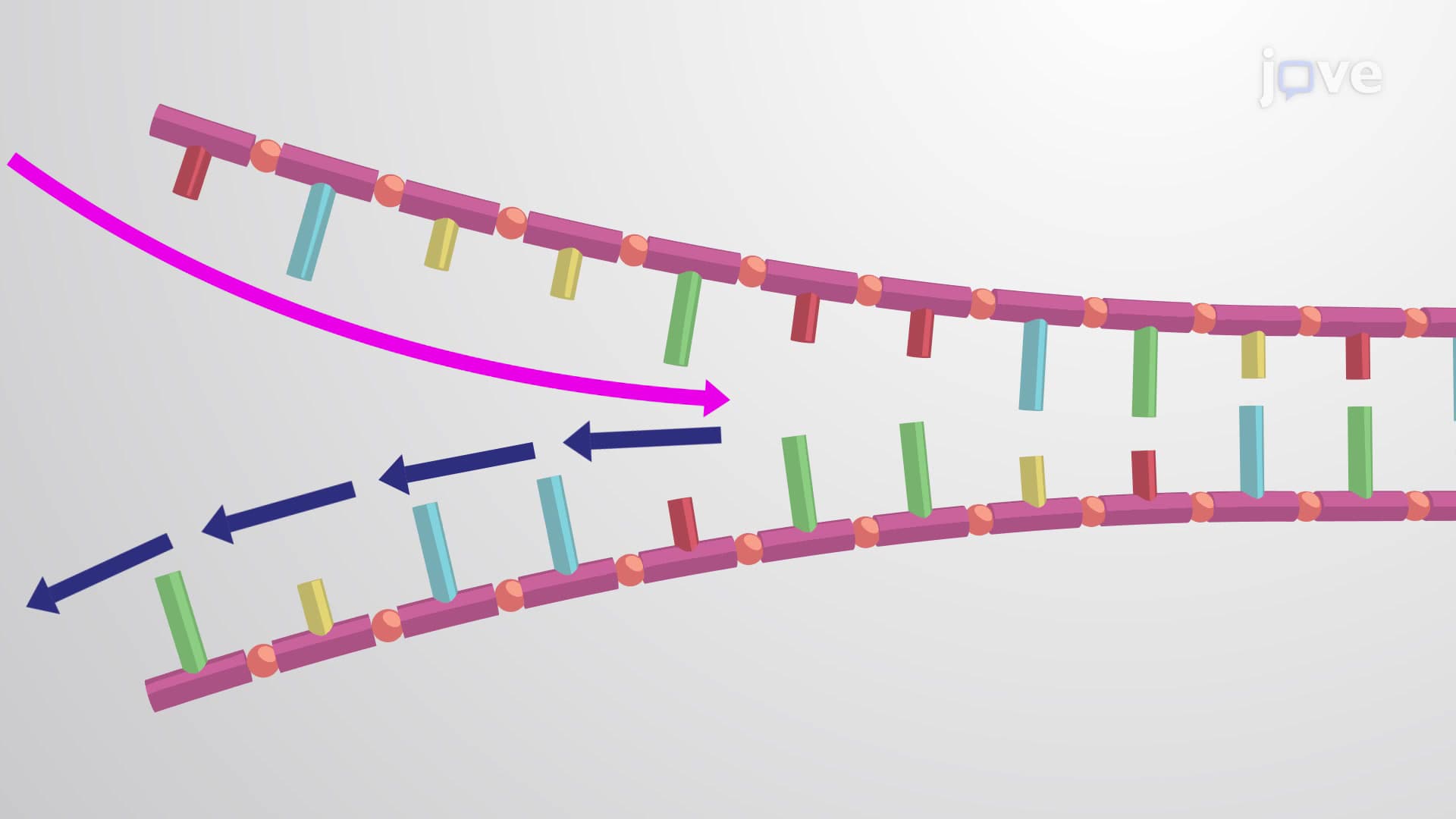
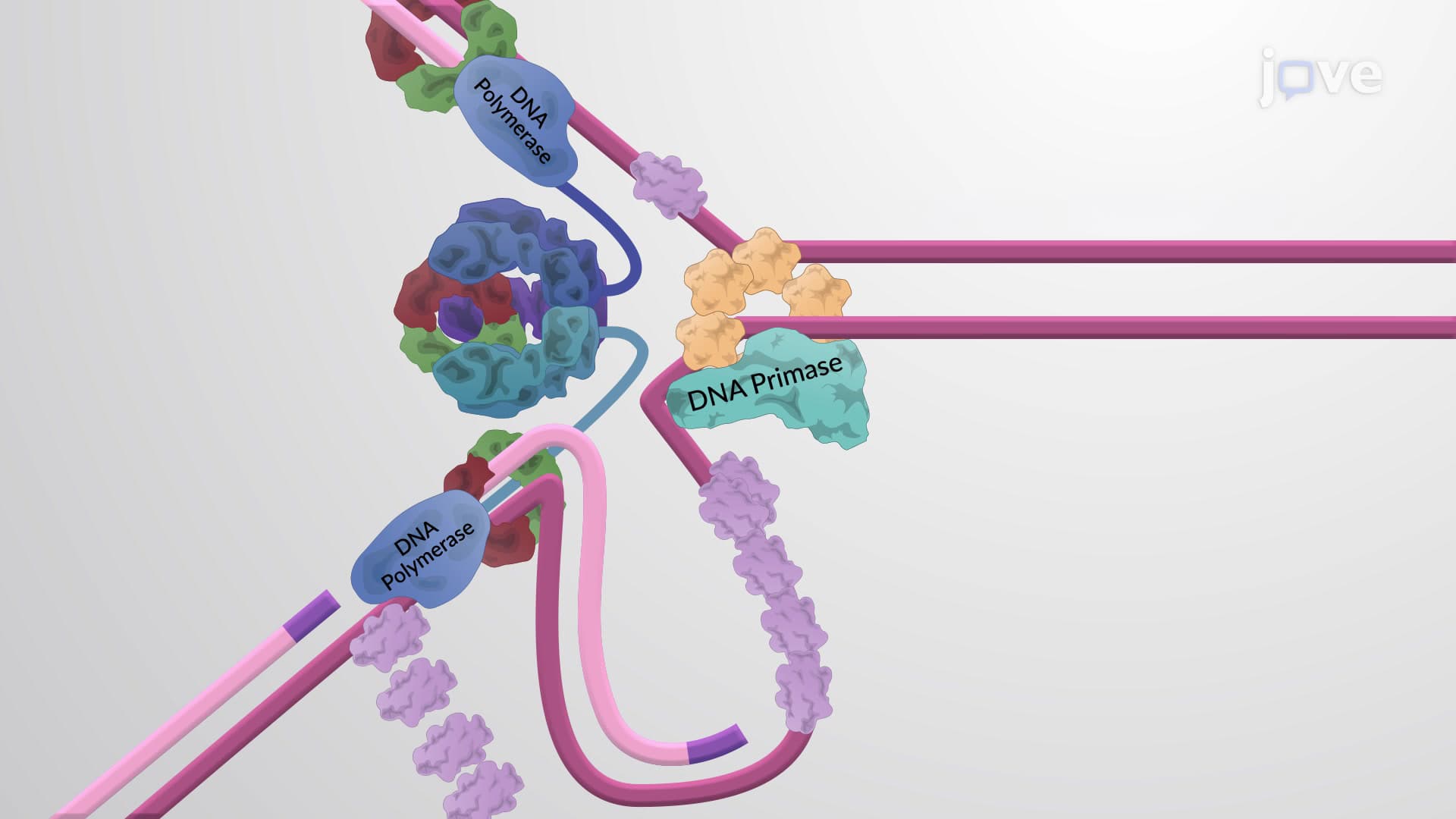
Every second, millions of cells in your body replicate their entire genome with stunning precision, but how does this molecular machinery actually work? The DNA replication fork serves as the cellular assembly line where this remarkable process unfolds, creating two identical copies from one original DNA molecule. Consider how pharmaceutical companies like Pfizer rely on understanding DNA replication mechanisms to develop targeted cancer therapies that disrupt tumor cell division. What is The DNA Replication Fork becomes clearer when we examine the Y-shaped structure formed when DNA helicase unwinds the double helix. Watch the full video on JoVE Coach to master this concept with expert-led visuals and step-by-step explanations.
The DNA replication fork represents one of biology's most elegant molecular machines, a dynamic structure where genetic information transfers from parent to daughter cells with extraordinary fidelity. Unlike a simple copying process, DNA replication involves sophisticated molecular choreography that ensures each new cell receives an exact genetic blueprint.
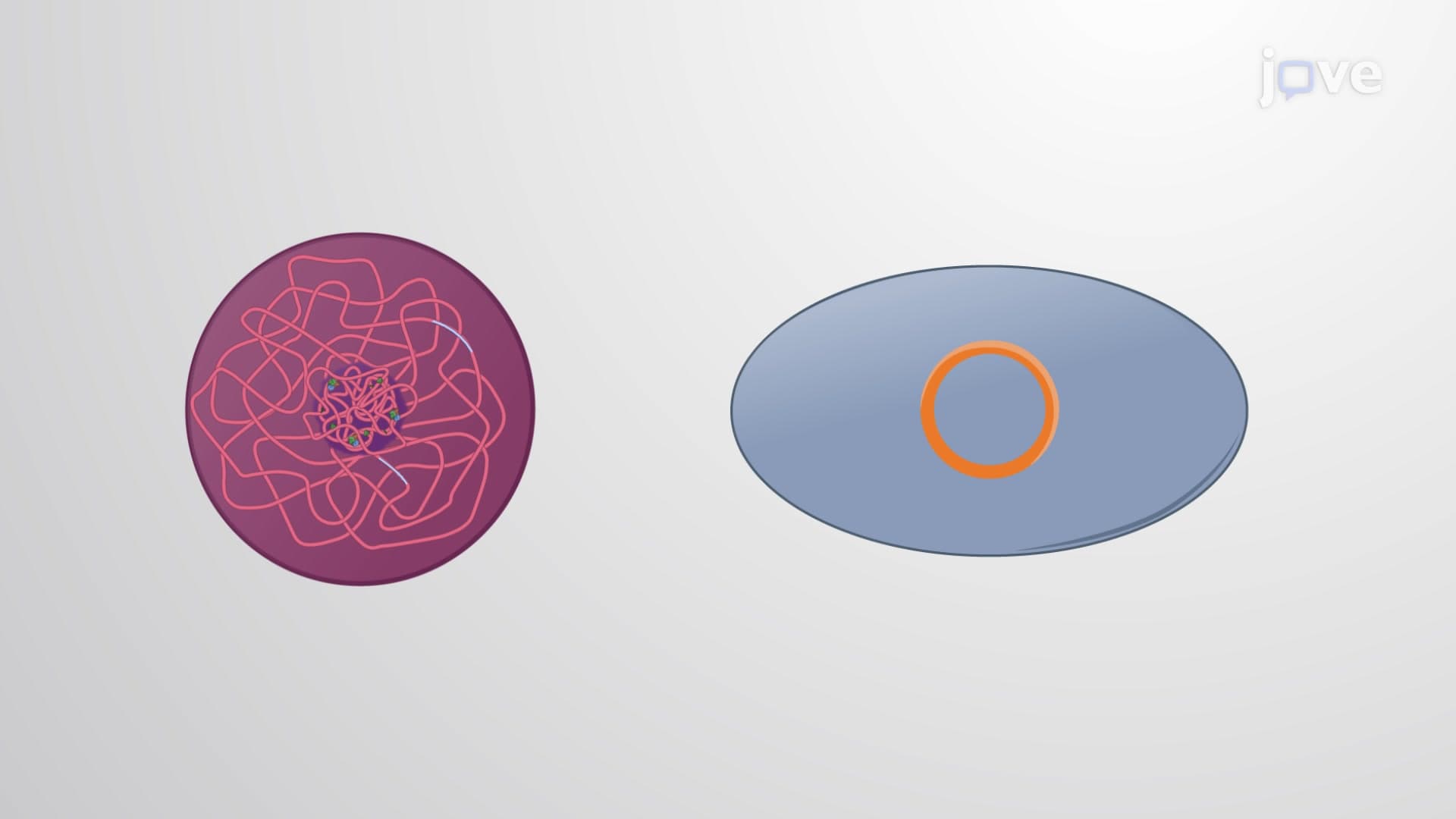
DNA replication doesn't start randomly. Specific DNA sequences called origins of replication serve as designated starting points, much like designated construction zones on a highway project. In human cells, thousands of these origins activate simultaneously across each chromosome, preventing replication from taking an impossibly long time. The Mayo Clinic's genetic research has shown that defects in origin recognition can lead to developmental disorders, highlighting this process's clinical significance.
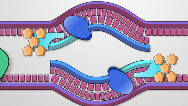
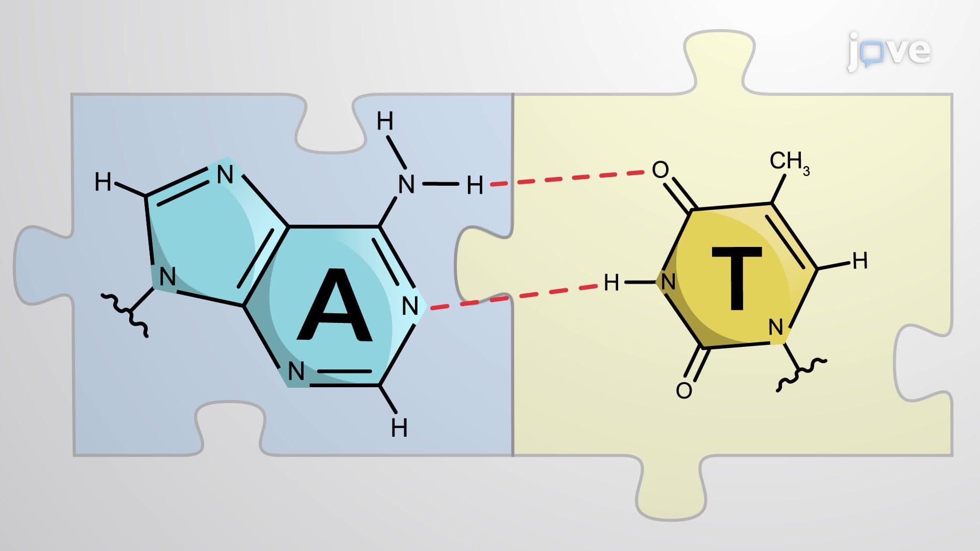
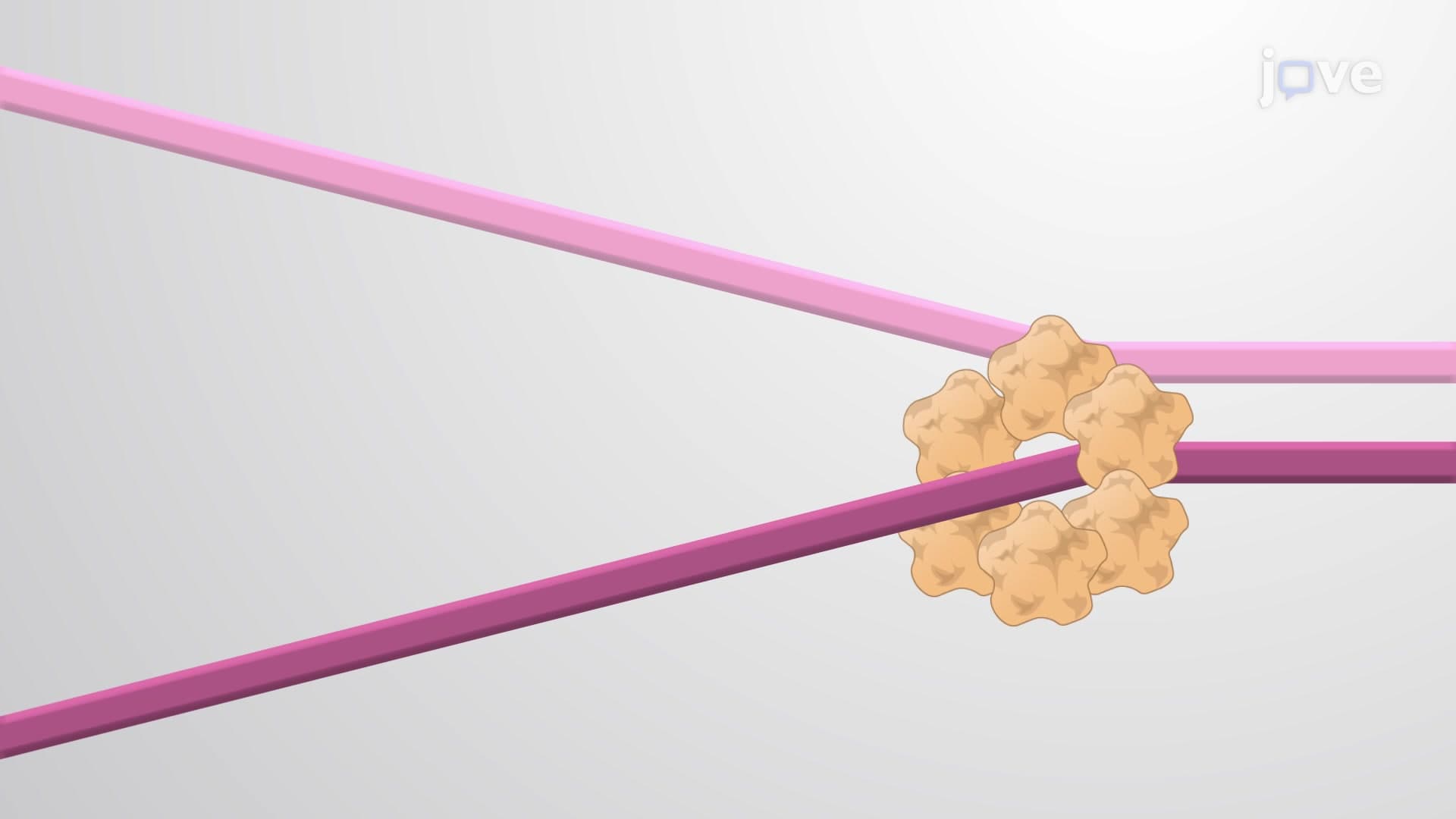
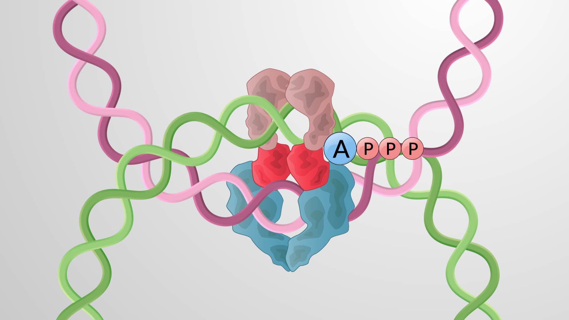
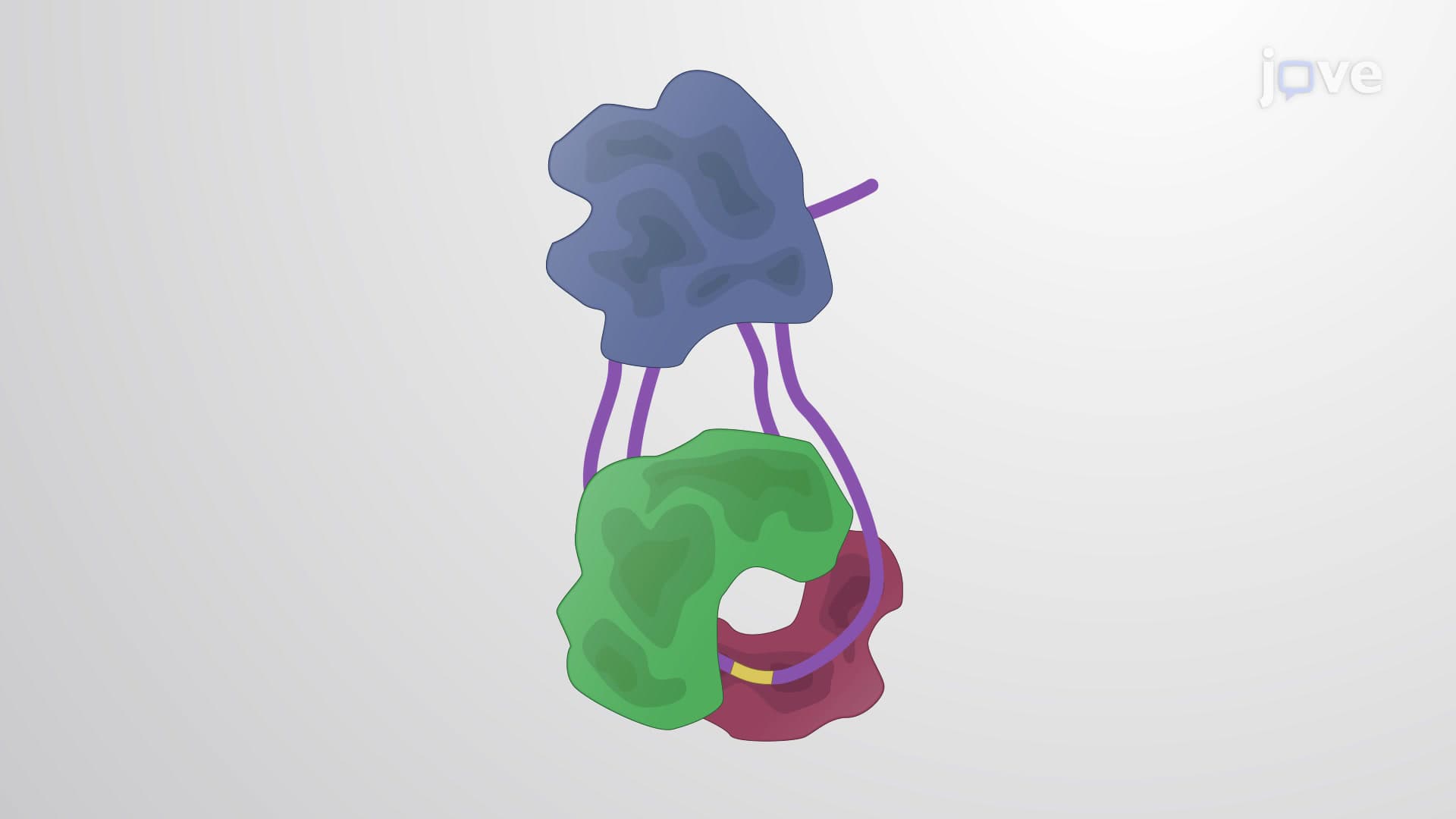
DNA helicase acts as the molecular "unzipper," using ATP energy to break hydrogen bonds between complementary base pairs. As helicase moves along the DNA, it creates the characteristic Y-shaped structure we call the replication fork. This enzyme moves at approximately 1000 base pairs per second in human cells, fast enough to complete chromosome replication within hours rather than days.
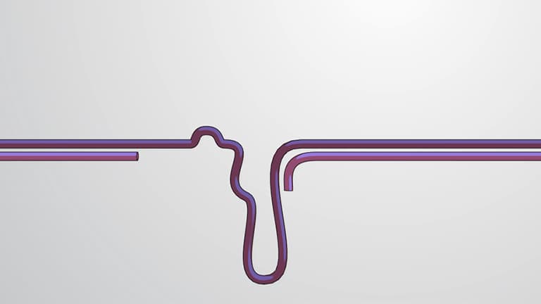
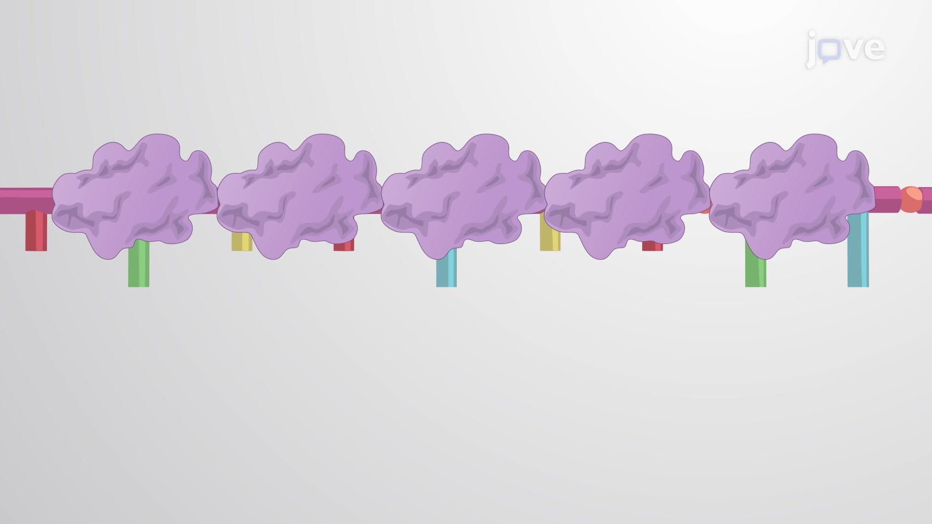
Once DNA strands separate, they face immediate challenges. Single-stranded DNA tends to fold back on itself, forming hairpin loops, or reanneal with its complement, both scenarios that would halt replication. Single-strand DNA binding proteins (SSBs) solve this problem by coating the separated strands like molecular scaffolding, keeping them accessible for replication machinery.
Understanding replication fork dynamics proves essential for students preparing for AP Biology exams, where questions frequently test knowledge of enzyme functions and structural relationships. Medical students taking the MCAT encounter similar concepts when analyzing how chemotherapy drugs target rapidly dividing cancer cells by disrupting replication fork stability. At institutions like Stanford Medical School, students learn how understanding these mechanisms directly relates to developing new cancer treatments that selectively target tumor cells while sparing healthy tissue.
Related Micro-courses