Loading video…


 21 Concepts
 74.4K views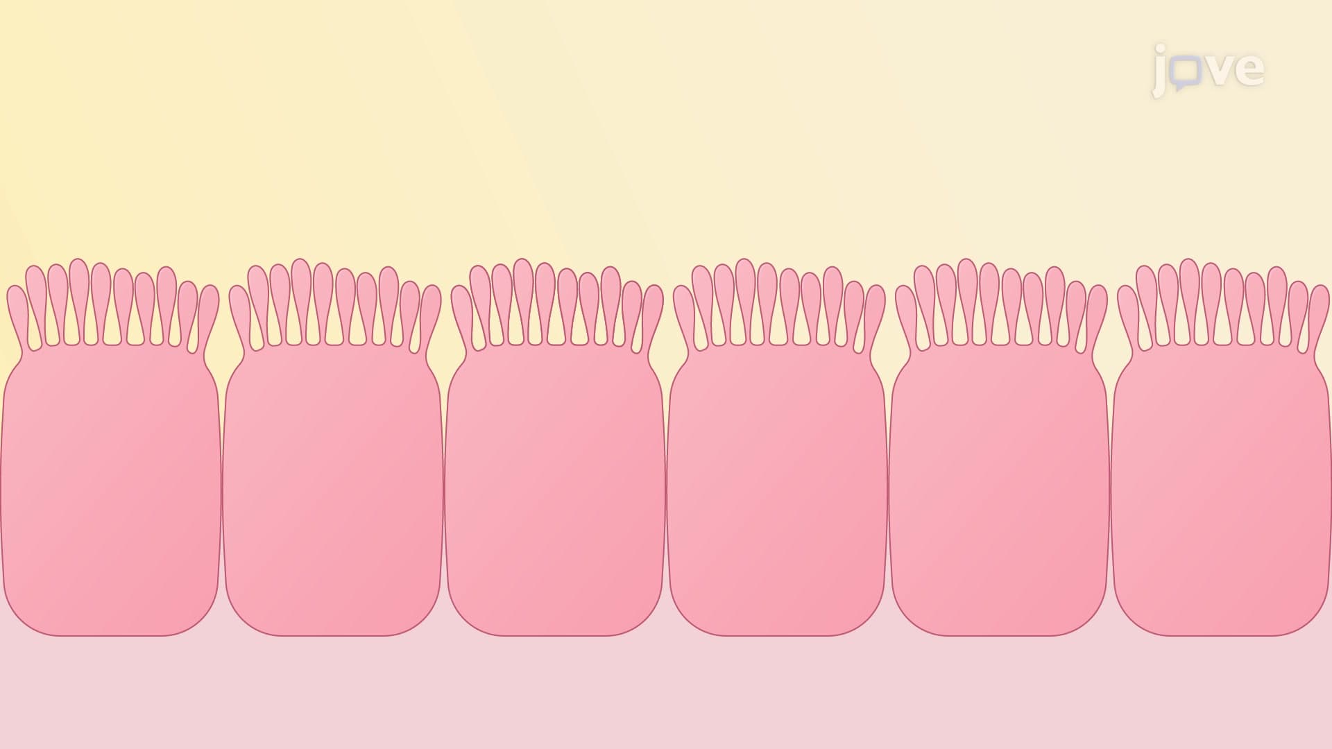

Video Summary: What Is Microvilli
Ever wondered how your small intestine, roughly the size of a tennis court when flattened, fits inside your abdomen? Microvilli explained biology reveals these microscopic finger-like projections that dramatically increase surface area for absorption. In the human small intestine, millions of microvilli create a brush border that boosts nutrient absorption twenty-five-fold. What is microvilli becomes clear when you consider how these cellular structures enable efficient digestion in organs like those studied at Johns Hopkins Medical School. Watch the full video on JoVE Coach to master this concept with expert-led visuals and step-by-step explanations.
Microvilli structure and function represents one of biology's most elegant solutions to a fundamental problem: maximizing surface area within limited space. These microscopic projections, measuring approximately 0.1 micrometers in diameter and 1-2 micrometers in length, transform flat cellular surfaces into highly efficient absorption interfaces.
The internal framework of each microvillus consists of a precisely organized actin core microvilli system. Twenty to thirty actin microfilaments run parallel through each projection, bundled together by specialized proteins including villin and fimbrin. This cytoskeletal arrangement provides both structural support and flexibility, allowing microvilli to maintain their shape while accommodating cellular movements.
The plasma membrane tightly wraps around this actin core, creating the characteristic finger-like appearance that gives brush border microvilli their name. When viewed under electron microscopy, thousands of these projections create a fuzzy border resembling a bottle brush, hence the term "brush border."
Intestinal microvilli absorption exemplifies the structure-function relationship in cellular biology. In the small intestine alone, microvilli increase the absorptive surface area from approximately 300 square meters to over 7,500 square meters-equivalent to a tennis court's surface area folded into your abdomen. This expansion proves crucial for absorbing nutrients from the 9-10 liters of fluid that pass through the digestive system daily.
Beyond the intestines, microvilli appear in kidney tubules, where they facilitate water and electrolyte reabsorption, and in the inner ear, where specialized microvilli called stereocilia enable hearing. Each location demonstrates how microvilli membrane projections adapt to specific physiological demands.
Understanding microvilli becomes essential for students preparing for medical school entrance exams like the MCAT, where questions often focus on cellular adaptations for absorption. In Advanced Placement Biology courses, microvilli frequently appear in questions about surface area-to-volume ratios and cellular specialization.
Medical conditions like celiac disease damage intestinal microvilli, dramatically reducing microvilli surface area and leading to malabsorption syndromes. This connection between structure and pathology illustrates why mastering microvilli concepts proves vital for healthcare professionals and students at institutions like Harvard Medical School or UCLA's medical program.
Related Micro-courses