Loading video…


 37 Concepts
 141K views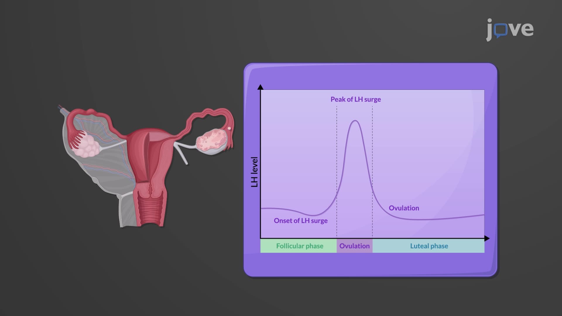

Video Summary: What Is Oogenesis
Did you know that by the time a female baby is born, she already has all the egg cells she'll ever produce in her lifetime? Oogenesis biology explained reveals this fascinating process where egg cell formation begins during fetal development and continues through a woman's reproductive years. Unlike sperm production which occurs continuously, American women are born with approximately 400,000 primary oocytes that must last their entire reproductive lifespan. The complex process of oogenesis involves multiple stages of cell division, developmental arrest, and hormonal regulation that determines female fertility. Watch the full video on JoVE Coach to master this concept with expert-led visuals and step-by-step explanations.
Oogenesis biology explained encompasses the intricate process by which female gametes develop within the ovaries, representing one of the most remarkable examples of long-term cellular development in human biology. Unlike the continuous production of sperm in males, oogenesis follows a unique timeline that spans from fetal development through menopause, making it essential for students preparing for AP Biology, college-level reproductive biology courses, and pre-health professional exams like the MCAT.
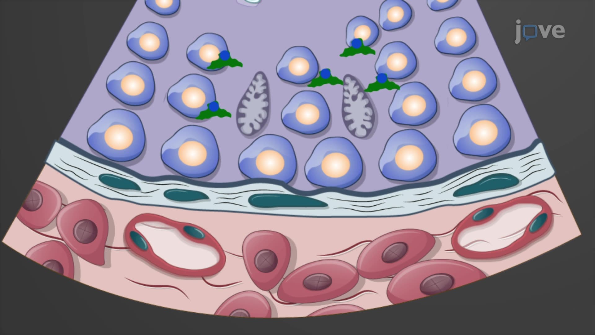
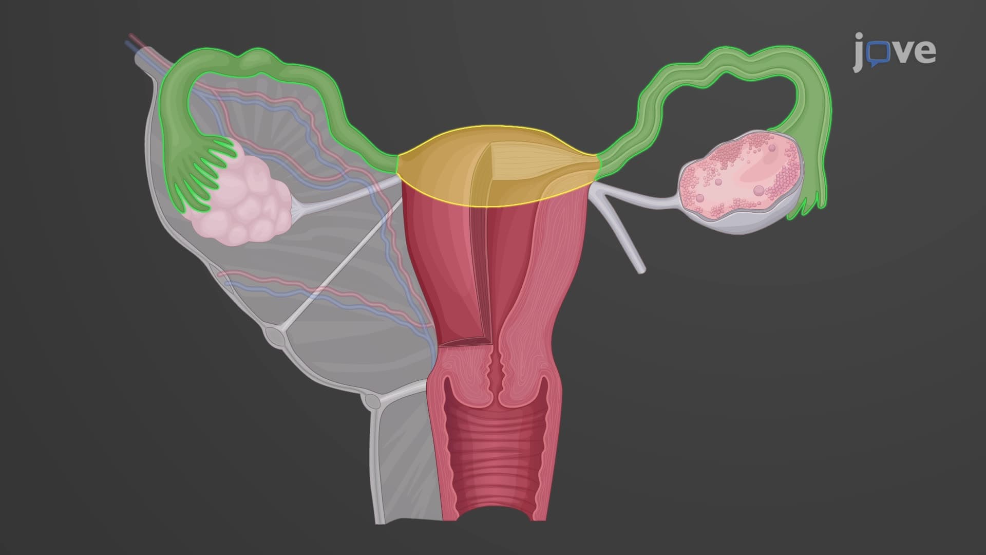
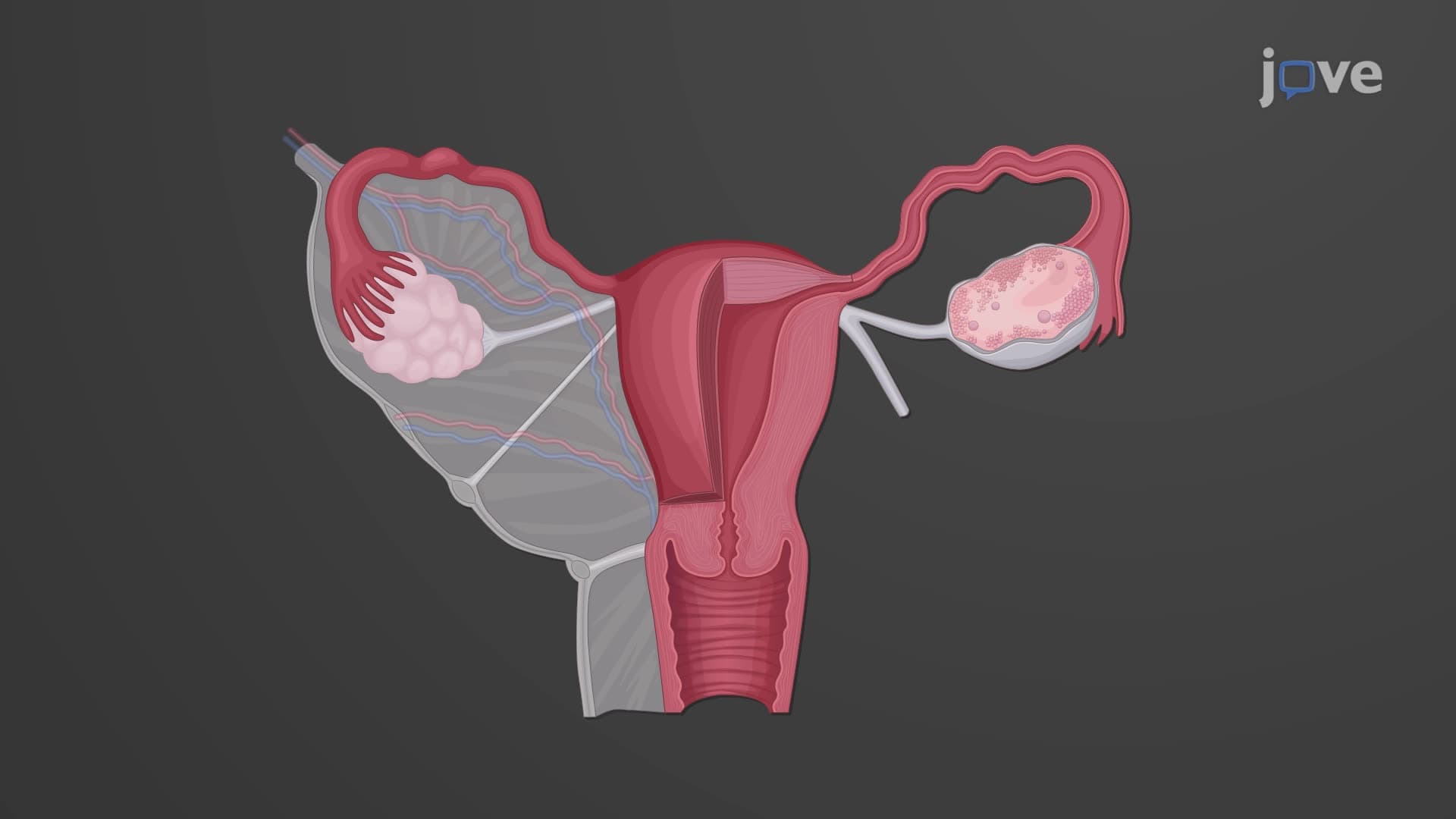
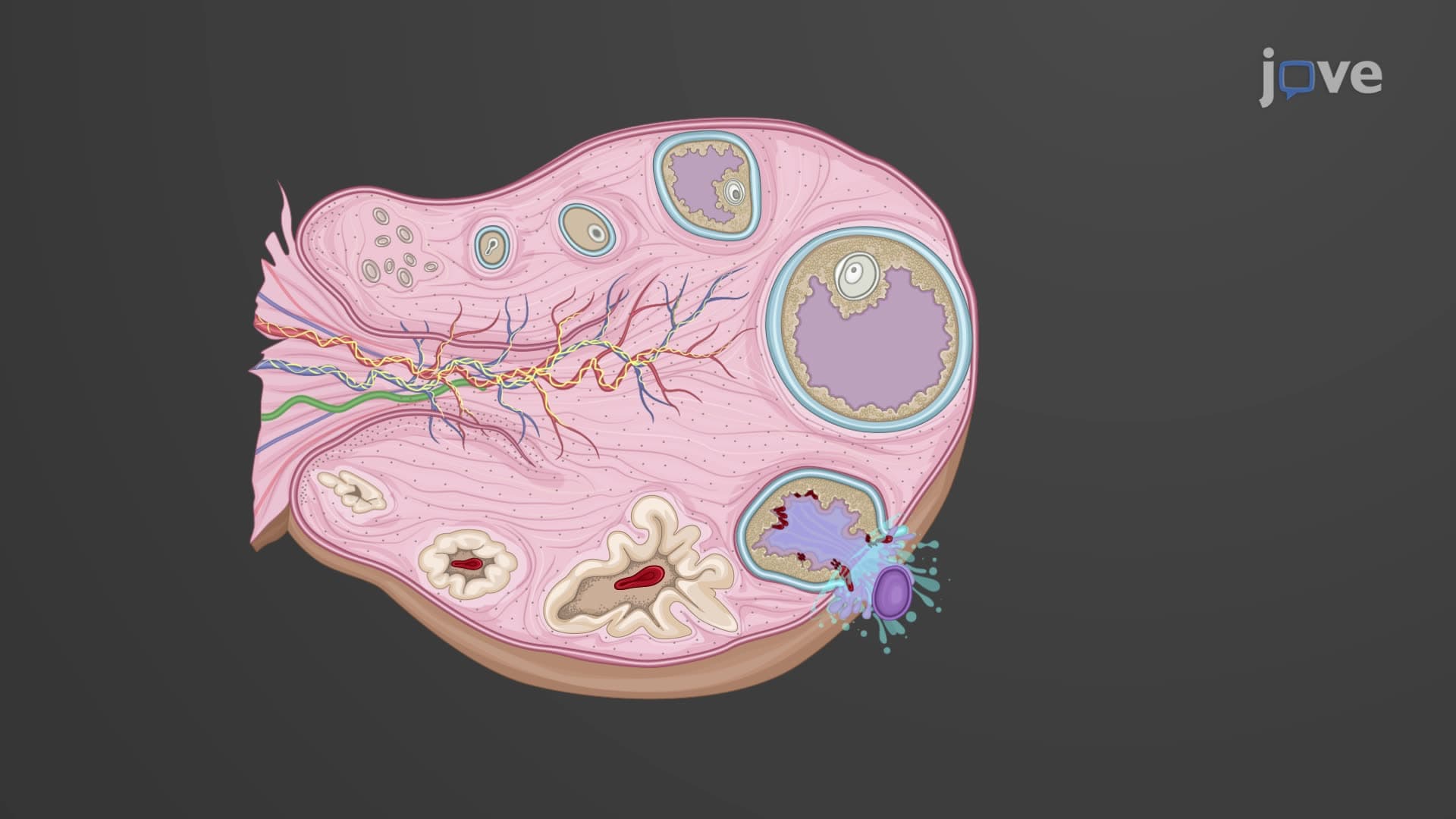
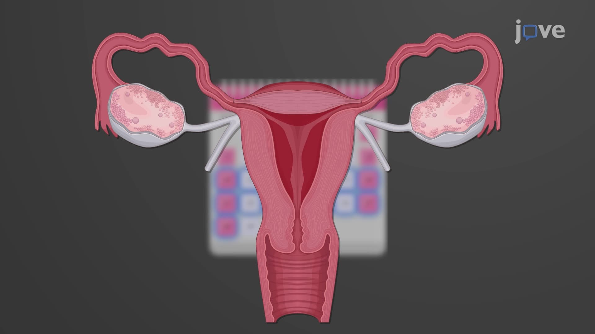
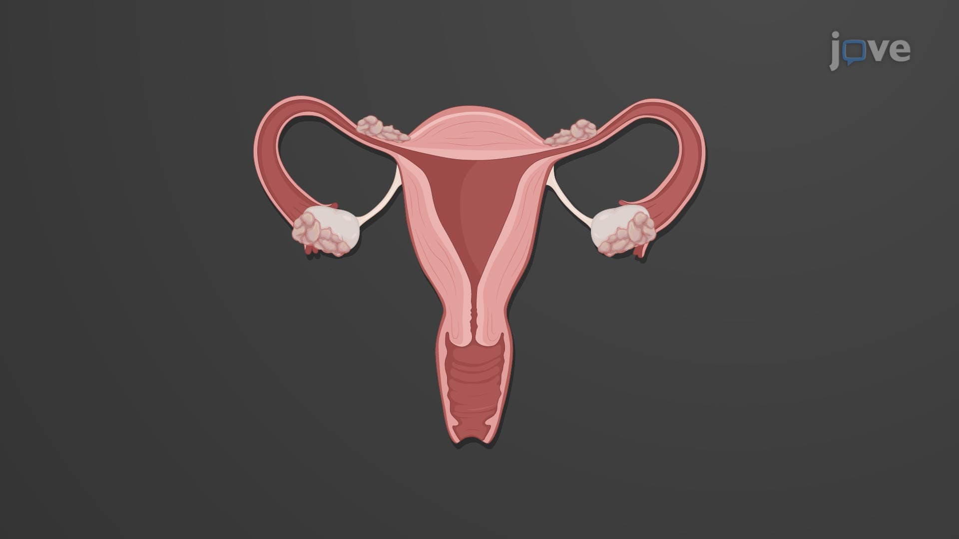
Egg cell development explained begins surprisingly early in human development. During the second trimester of pregnancy, diploid oogonia within the developing ovaries undergo rapid mitotic divisions, multiplying from thousands to millions of cells. These oogonia then differentiate into primary oocytes and begin meiosis I, but remarkably, they arrest in prophase I-a phase that can last decades. This arrest mechanism, controlled by cyclic adenosine monophosphate (cAMP) levels, ensures that oocytes remain viable until ovulation occurs. By birth, a female infant possesses all the oocytes she will ever have, approximately 400,000 primary oocytes surrounded by pre-granulosa cells forming primordial follicles.
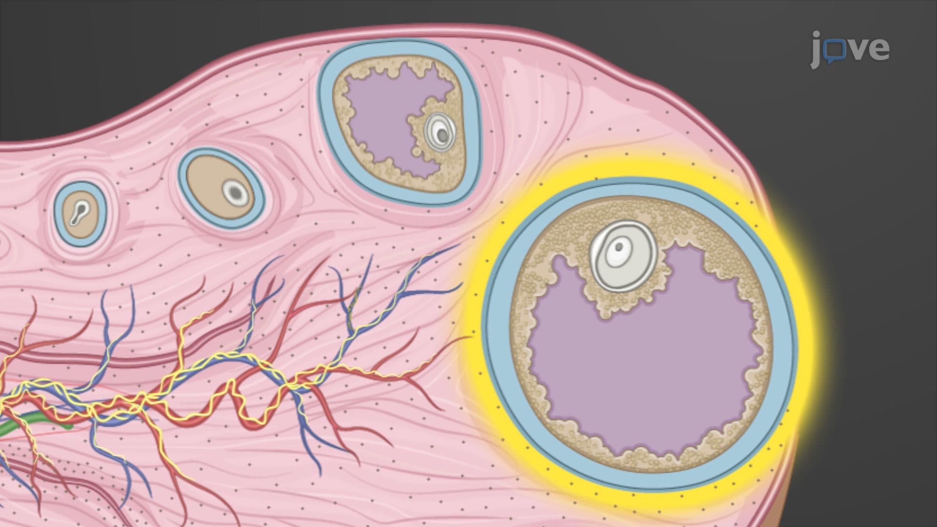
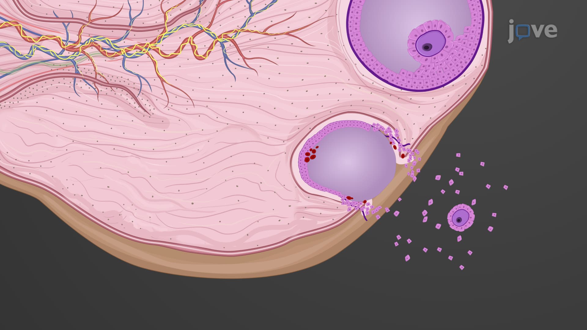
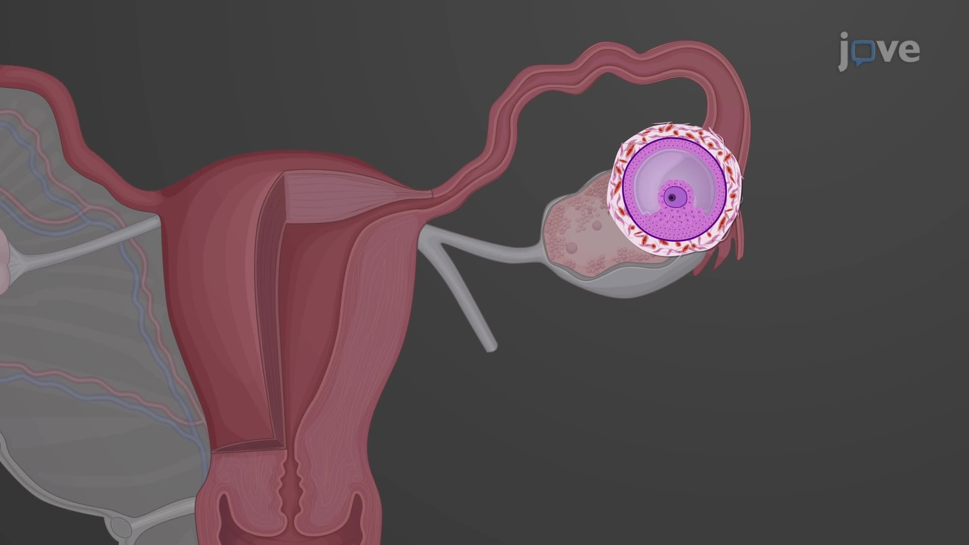
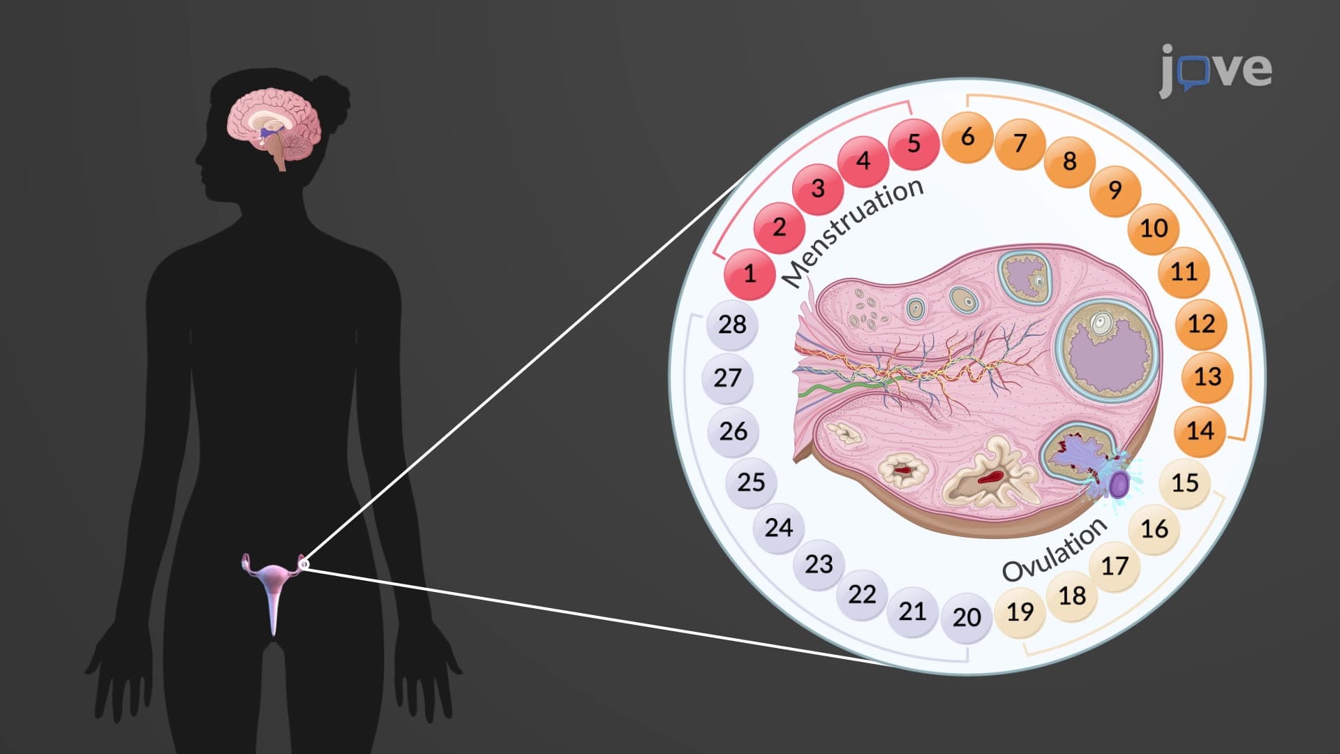
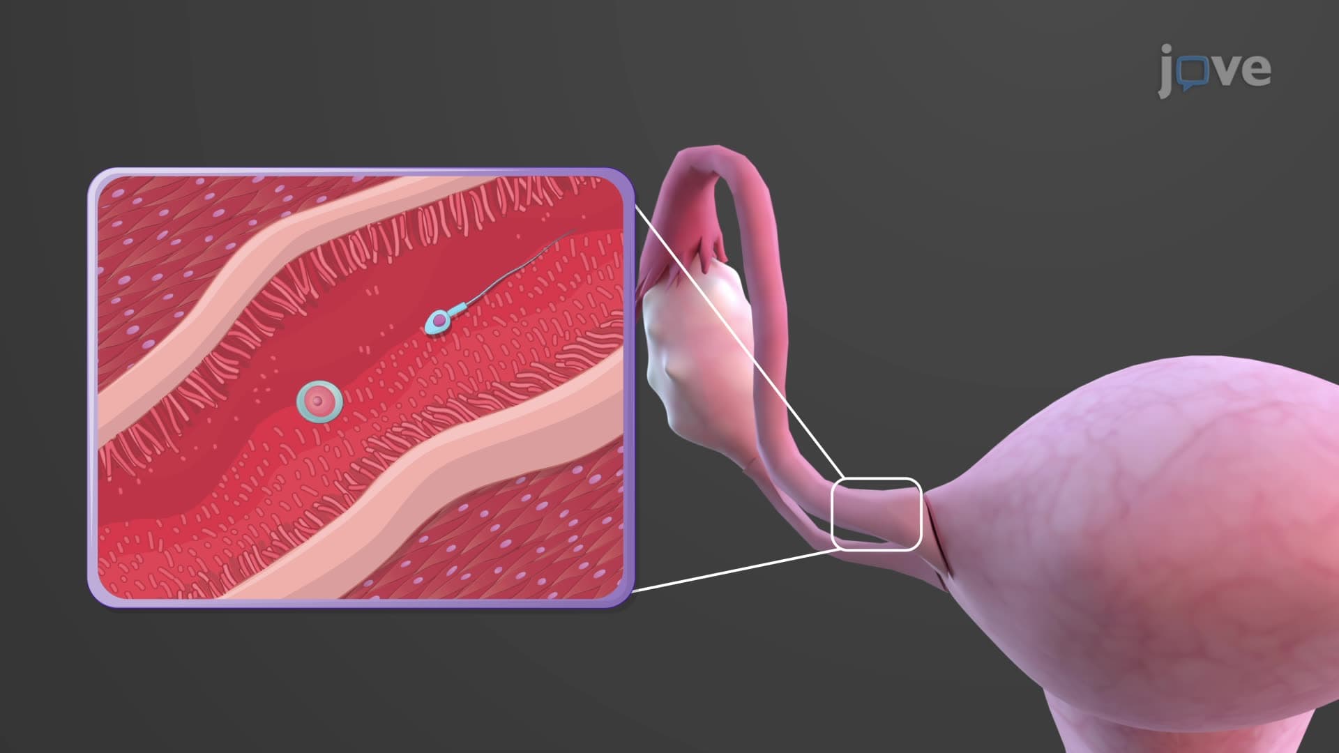
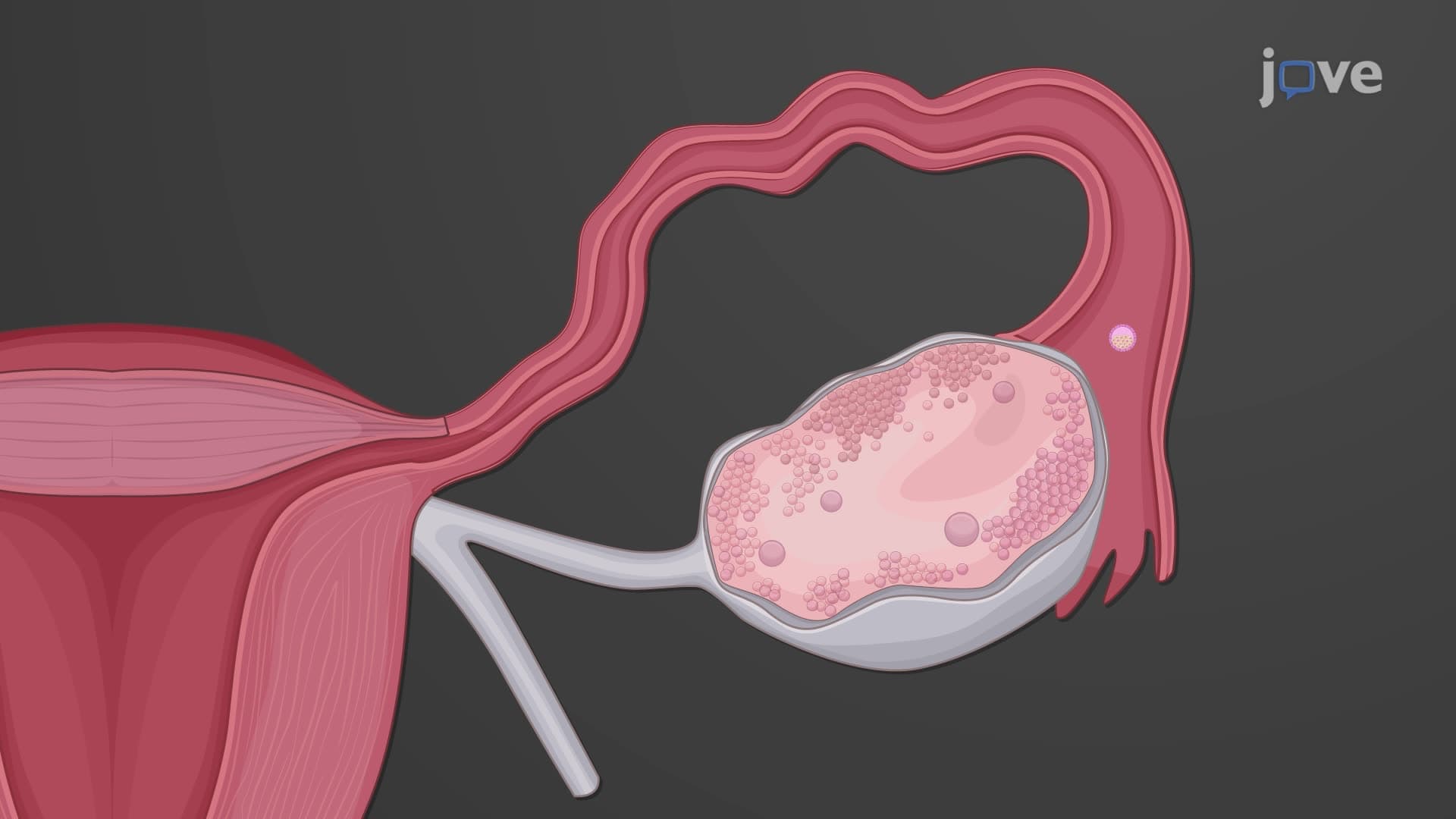
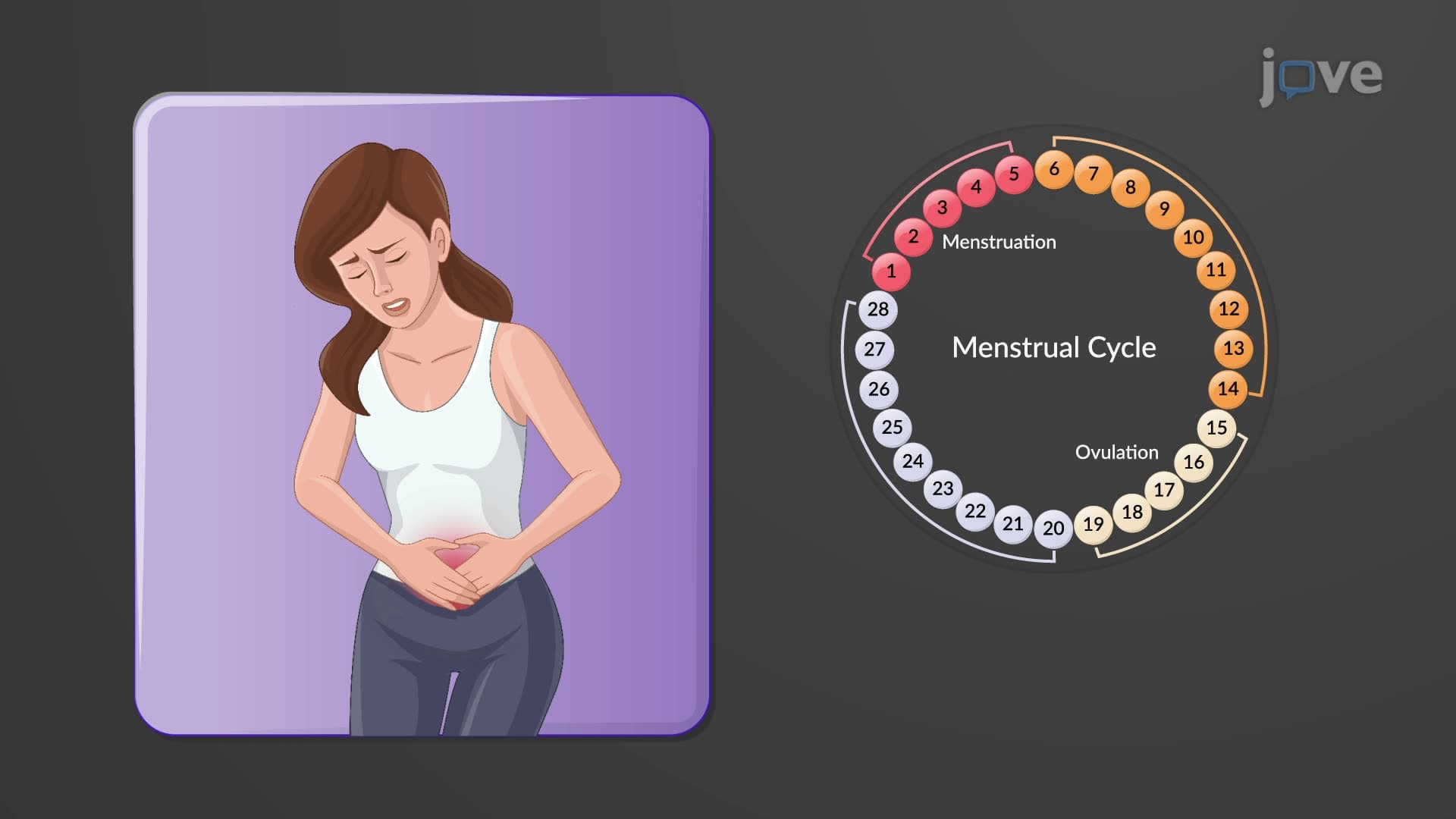
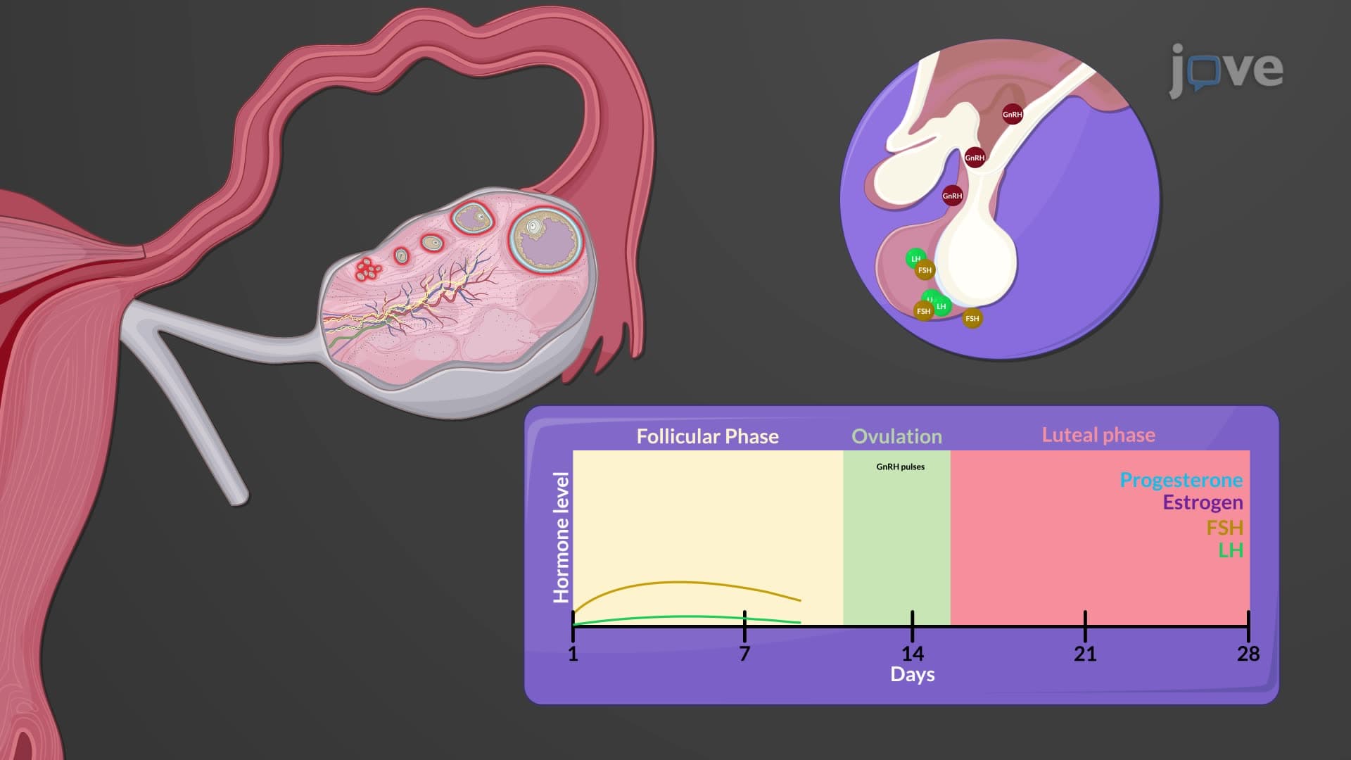
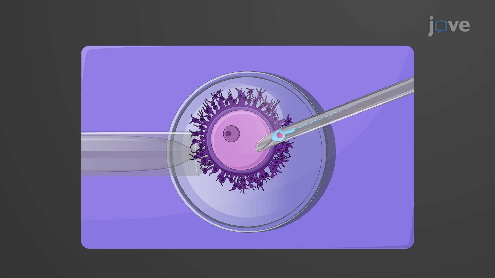
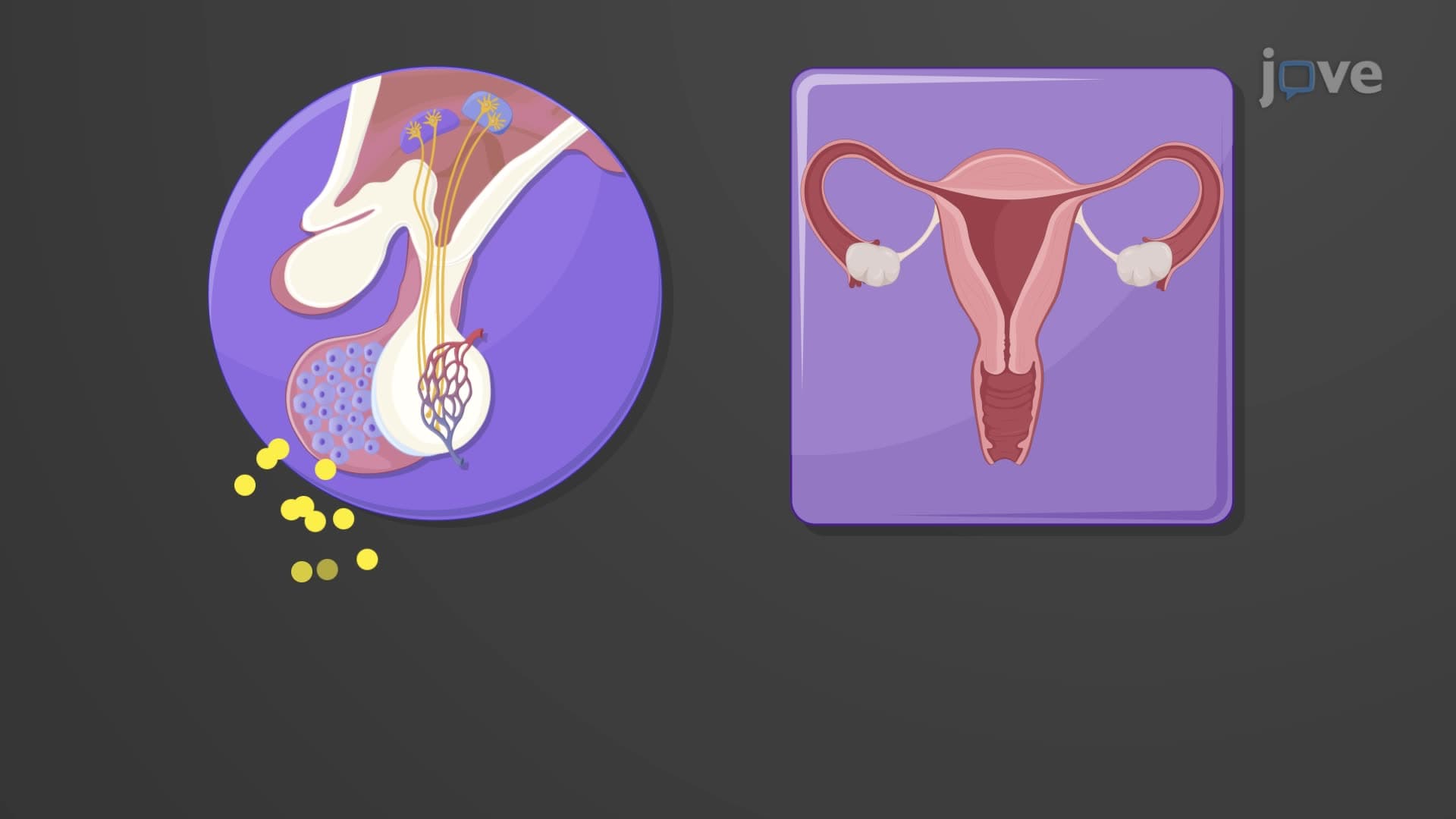
What is oogenesis in female biology becomes particularly relevant during puberty when hormonal changes reactivate the arrested oocytes. Each menstrual cycle, follicle-stimulating hormone (FSH) from the anterior pituitary stimulates several primordial follicles to resume development. However, typically only one follicle becomes dominant and completes meiosis I, producing a large secondary oocyte and a much smaller first polar body. This unequal division ensures that the oocyte retains maximum cytoplasm and organelles necessary for early embryonic development. The secondary oocyte then arrests in metaphase II until potential fertilization occurs.
Understanding oogenesis has profound implications for reproductive medicine practiced across American healthcare systems. The concept of ovarian reserve-the number of viable oocytes remaining in a woman's ovaries-directly relates to fertility potential and is routinely assessed in fertility clinics from Boston to Los Angeles. Students studying for the MCAT or NCLEX examinations must understand how oogenesis timing explains age-related fertility decline, as oocytes that have been arrested in meiosis I for 35-40 years show increased rates of chromosomal abnormalities like Down syndrome. This knowledge also underlies assisted reproductive technologies like in vitro fertilization (IVF), where understanding oocyte maturation stages is crucial for timing egg retrieval procedures.
Related Micro-courses