Loading video…


 15 Concepts
 974.8K views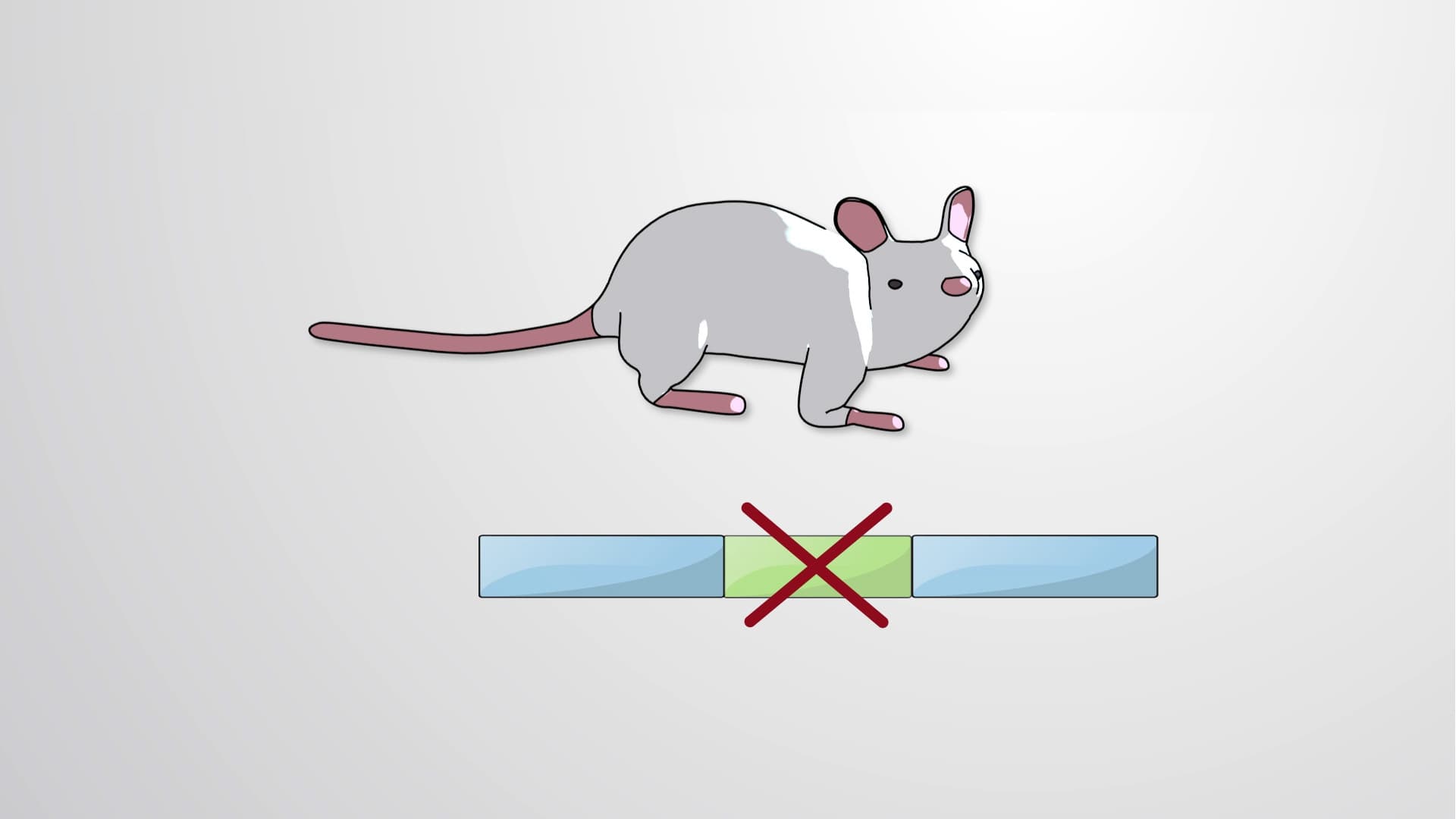

Video Summary: What Is in Vitro Mutagenesis
Did you know that scientists at Harvard Medical School have created mice without specific genes to understand diseases like cystic fibrosis? In vitro mutagenesis allows researchers to deliberately modify or "knock out" genes in laboratory settings, providing crucial insights into gene function and human health. This powerful technique involves replacing target genes with engineered DNA sequences through a process called homologous recombination, ultimately creating organisms with specific genetic modifications for research purposes. Watch the full video on JoVE Coach to master this concept with expert-led visuals and step-by-step explanations.
In vitro mutagenesis represents one of the most revolutionary techniques in modern molecular biology, allowing scientists to deliberately alter genes outside of living organisms before introducing these changes into biological systems. This method has transformed our understanding of gene function and enabled groundbreaking research at institutions like the National Institutes of Health and major university research centers across the United States. Unlike natural mutations that occur randomly, in vitro mutagenesis provides precise control over genetic modifications, making it an essential tool for studying everything from cancer mechanisms to neurological disorders.
The success of in vitro mutagenesis depends on carefully designed targeting vectors-engineered DNA constructs that serve as molecular scissors and replacement parts. These vectors contain three critical components that work together like a sophisticated delivery system. The homologous sequences act as GPS coordinates, directing the vector to the exact chromosomal location where gene replacement should occur. The positive selection marker, typically the neomycin resistance gene (NeoR), functions like a survival badge-only cells that successfully incorporate the vector can survive in neomycin-containing culture medium. The negative selection marker, such as thymidine kinase (TK), acts as a quality control mechanism, eliminating cells where the vector inserted incorrectly.
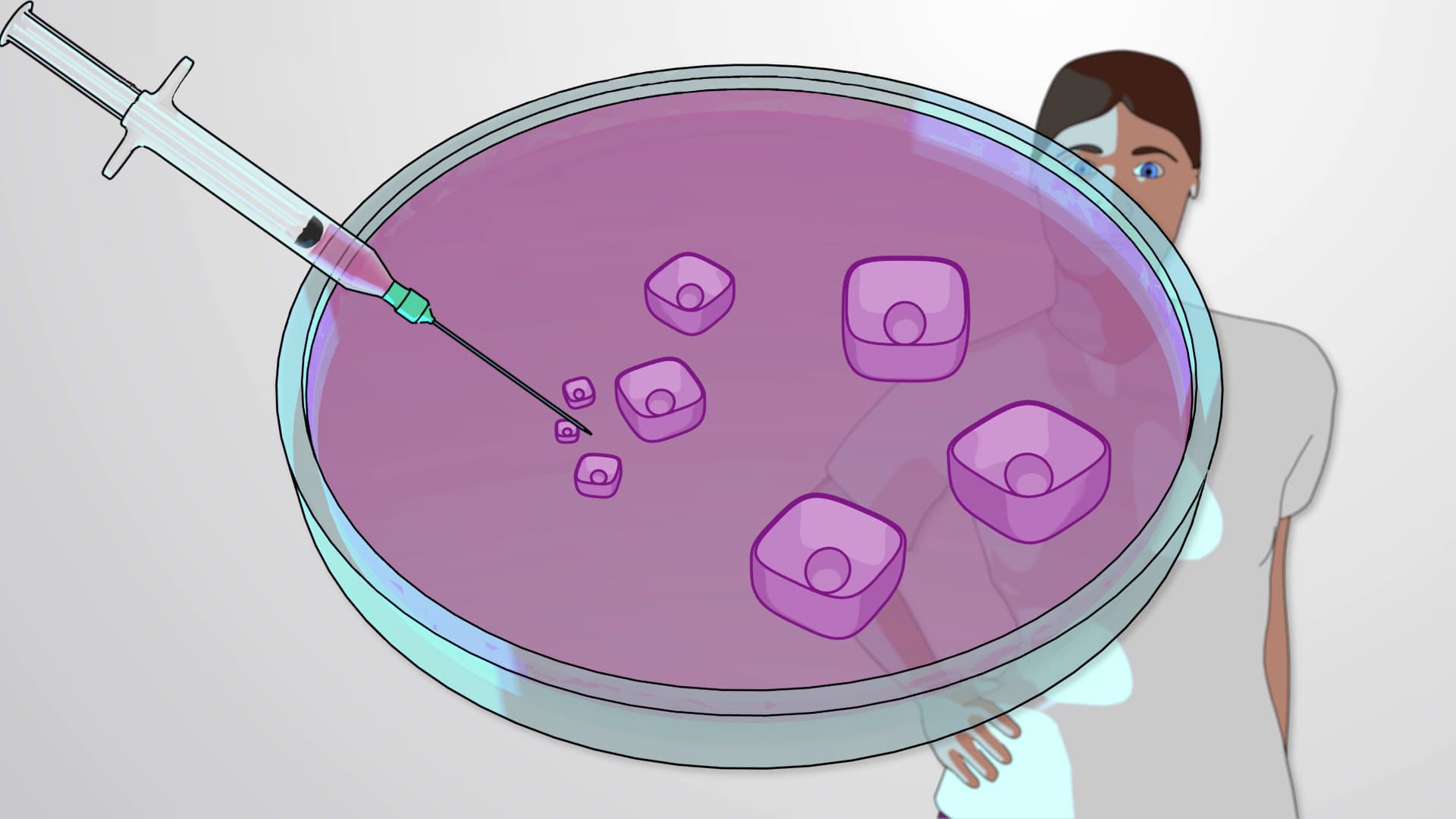
The journey from laboratory manipulation to living organisms involves multiple sophisticated steps that demonstrate the power of developmental biology. Embryonic stem cells serve as the perfect starting material because they retain the ability to develop into any cell type while remaining relatively easy to manipulate in culture. When these modified stem cells are injected into mouse blastocysts (early embryos), they integrate with the developing organism, creating chimeric mice that contain both normal and genetically modified cells. This process, perfected at research institutions like the Jackson Laboratory in Maine, requires precise timing and sterile techniques that students often encounter in advanced AP Biology courses.
Knockout mouse models created through in vitro mutagenesis have been instrumental in developing treatments for numerous human diseases. For example, researchers at the University of Pennsylvania used knockout mice to understand BRCA1 gene function, leading to better breast cancer screening and treatment protocols. These applications frequently appear on MCAT exam questions, where students must analyze experimental designs involving gene knockout studies. The technique has also proven valuable in pharmaceutical research, where companies like Pfizer and Merck use knockout models to test drug safety and efficacy before human trials.
Related Micro-courses