Loading subject page…

- Anatomy and Physiology
- Cells and Their Components
- Introduction to Cytoskeleton
Micro-course: Cells and their Components
74,359 views
Learn key concepts and earn your certificate.


Introduction to Cytoskeleton

00:00
01:33
Video Summary: What Is Cytoskeleton
Ever wondered how a single human cell maintains its shape while constantly moving and dividing? The cytoskeleton explained biology reveals a fascinating protein highway system that gives cells their structural integrity and enables complex movements. Just like the steel framework supporting Chicago's Willis Tower, the cytoskeleton provides the internal scaffolding that keeps cells organized and functional. This dynamic network of microfilaments, intermediate filaments, and microtubules orchestrates everything from muscle contraction in athletes to the precise cell divisions occurring in your body right now. Watch the full video on JoVE Coach to master this concept with expert-led visuals and step-by-step explanations.
- Understand what is cytoskeleton and its three primary protein filament types that maintain cellular structure and enable movement
- Identify the distinct functions of microfilaments (actin), intermediate filaments, and microtubules in cellular processes
- Analyze how cytoskeletal components work together to facilitate intracellular transport and organelle positioning
- Compare the mechanical properties and roles of different cytoskeletal elements in maintaining cell shape and integrity
- Evaluate the clinical significance of cytoskeletal dysfunction in cancer metastasis and neurodegenerative diseases
- Apply knowledge of motor proteins like kinesin and dynein to explain cellular cargo transport mechanisms
- Recognize the dynamic nature of cytoskeletal assembly and disassembly in cellular adaptation
- Explore connections between cytoskeletal abnormalities and human diseases studied in medical research
Understanding What is Cytoskeleton: The Cell's Internal Framework
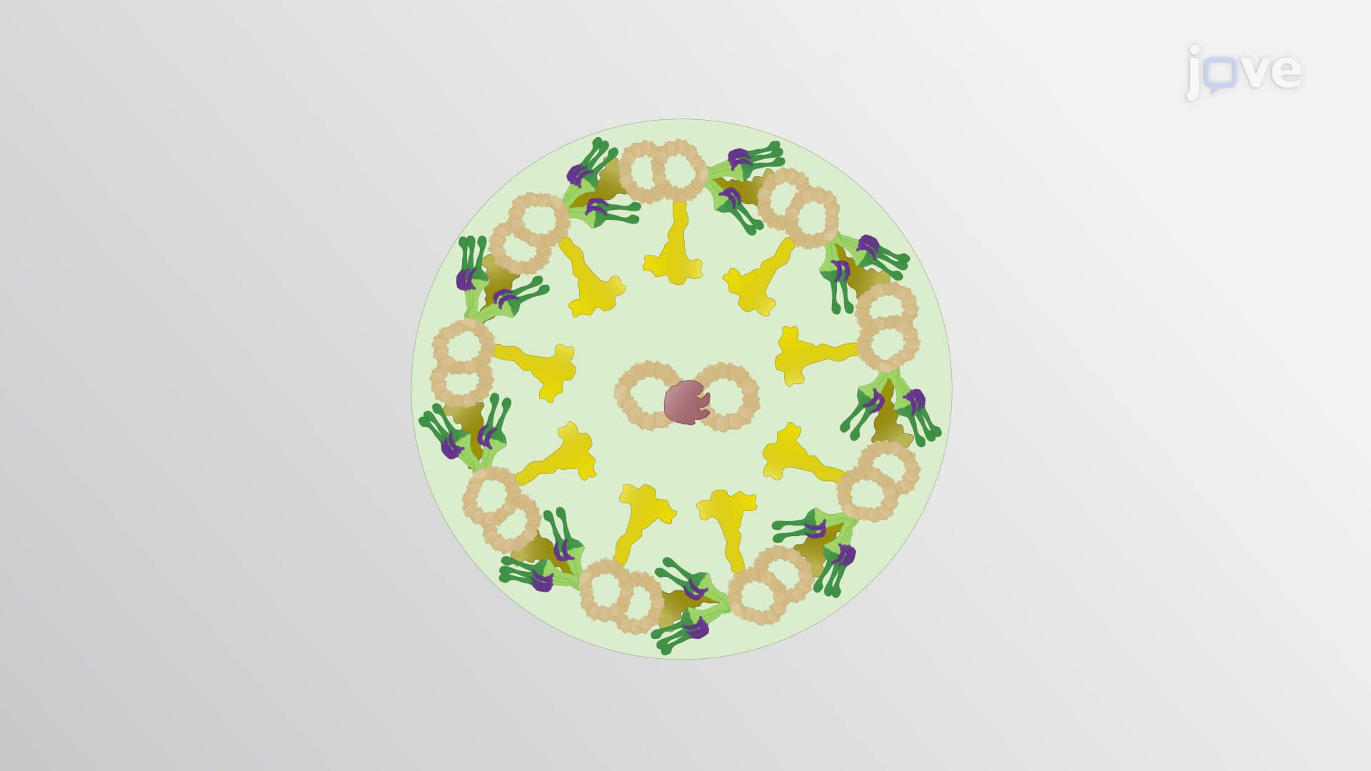
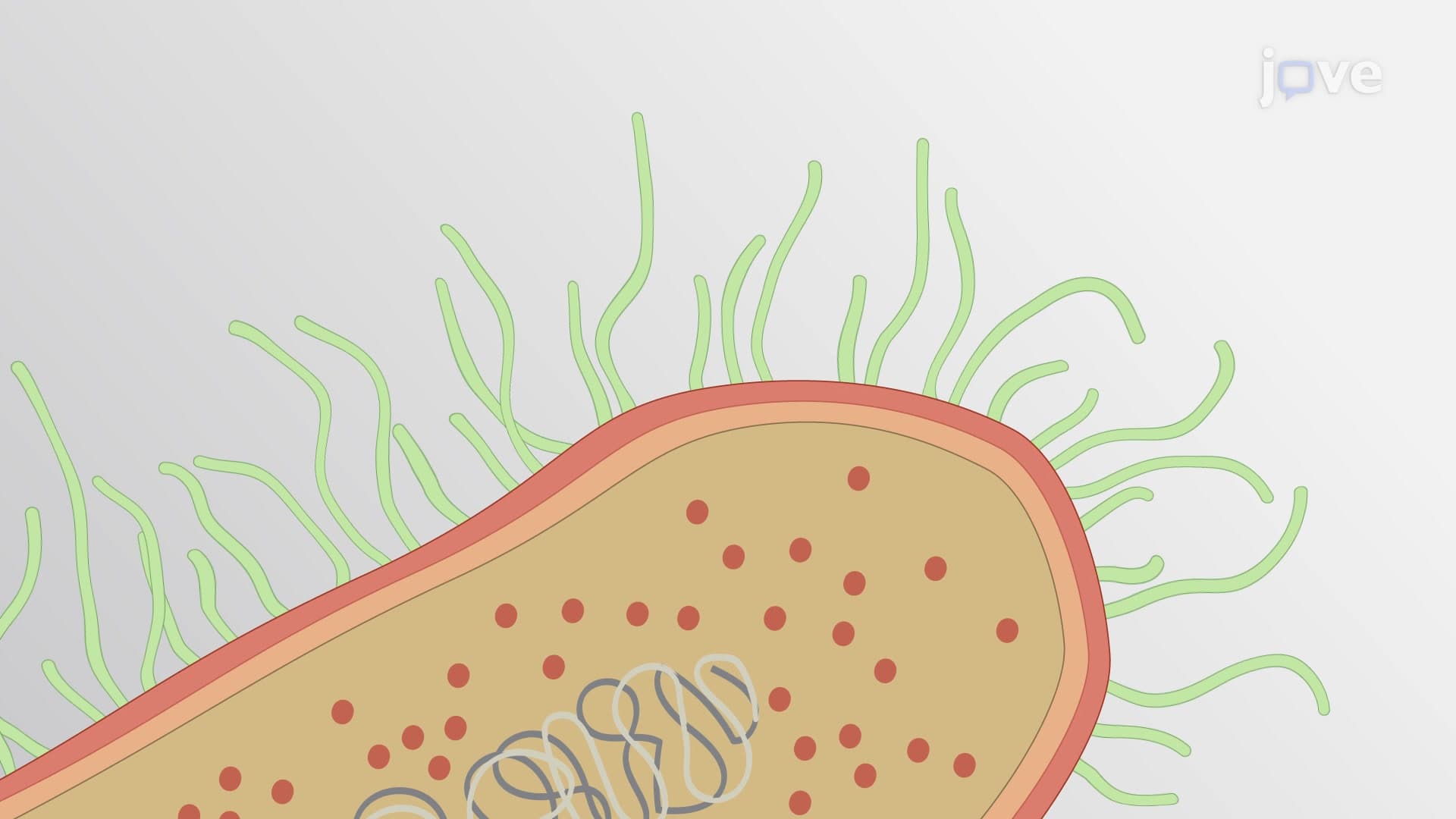
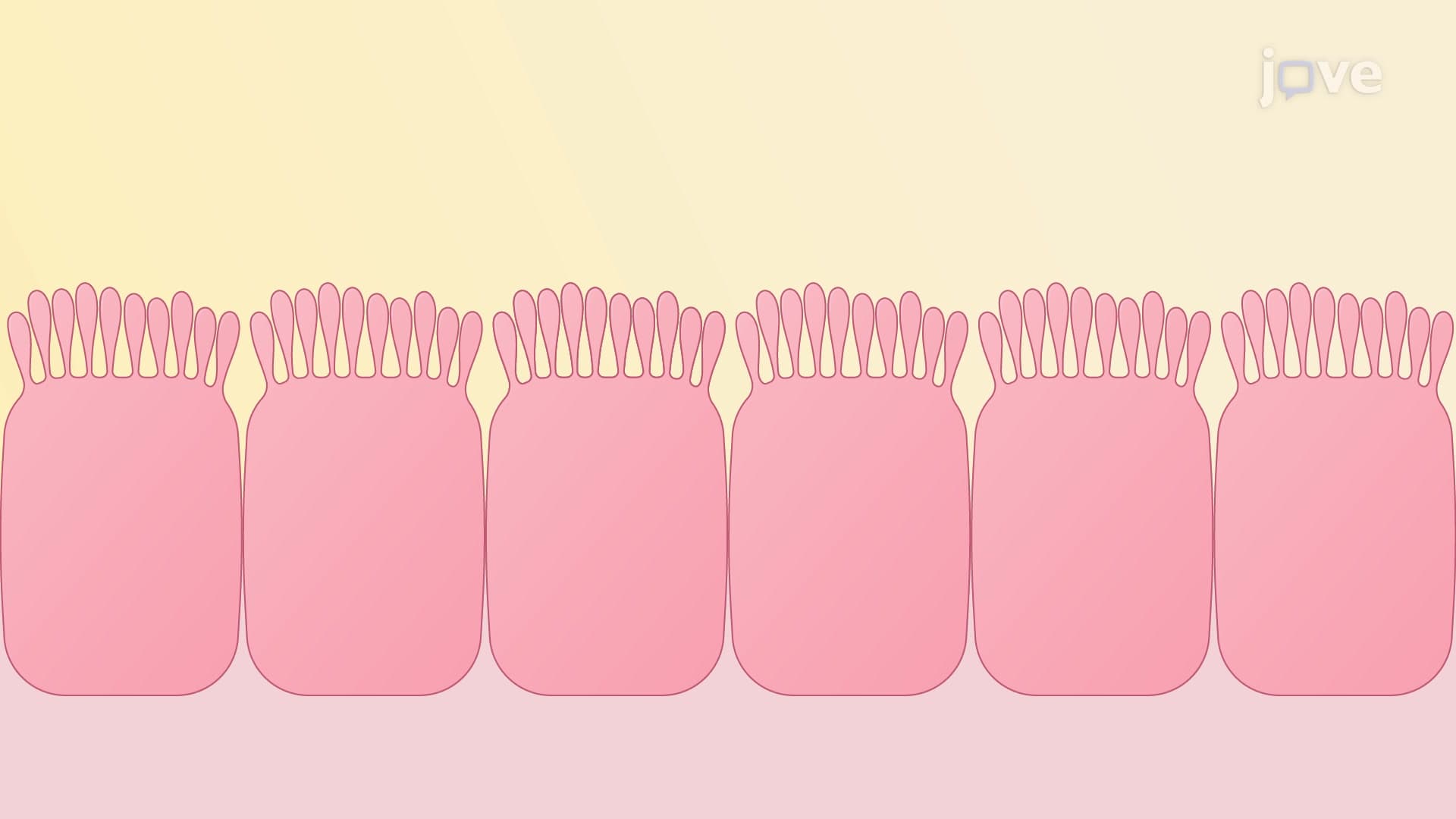
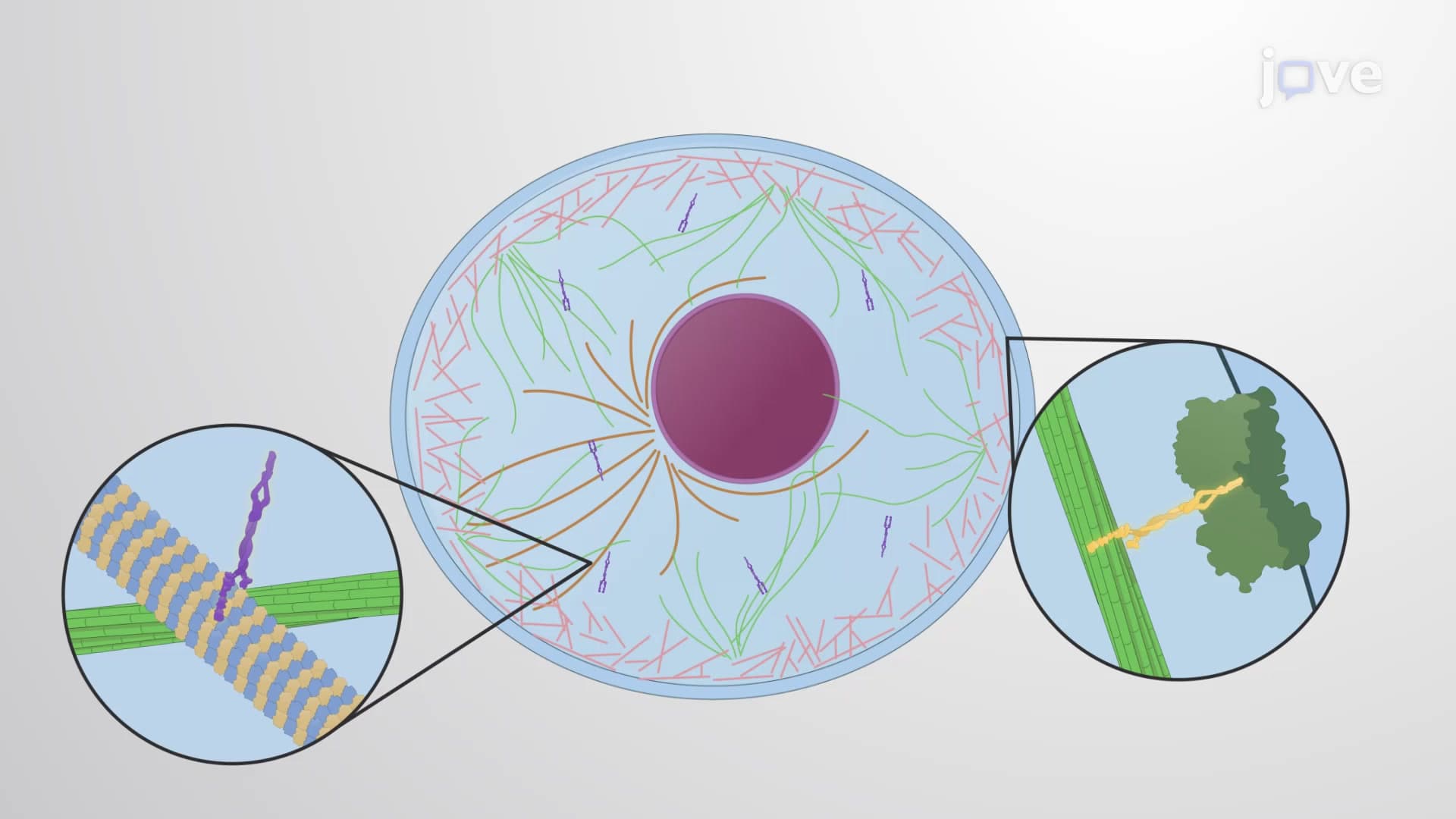
The cytoskeleton structure and function represents one of biology's most elegant solutions to cellular organization. Unlike the static framework of a building, this protein network continuously remodels itself, adapting to cellular needs in real-time. Three distinct filament systems work in concert: actin microfilaments cytoskeleton components handle rapid shape changes and movement, intermediate filaments cytoskeleton structures provide tensile strength, and microtubule cytoskeleton elements serve as highways for intracellular transport.
Microfilaments: The Cell's Muscle System
Actin microfilaments cytoskeleton networks drive cellular motility through sophisticated polymerization dynamics. These 7-nanometer filaments power muscle contraction in cardiac patients undergoing stress tests at Johns Hopkins Hospital, where actin-myosin interactions generate the force needed for each heartbeat. In non-muscle cells, actin filaments create the contractile ring during cell division and form the core of cellular projections called filopodia, essential for nerve growth during embryonic development.
Intermediate Filaments: Structural Resilience
Intermediate filaments cytoskeleton components provide mechanical stability that prevents cellular damage under stress. Keratin filaments in skin cells withstand daily mechanical forces, while neurofilaments in neurons maintain axonal structure over distances spanning from your spinal cord to your fingertips. Mutations in intermediate filament proteins cause muscular dystrophies, highlighting their clinical importance in conditions treated at specialized centers like the NIH Clinical Center.
Microtubules: Cellular Transportation Networks
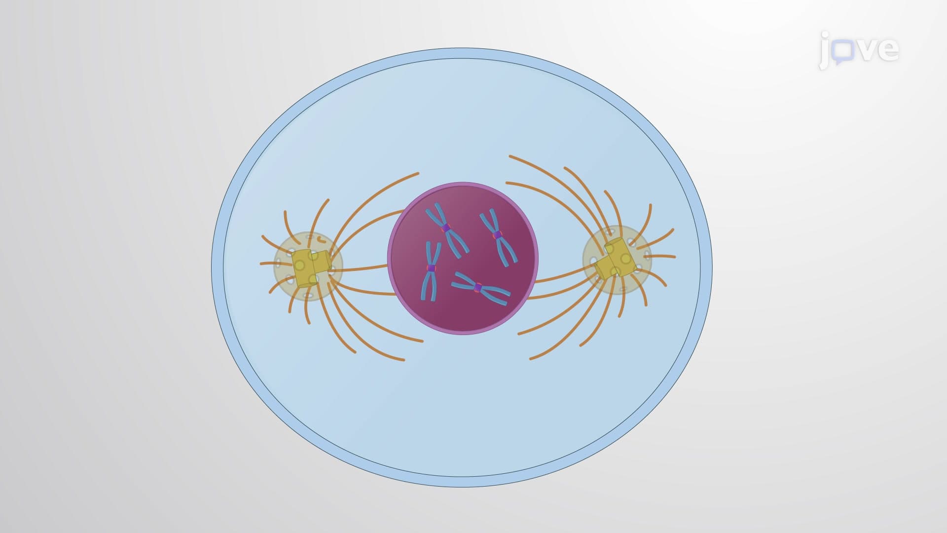
The microtubule cytoskeleton forms dynamic tracks radiating from the cell's organizing center, facilitating long-distance transport via motor proteins. Kinesin motors carry cargo toward cell peripheries, while dynein motors transport materials toward the nucleus. During mitosis, microtubules form the mitotic spindle, ensuring accurate chromosome segregation-a process frequently tested on AP Biology exams and college cell biology midterms.
Clinical and Research Applications
Understanding cytoskeletal network function proves crucial for medical advances at institutions like the Mayo Clinic, where researchers investigate how cancer cells exploit cytoskeletal remodeling for metastasis. Alzheimer's research at Stanford focuses on tau protein dysfunction, which disrupts microtubule stability in neurons. These connections between basic cell shape cytoskeleton mechanisms and human disease make cytoskeleton knowledge essential for pre-med students preparing for the MCAT.
Frequently Asked Questions
The cytoskeleton is a dynamic network of protein filaments that provides structural support, maintains cell shape, and enables cellular movement and transport. It's crucial because it acts as the cell's internal framework, organizing organelles and facilitating essential processes like cell division, muscle contraction, and intracellular transport that keep all living organisms functioning.
The cytoskeleton consists of three main protein filament types: microfilaments (actin filaments) at 7nm diameter for movement and shape changes, intermediate filaments at 10nm for mechanical strength, and microtubules at 25nm that serve as tracks for organelle transport. Each type has distinct proteins and functions but works together as an integrated system.
AP Biology commonly tests cytoskeleton concepts through questions about cell structure-function relationships, protein interactions, and cellular transport mechanisms. Students should understand how actin enables muscle contraction, how microtubules facilitate mitosis, and how cytoskeletal dysfunction relates to diseases-knowledge that appears in both multiple-choice and free-response sections.
MCAT questions emphasize cytoskeletal motor proteins (kinesin, dynein, myosin), polymerization dynamics, and disease connections like cancer metastasis and neurodegeneration. Focus on understanding how ATP powers motor protein movement, how cytoskeletal drugs work, and the relationship between cytoskeletal structure and cellular function in different tissue types.
Cytoskeletal defects lead to serious medical conditions treated in US hospitals daily: muscular dystrophies from intermediate filament mutations, Alzheimer's disease from microtubule-associated tau protein dysfunction, and cancer metastasis from abnormal actin dynamics. Understanding these connections helps explain why cytoskeleton research receives significant NIH funding for therapeutic development.
Not at all-cytoskeleton concepts build logically from basic protein structure knowledge taught in introductory biology courses. Start with the analogy of cellular scaffolding, then explore each filament type's specific role. The key is connecting structure to function and relating abstract concepts to familiar processes like muscle movement.
Create associations between filament size, protein composition, and function: "Actin=Action" for microfilaments and movement, "Intermediate=In-between" for structural support, and "Micro-tubes=Transport tubes" for microtubules. Practice drawing diagrams showing all three systems in a typical cell, labeling motor proteins and their directions of movement.
Explore cytoskeletal regulation through signaling pathways, advanced motor protein mechanisms, and cutting-edge research on cytoskeletal drugs in cancer therapy. These topics prepare you for upper-level cell biology courses and research opportunities at universities with strong biomedical programs.
Related Micro-courses

15 Concepts
16 Concepts
26 Concepts