Loading subject page…


74,364 views

Video Summary: What Is Mechanism of Ciliary Motion
Every breath you take depends on millions of microscopic hair-like structures called cilia beating 1,000 times per minute in your respiratory tract. The ciliary motion mechanism relies on a sophisticated molecular motor system within each cilium's axoneme structure. In patients with primary ciliary dyskinesia at Johns Hopkins Hospital, defective ciliary motion leads to chronic respiratory infections because mucus cannot be properly cleared from airways. Understanding what is the mechanism of ciliary motion reveals how dynein proteins create the coordinated beating patterns essential for human health. Watch the full video on JoVE Coach to master this concept with expert-led visuals and step-by-step explanations.
The ciliary motion mechanism represents one of biology's most elegant molecular machines, converting chemical energy into coordinated mechanical movement. This process enables critical physiological functions from clearing mucus in your lungs to propelling sperm cells during fertilization. For students preparing for AP Biology or college-level cell biology courses, understanding ciliary motion provides essential insights into how cells generate movement and maintain homeostasis.
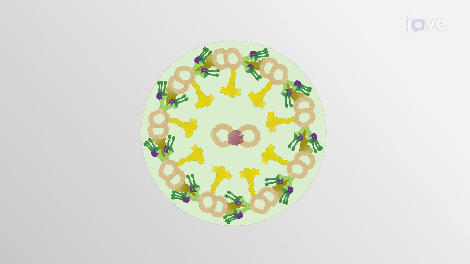
At the heart of every motile cilium lies the axoneme, a precisely organized cytoskeletal structure following the universal 9+2 microtubule cilium pattern. Nine peripheral microtubule doublets surround two central singlet microtubules, creating a framework that transforms molecular forces into macroscopic motion. Each peripheral doublet consists of A and B tubules, with the A tubule bearing dynein arms that extend toward neighboring doublets. This arrangement, first discovered through electron microscopy at Harvard Medical School, remains consistent across species from algae to humans, demonstrating evolution's conservation of effective molecular designs.
Dynein ciliary motion depends on massive protein complexes that walk along microtubules using ATP hydrolysis. Outer dynein arms generate the primary power stroke cilia force, while inner dynein arms provide fine-tuning control. During each cycle, dynein heads bind to adjacent B tubules, undergo conformational changes powered by ATP, then release and reset. This creates sliding forces between microtubule doublets that would cause axoneme extension if unconstrained. However, nexin links and radial spokes convert linear sliding into the bending motions characteristic of ciliary beating.
Understanding the mechanism of cilia movement proves crucial for medical diagnosis and treatment. Primary ciliary dyskinesia affects approximately 1 in 15,000 Americans, causing chronic sinusitis, bronchitis, and male infertility due to defective sperm flagella. At institutions like the Mayo Clinic, specialists use electron microscopy to examine ciliary ultrastructure, identifying missing dynein arms or structural abnormalities. Students encountering this topic on MCAT examinations should recognize how ciliary dysfunction exemplifies the connection between molecular defects and organ-system pathology, illustrating why cellular biology knowledge translates directly to clinical practice.
Related Micro-courses