Loading subject page…


74,363 views
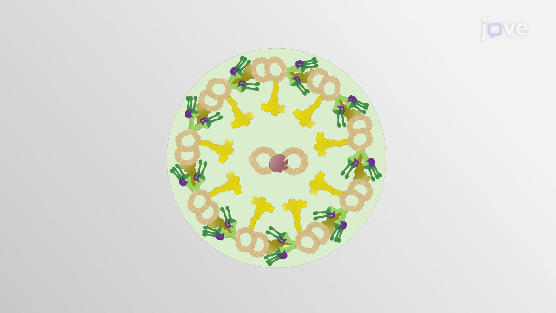

Video Summary: What Is Microtubules in Cell Motility
Ever wonder how cancer cells spread throughout the body or how nerve cells extend over distances up to three feet in humans? Microtubules cell motility represents one of biology's most sophisticated transportation networks, where microscopic protein highways enable cellular movement and cargo transport. From the migration of immune cells responding to infections at Mayo Clinic research labs to the precise division of cells during wound healing, microtubule role in cell movement drives countless biological processes. These dynamic protein structures work alongside motor proteins like dynein and kinesin to orchestrate everything from basic cell migration to complex neuronal development. Watch the full video on JoVE Coach to master this concept with expert-led visuals and step-by-step explanations.
How microtubules contribute to cell motility represents a masterpiece of cellular engineering that rivals any human-designed transportation system. These hollow, cylindrical structures-composed of α- and β-tubulin protein dimers-form the backbone of cellular movement processes that keep us alive and healthy. From the moment a white blood cell detects an infection signal to the complex choreography of cell division, microtubules orchestrate movement with remarkable precision.
The concept of dynamic instability sets microtubules apart from static structural components. This phenomenon, discovered by researchers at the University of California, involves rapid cycles of polymerization (growth) and depolymerization (shrinkage) that allow cells to quickly reorganize their internal architecture. During cell migration, microtubules can rapidly extend toward the leading edge while simultaneously retracting from the trailing edge, creating a coordinated movement pattern essential for processes like immune surveillance.
This dynamic behavior proves crucial for students preparing for advanced biology exams like the AP Biology test, where understanding cytoskeletal dynamics frequently appears in free-response questions about cell structure and function. College students taking cell biology courses often encounter this concept in laboratory exercises examining live cell imaging, where fluorescently-labeled microtubules reveal their constant growth and shrinkage patterns.
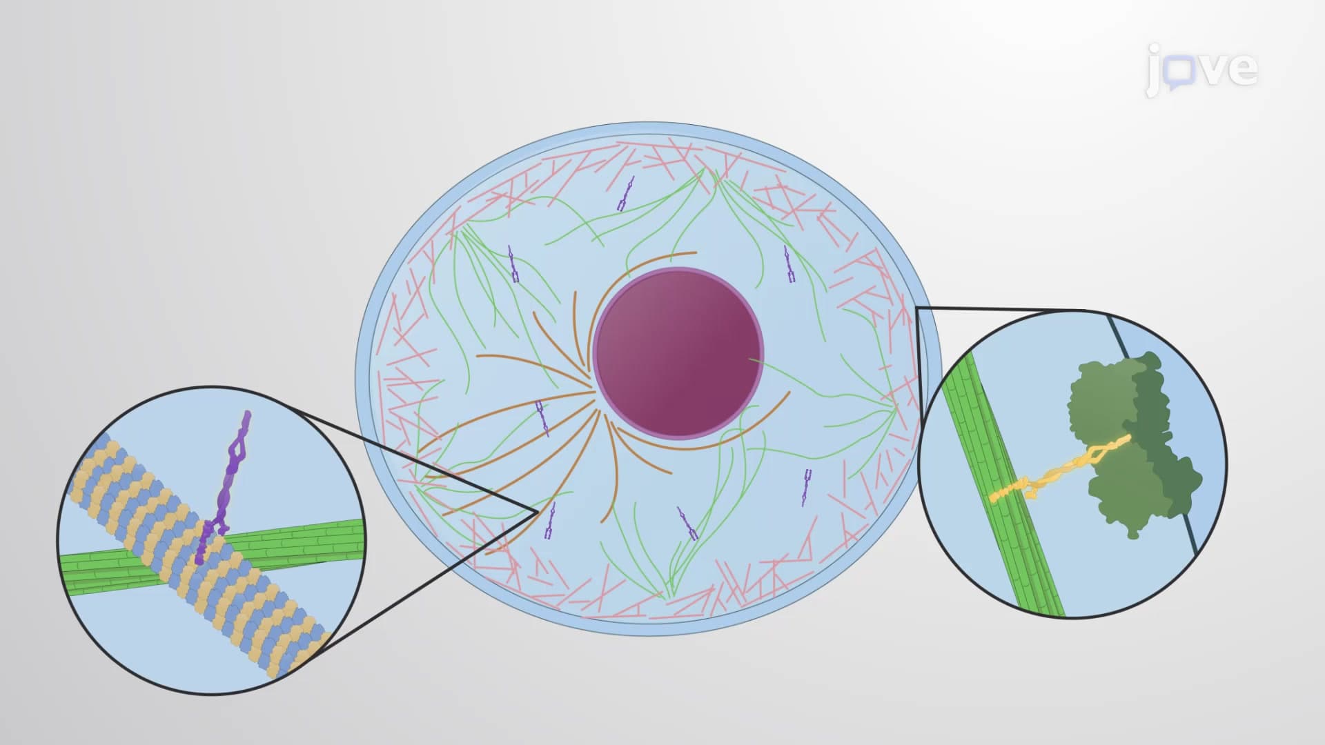
Dynein kinesin microtubule interactions represent one of biology's most elegant transportation systems. Kinesin motors typically move cargo toward the cell's periphery (plus-end directed), while dynein motors transport materials toward the cell center (minus-end directed). This bidirectional transport system enables everything from the delivery of synaptic vesicles in neurons to the positioning of mitochondria where energy is most needed.
Research at institutions like Johns Hopkins University has revealed how disruptions in axonal transport microtubule function contribute to neurodegenerative diseases. In Alzheimer's disease, for example, tau protein abnormally binds to microtubules, disrupting the normal cargo transport that keeps neurons healthy. This connection between basic cell biology and human disease makes microtubule motility a high-yield topic for pre-med students preparing for the MCAT.
Understanding cytoskeletal motility microtubule mechanisms has revolutionized cancer treatment through drugs like paclitaxel (Taxol), originally derived from Pacific yew trees. These medications work by stabilizing microtubules, preventing the dynamic instability required for cancer cell division and migration. Research teams at cancer centers like MD Anderson continue investigating how cell migration microtubule pathways might be targeted to prevent metastasis.
Current research at major US universities focuses on how microtubule motor protein systems coordinate with other cellular components during wound healing, tissue regeneration, and immune responses-areas of study that frequently appear in graduate school entrance examinations and advanced undergraduate coursework.
Related Micro-courses