Loading subject page…

Micro-course: The Reproductive System
141,004 views
Learn key concepts and earn your certificate.


Ovaries

00:00
01:26
Video Summary: What are Ovaries
Did you know that ovaries anatomy biology reveals these tiny organs produce over 400 different types of eggs throughout a woman's lifetime? Understanding what are the ovaries in female anatomy is crucial for grasping human reproduction-from fertility treatments at major US medical centers like Mayo Clinic to AP Biology exam success. The ovary structure function explained shows how these paired glands control hormonal cycles and reproduction through specialized tissue layers and follicular development. Watch the full video on JoVE Coach to master this concept with expert-led visuals and step-by-step explanations.
- Understand what are ovaries and their essential role in the female reproductive system, including their anatomical position and ligamentous attachments
- Identify the distinct histological layers of ovarian anatomy, from the outer mesothelium through the tunica albuginea to the cortex and medulla
- Analyze the ovarian cortex medulla organization and explain how follicular development progresses from primordial to mature Graafian follicles
- Compare the structural and functional differences between corpus luteum and corpus albicans in post-ovulation ovary function
- Evaluate the hormonal contributions of ovarian structures, particularly progesterone, estrogen, relaxin, and inhibin production
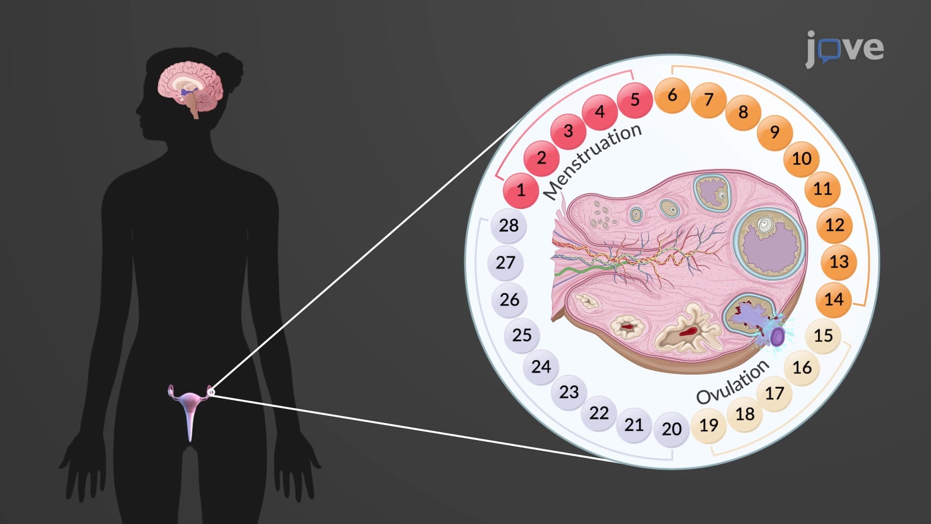
- Apply knowledge of follicle corpus luteum ovary relationships to understand menstrual cycle regulation and ovulation ovary function
- Recognize the clinical significance of ovarian ligament anatomy in surgical procedures and reproductive health assessments
- Explore connections between ovary female reproductive anatomy and broader endocrine system interactions
Understanding What are Ovaries: Structural Foundation
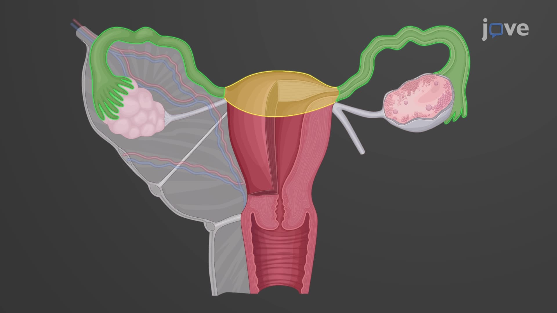
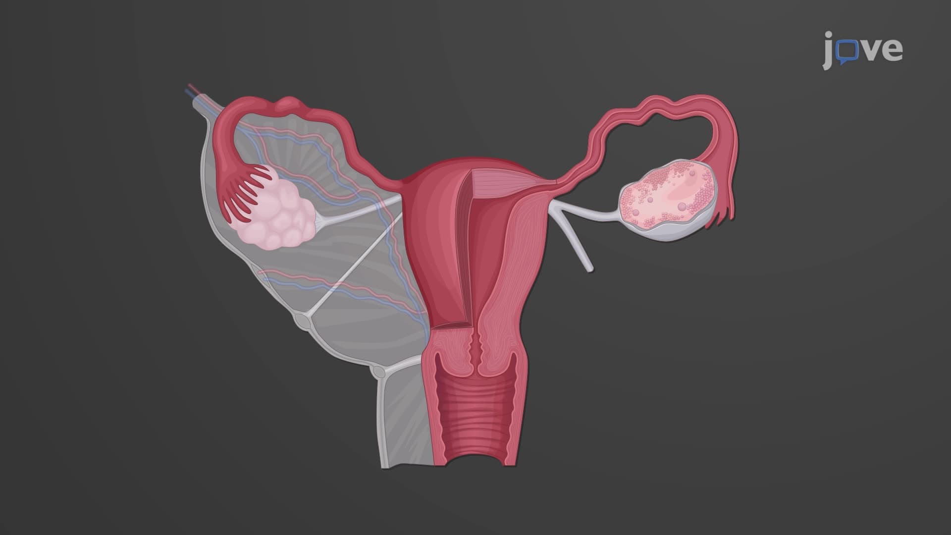
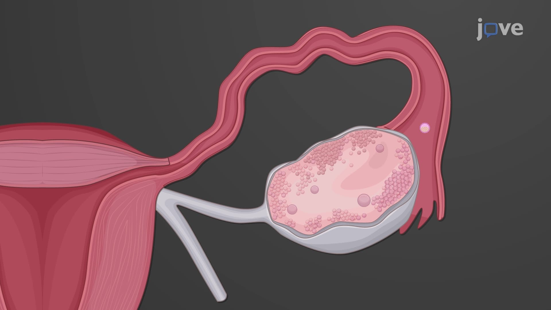
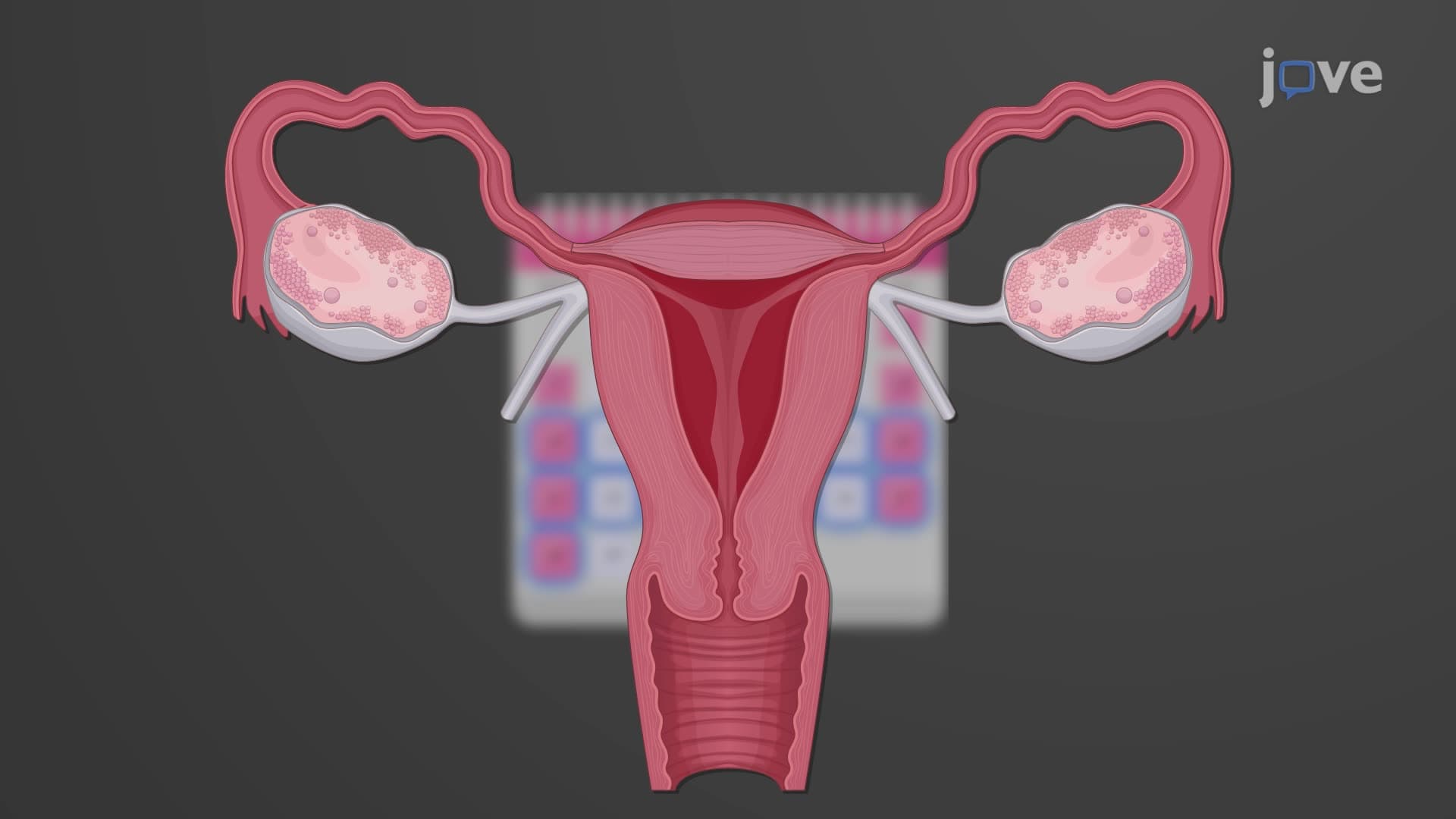
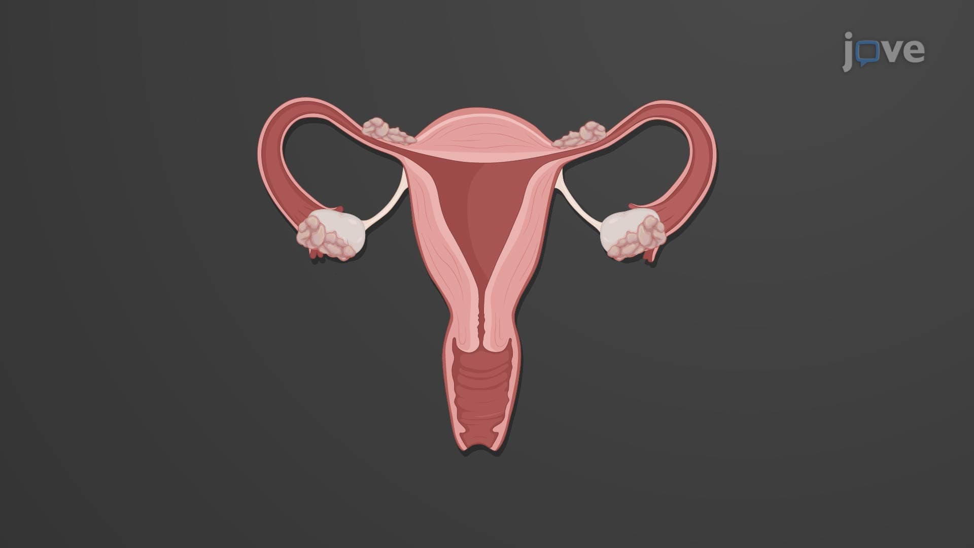
The ovaries represent masterpieces of biological engineering within ovaries anatomy biology. These almond-sized organs, weighing approximately 14 grams each, anchor to the uterus through three critical ligament systems. The broad ligament provides primary support, while the suspensory ligament carries vital blood supply from the ovarian artery. The ovarian ligament creates direct connection to the uterus, forming the structural framework essential for ovary female reproductive anatomy.
Each ovary's hilum serves as the entry point for the ovarian artery (branching from the abdominal aorta) and ovarian vein, plus sympathetic nerve fibers from the ovarian plexus. This vascular arrangement becomes clinically significant during procedures like oophorectomy, commonly performed at US medical centers for conditions ranging from ovarian cysts to preventive surgery in BRCA mutation carriers.
Histological Architecture: From Surface to Core
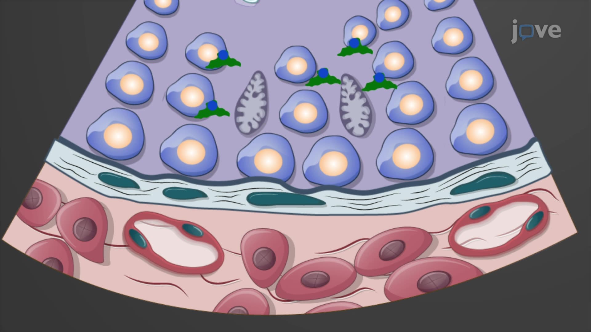
The ovary structure function explained reveals four distinct tissue layers working in harmony. The outermost ovarian mesothelium consists of simple cuboidal epithelium-often mistakenly called "germinal epithelium" in older textbooks, though oocytes don't originate here. Beneath lies the tunica albuginea, a dense connective tissue capsule providing structural integrity during the monthly follicular rupture of ovulation ovary function.
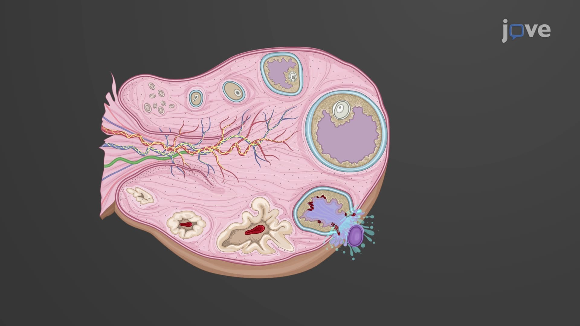
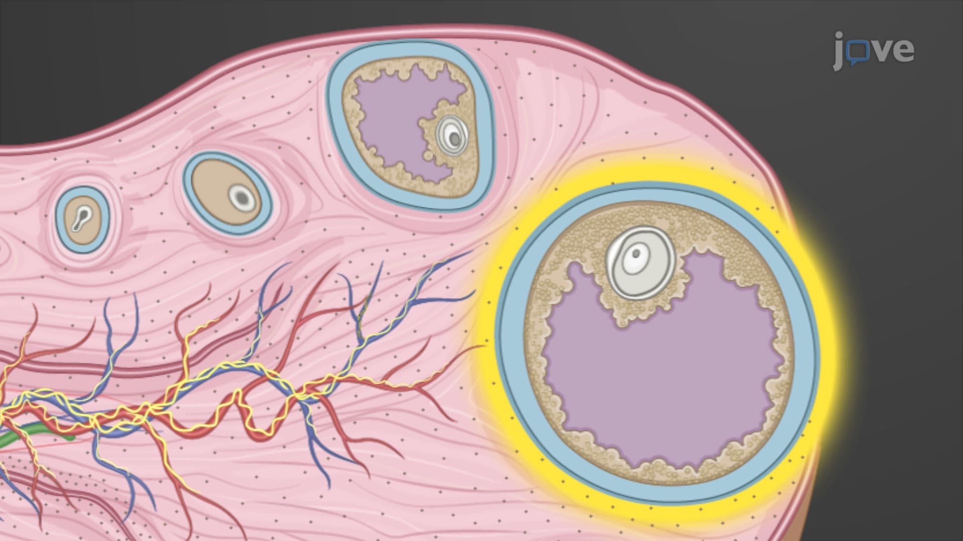
The ovarian cortex medulla boundary, while histologically indistinct, represents a functional divide crucial for understanding reproductive physiology. The cortex houses all follicular development stages, from primordial follicles (formed during fetal development) through mature Graafian follicles reaching 20mm diameter before ovulation. Meanwhile, the medulla contains the organ's vascular and neural networks, supporting the metabolically active follicular tissues above.
Follicular Development and Hormonal Control
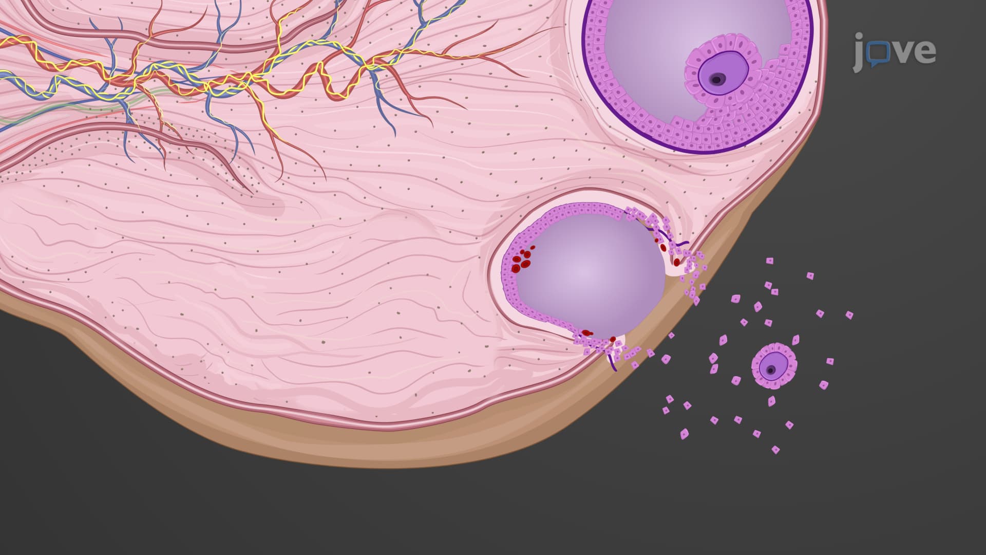
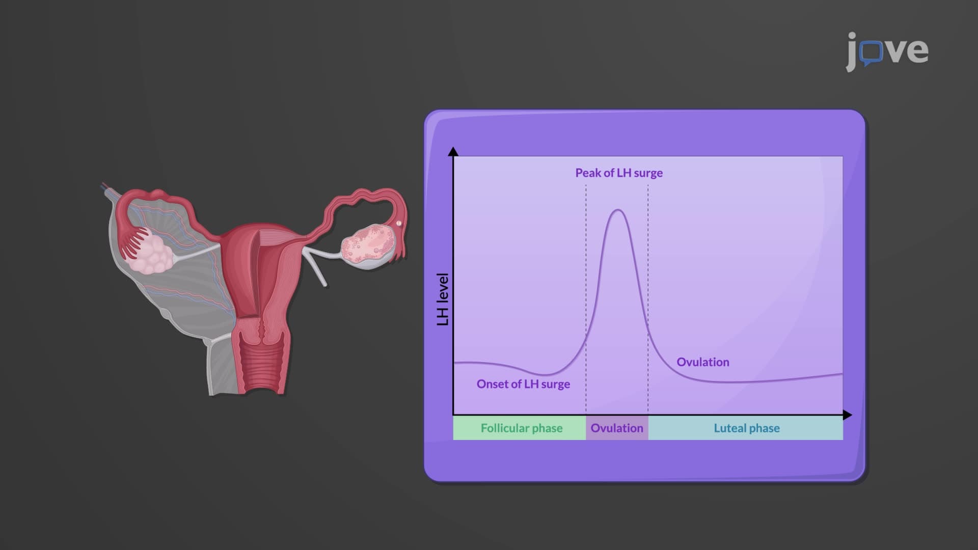
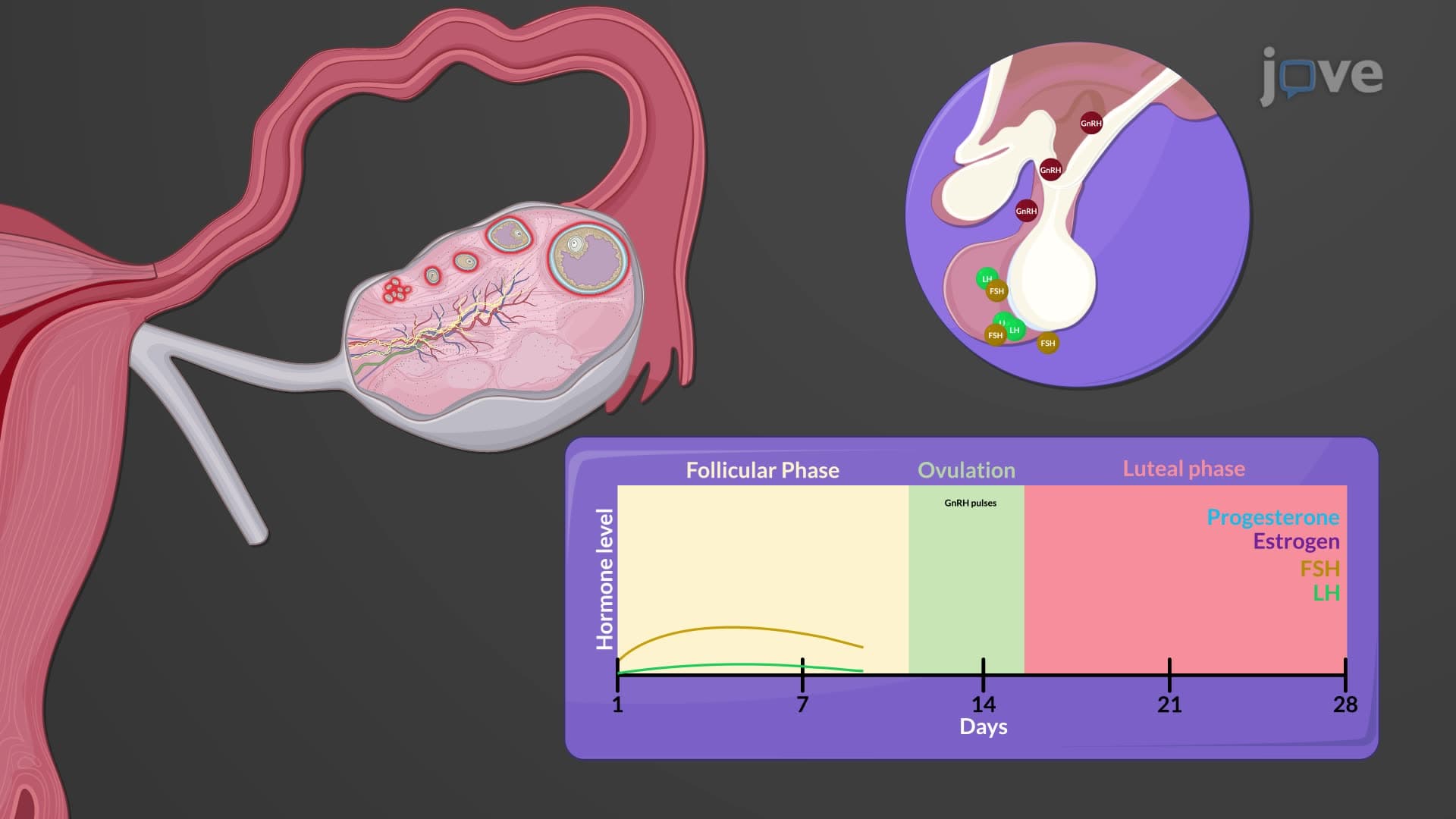
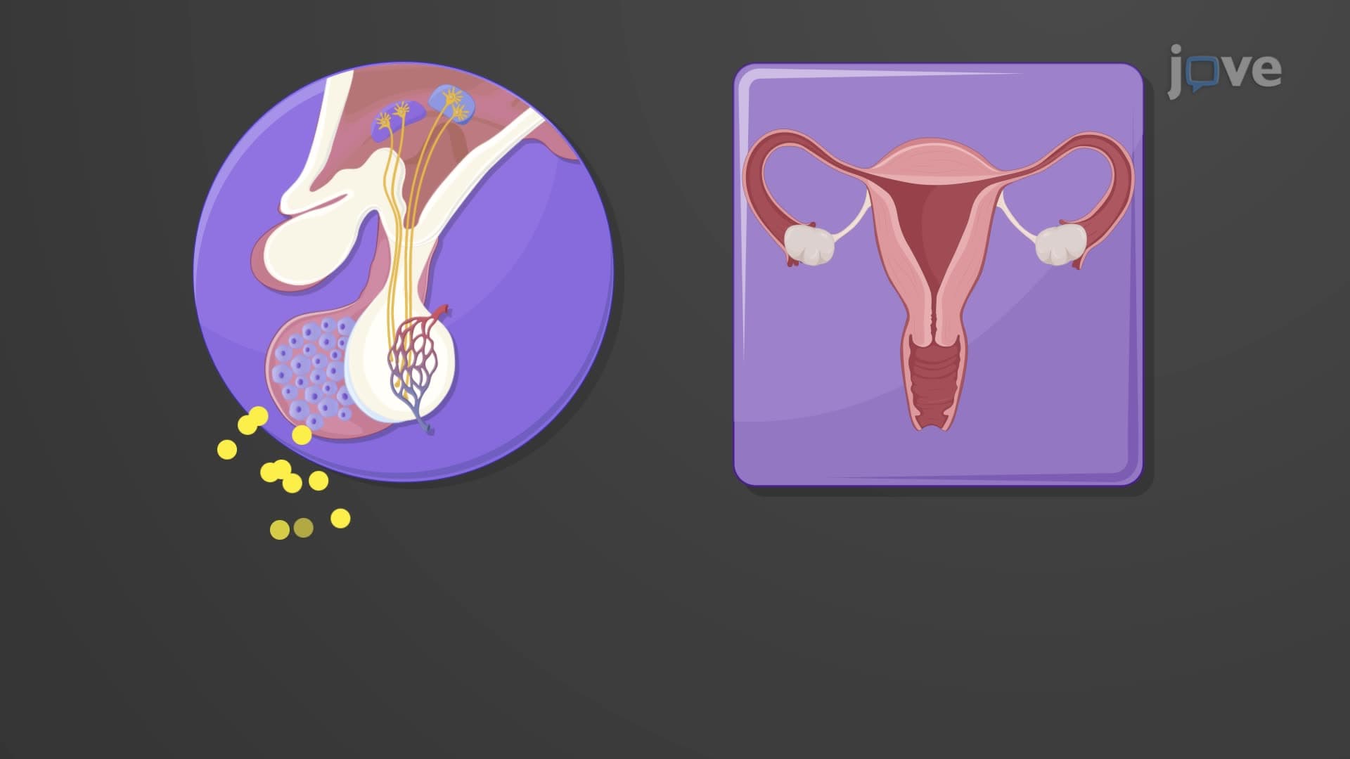
Understanding follicle corpus luteum ovary relationships proves essential for MCAT success and clinical applications. During each menstrual cycle, typically one dominant follicle emerges from the monthly cohort, driven by FSH stimulation. This mature follicle produces estradiol, triggering the LH surge that initiates ovulation approximately 34-36 hours later.
Post-ovulation, the collapsed follicle transforms into the corpus luteum, a temporary endocrine gland producing progesterone (essential for endometrial preparation), estrogen, relaxin (important during pregnancy), and inhibin (providing negative feedback to the pituitary). Without pregnancy, luteolysis occurs after 14 days, leaving the fibrous corpus albicans scar. This cyclical process, repeated roughly 400 times during a woman's reproductive years, demonstrates the remarkable regenerative capacity within ovarian ligament anatomy.
Clinical Applications and Exam Relevance
For AP Biology students, ovaries anatomy biology frequently appears in reproductive system questions, particularly regarding hormonal feedback loops and follicular development. College-level courses emphasize the relationship between ovarian structure and function, especially in endocrinology and reproductive physiology contexts. MCAT preparation should focus on integrating ovarian function with hypothalamic-pituitary axis regulation and menstrual cycle coordination.
Frequently Asked Questions
The ovaries serve two essential functions: producing mature eggs (oocytes) for reproduction and secreting crucial hormones including estrogen, progesterone, and inhibin. These paired organs control menstrual cycles, maintain reproductive health, and support pregnancy when fertilization occurs. Their dual role as both reproductive glands and endocrine organs makes them central to female physiology and overall hormonal balance.
MCAT questions often focus on ovarian hormone feedback loops, follicular development stages, and integration with the hypothalamic-pituitary axis. AP Biology exams emphasize ovarian structure-function relationships, particularly corpus luteum formation and hormonal regulation of the menstrual cycle. Students should master follicular development timelines and understand how ovarian dysfunction affects reproductive health for both exam formats.
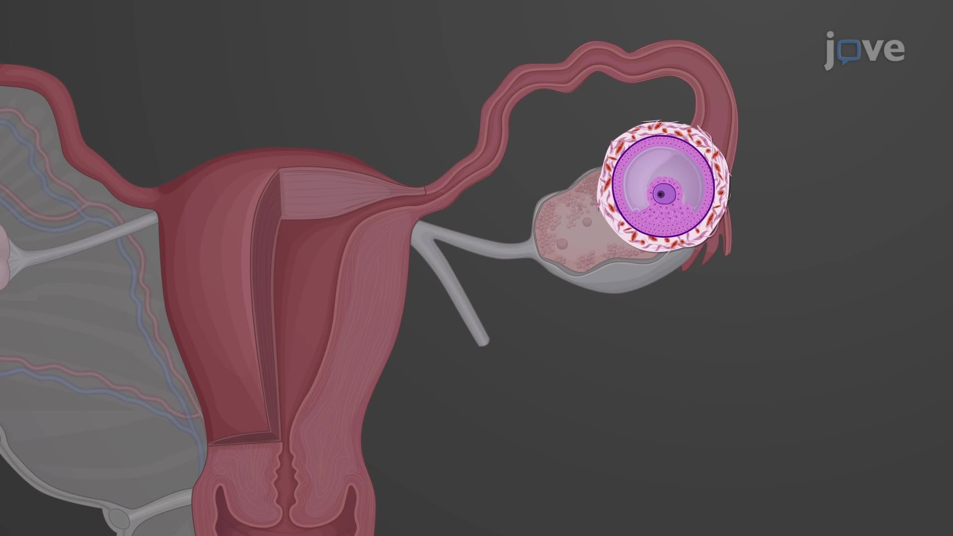
The mature Graafian follicle ruptures during ovulation to release the secondary oocyte. This large, fluid-filled structure develops over approximately 14 days under FSH and LH stimulation, reaching up to 20mm in diameter. After ovulation, the collapsed follicle transforms into the corpus luteum, which produces progesterone and other hormones essential for potential pregnancy support.
Reproductive endocrinologists at centers like Johns Hopkins and UCLA monitor follicular development using transvaginal ultrasound during IVF cycles. They track follicle size, count, and hormone production to optimize egg retrieval timing. Understanding ovarian blood supply through the ovarian and uterine arteries also guides surgical approaches for procedures like ovarian cyst removal while preserving fertility potential.
Basic understanding of steroid hormone structure helps, but isn't essential for grasping ovarian function. Focus on hormone names (estradiol, progesterone, inhibin), their target tissues, and feedback mechanisms rather than complex biosynthetic pathways. High school biology students can master ovarian physiology by emphasizing functional relationships between follicular development and hormonal changes throughout the menstrual cycle.
Create visual diagrams linking ovarian layers (mesothelium, tunica albuginea, cortex, medulla) with their specific functions. Use timeline charts showing follicular development from primordial through corpus albicans stages. Practice drawing the ovarian ligament attachments and blood supply, then quiz yourself on hormonal changes during different cycle phases. Flashcards work well for hormone names and their primary functions.
Explore ovarian pathophysiology including polycystic ovary syndrome (PCOS), ovarian cancer screening, and age-related changes during menopause. Advanced endocrinology courses cover steroidogenesis pathways and ovarian-hypothalamic feedback mechanisms in greater detail. Clinical rotations in obstetrics-gynecology provide hands-on experience with ovarian disorders and their treatment approaches used throughout US healthcare systems.
Ovarian knowledge provides the foundation for understanding menstrual cycle regulation, pregnancy hormone production, and fertility treatments. The corpus luteum's progesterone production connects directly to endometrial changes and early pregnancy support. Mastering ovarian follicular development also explains concepts like ovarian reserve testing and age-related fertility decline commonly discussed in reproductive health contexts.
Related Micro-courses

14 Concepts
6 Concepts
21 Concepts