Loading subject page…


141,004 views
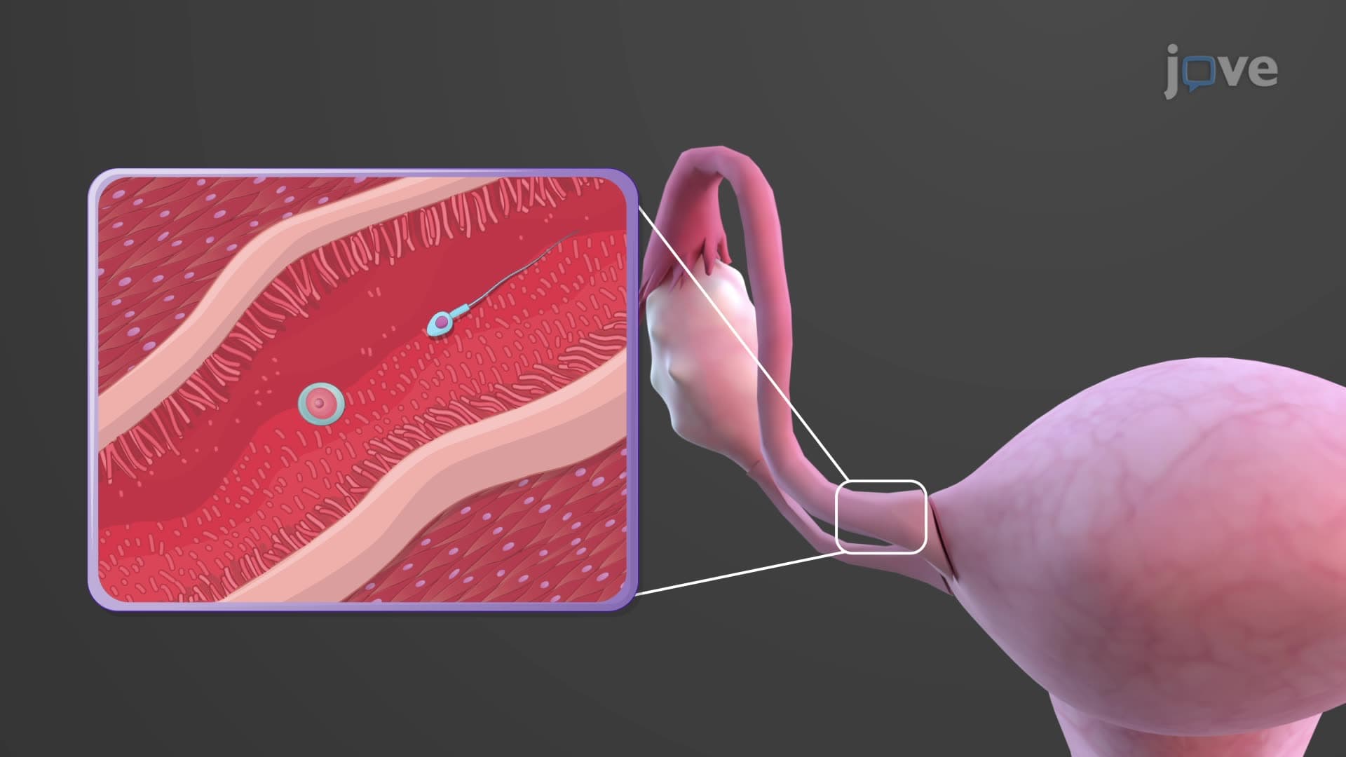

Video Summary: What are Uterine Tubes
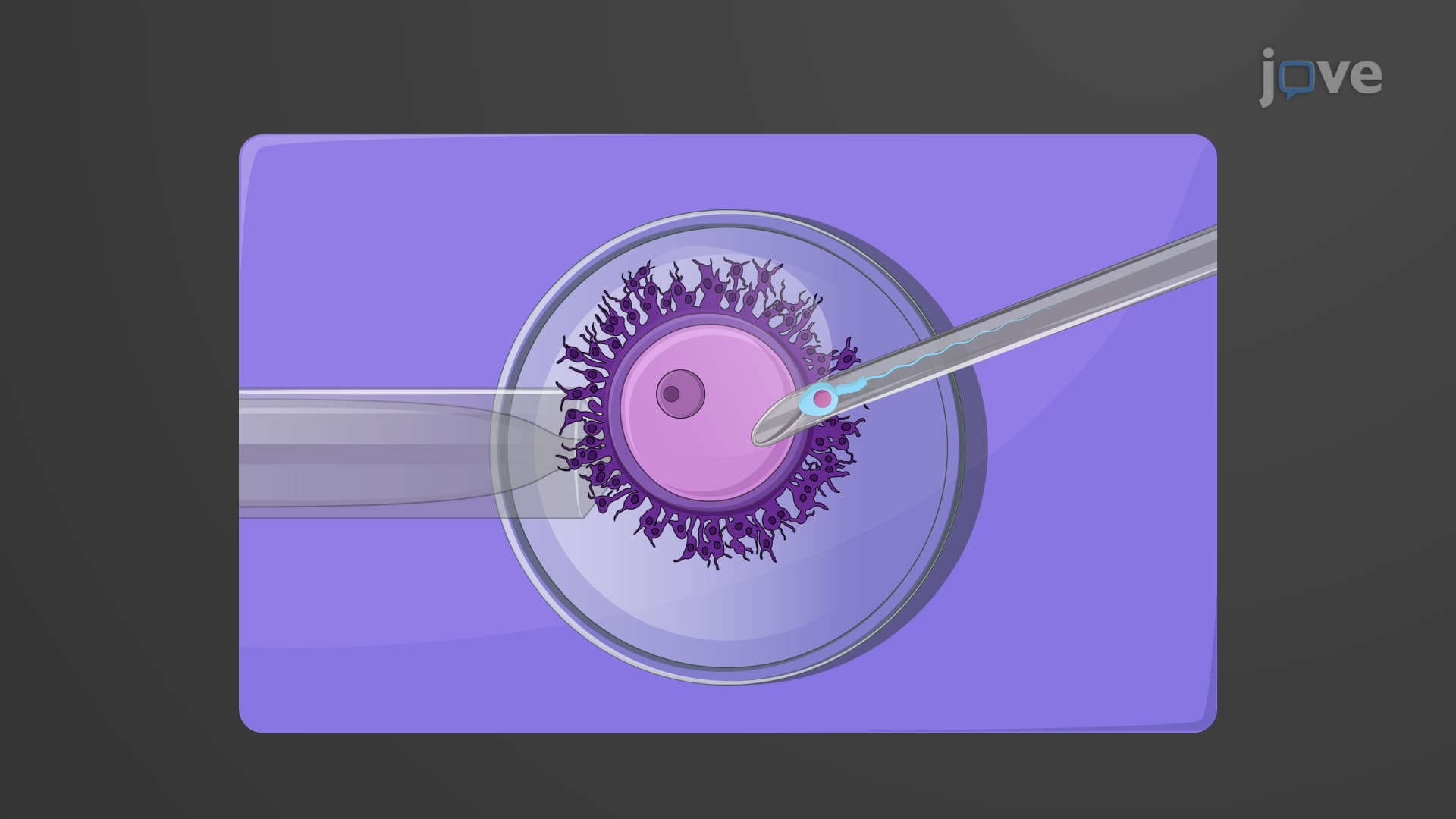
Every month, a remarkable 10-centimeter journey determines whether human life begins-and it all happens within the uterine tubes anatomy biology. These fallopian tubes create the perfect environment where 300 million sperm compete to fertilize a single egg, with success rates studied extensively at universities like Johns Hopkins and Stanford. Understanding what are the uterine tubes in female anatomy reveals how these specialized passageways orchestrate one of biology's most critical processes through their unique three-part structure and coordinated cellular mechanisms. Watch the full video on JoVE Coach to master this concept with expert-led visuals and step-by-step explanations.
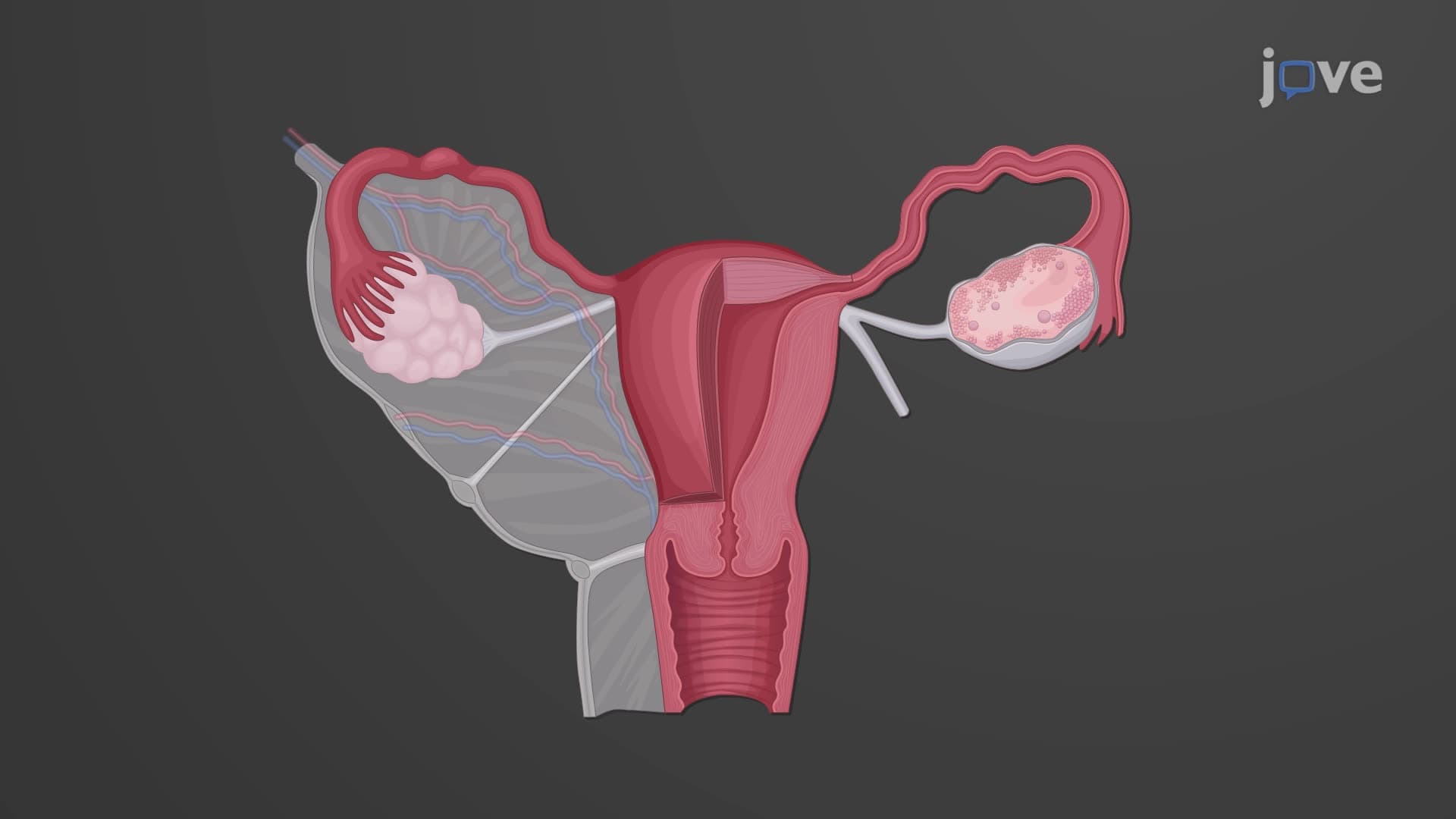
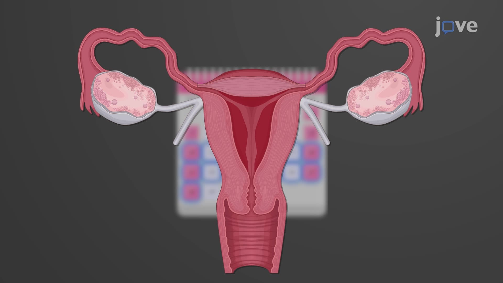
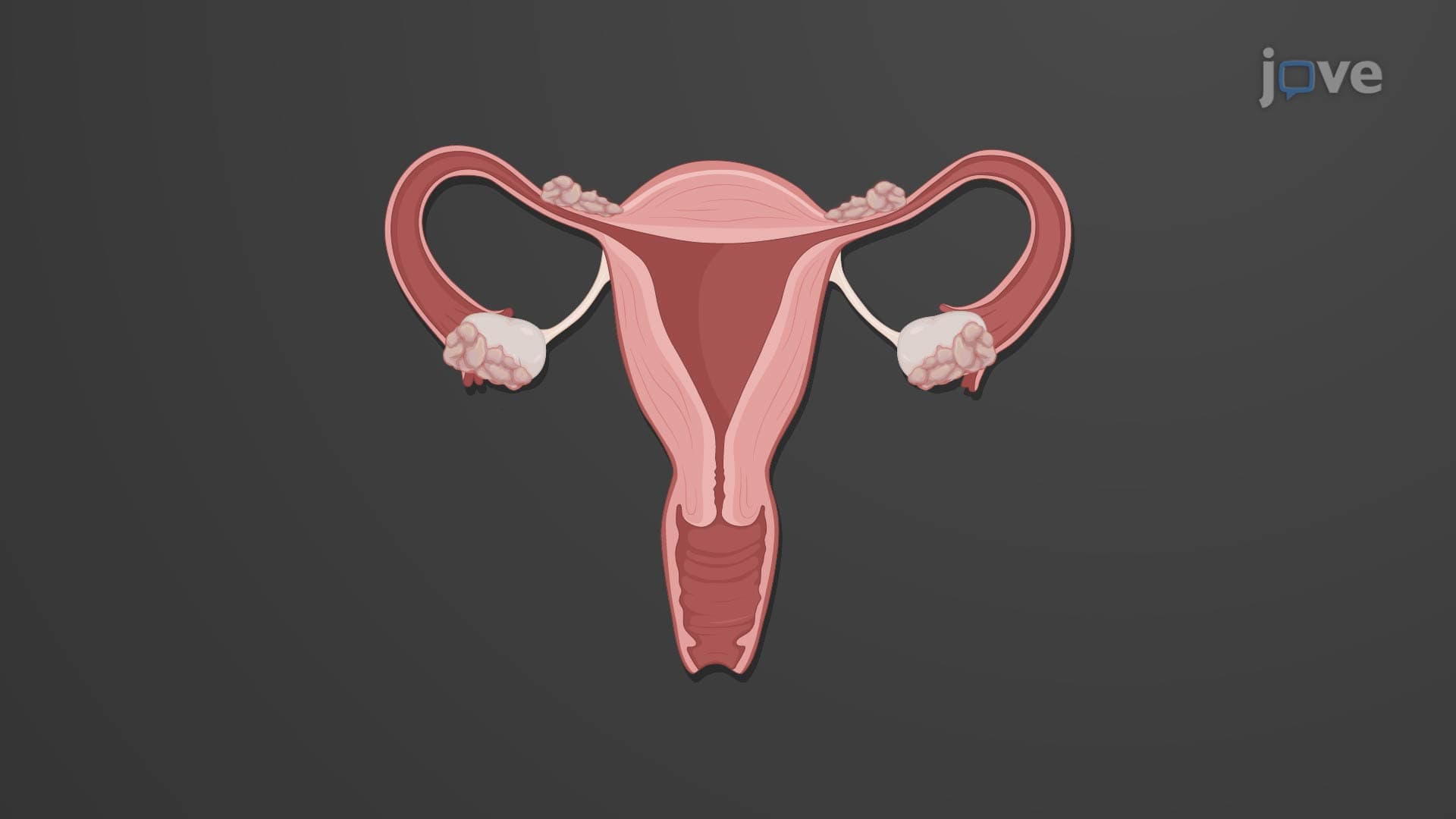
The uterine tubes anatomy biology represents one of the most sophisticated transport systems in the human body. These bilateral structures, measuring 10-13 centimeters in length, serve as the exclusive pathway connecting the ovaries to the uterus. Unlike simple tubes, they function as dynamic, living conduits that actively participate in reproduction through coordinated cellular and muscular activities.
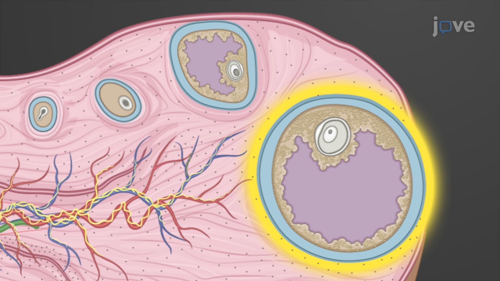
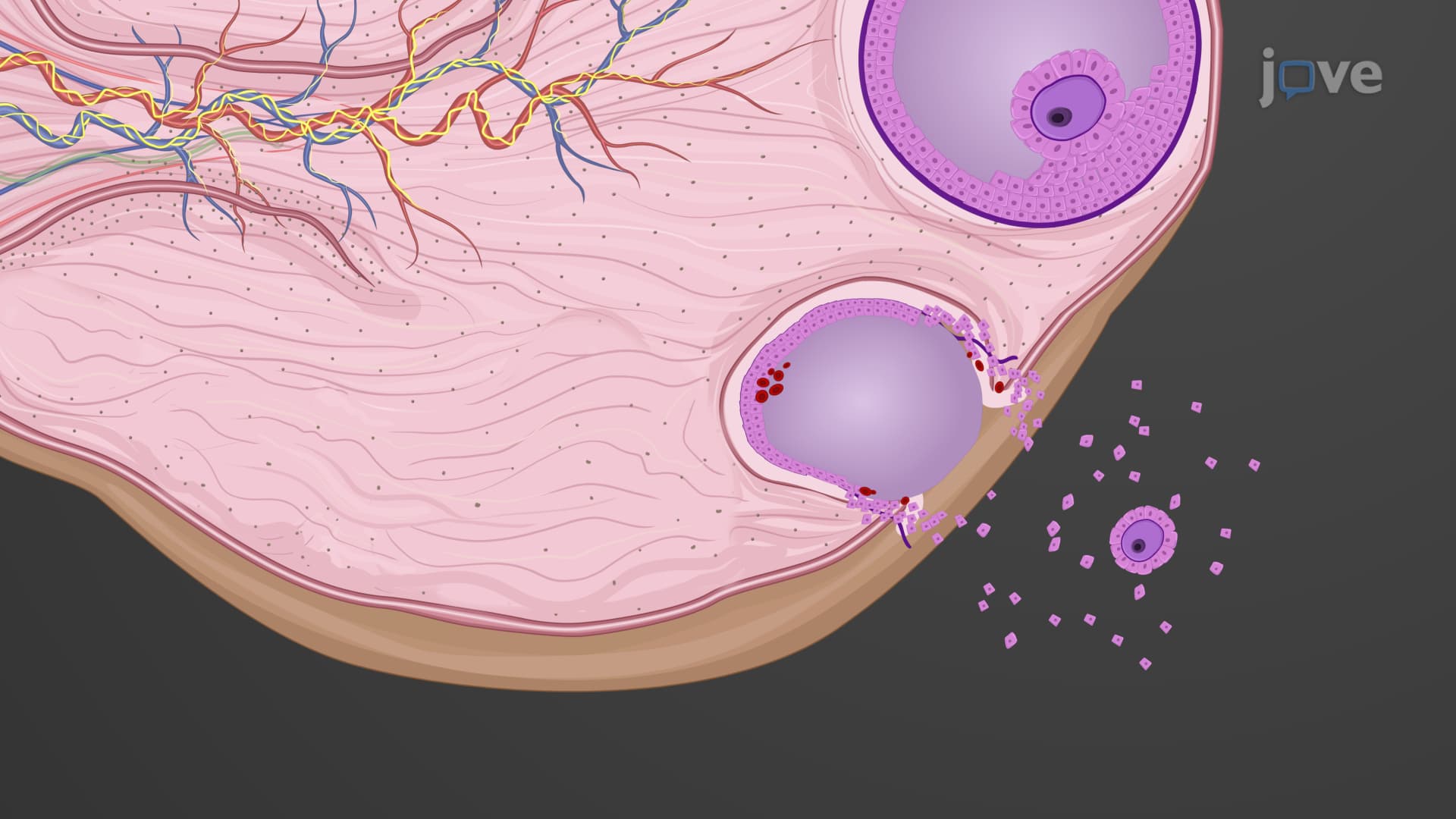
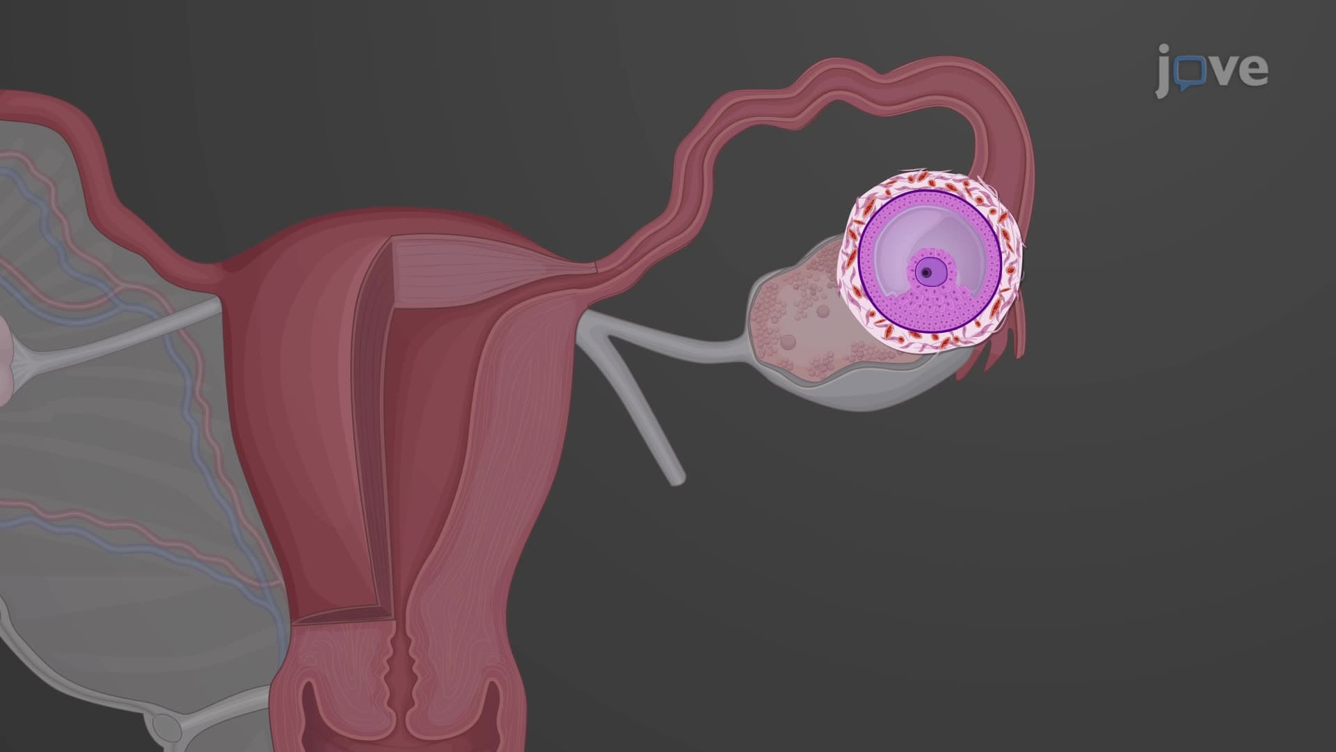
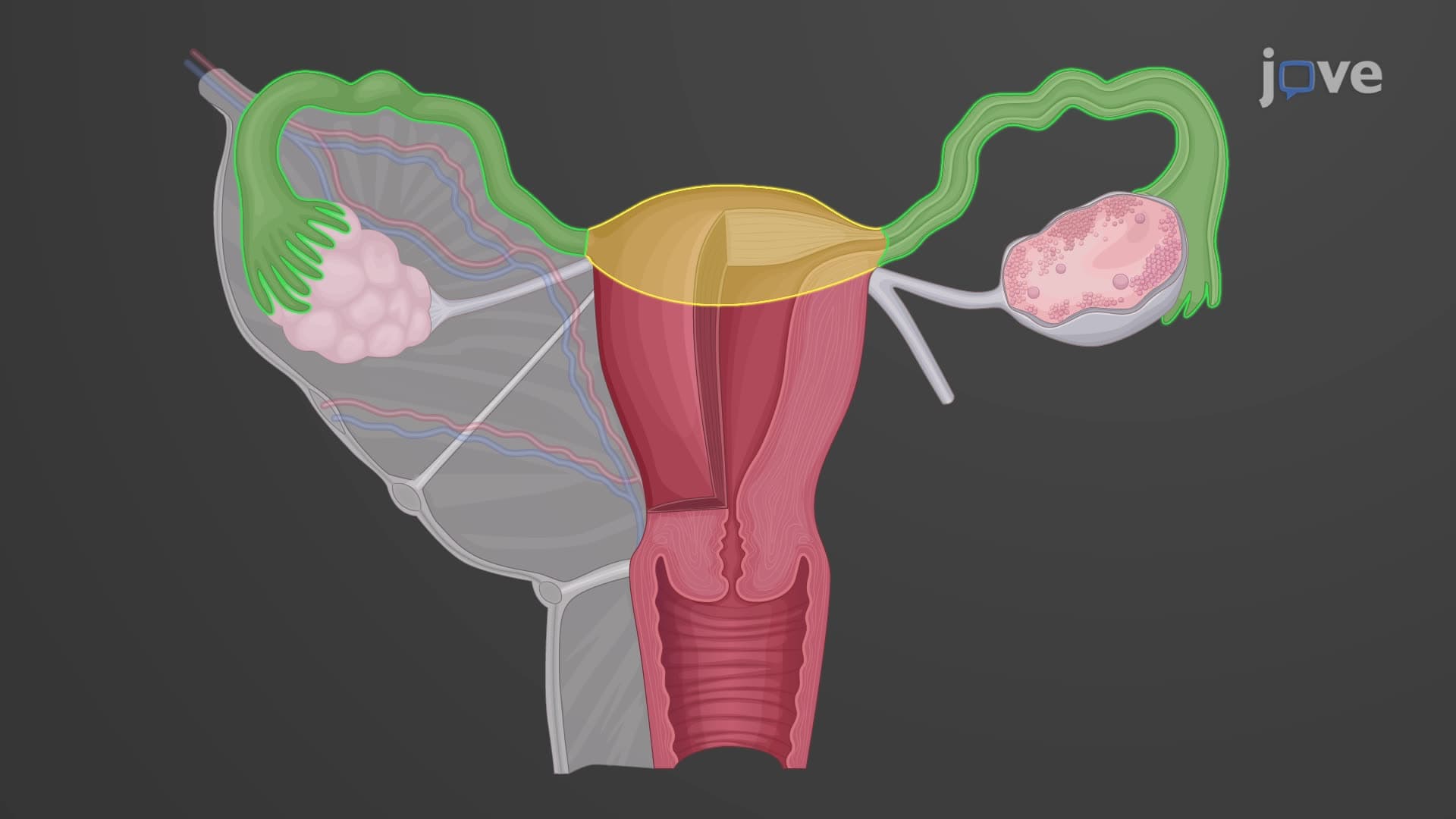
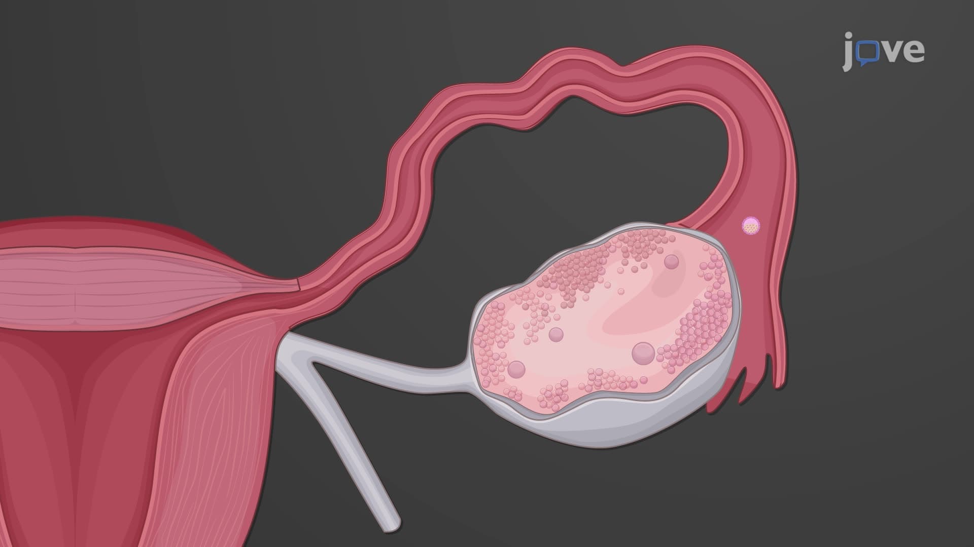
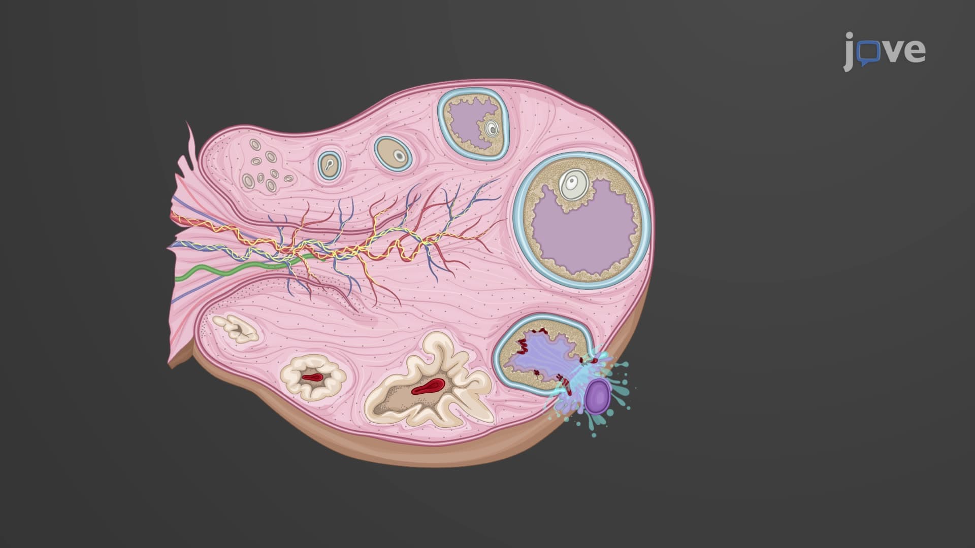
The fallopian tube structure explained reveals three specialized segments, each optimized for specific reproductive functions. The infundibulum forms the outermost funnel-shaped opening, crowned with delicate finger-like projections called fimbriae. These structures, containing thousands of ciliated cells, create sweeping motions across the ovarian surface during ovulation-a process observable in real-time using advanced imaging techniques developed at fertility centers like those at Mayo Clinic and Cleveland Clinic.
The ampulla constitutes the tube's widest segment, comprising over half its total length. This region's expanded diameter and specialized epithelial lining create optimal conditions for fertilization uterine tube events. Research conducted at institutions like Harvard Medical School demonstrates how the ampulla's unique microenvironment supports sperm capacitation and oocyte-sperm interaction.
The isthmus represents the narrowest portion, connecting directly to the uterus. Its thick muscular walls and smaller diameter create a natural regulatory checkpoint, controlling embryonic transport timing-a concept frequently tested on MCAT exams and featured in AP Biology curricula.
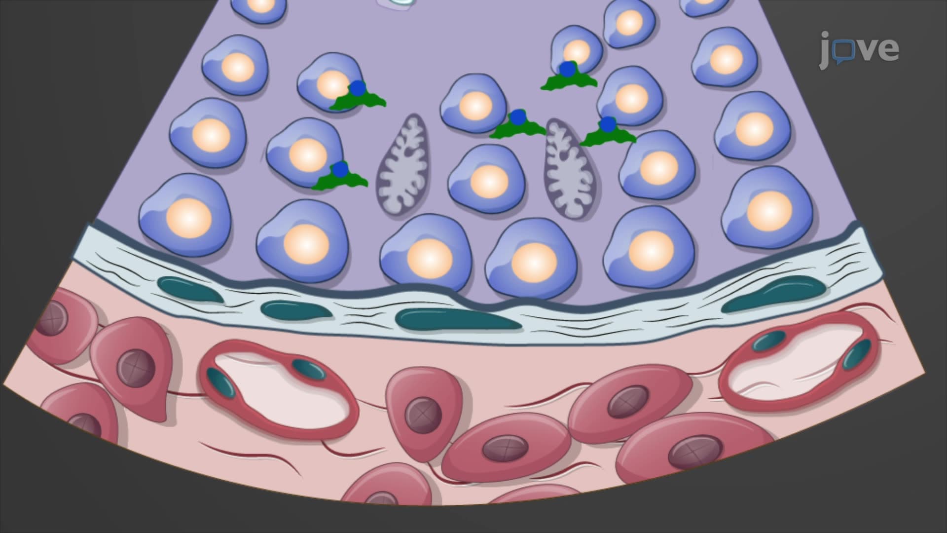
The oocyte transport uterine tube process involves remarkable cellular coordination. Ciliated epithelial cells generate synchronized beating patterns, creating fluid currents that guide reproductive cells. Non-ciliated secretory cells produce nutrient-rich tubular fluid containing proteins, electrolytes, and growth factors essential for oocyte viability and early embryonic development.
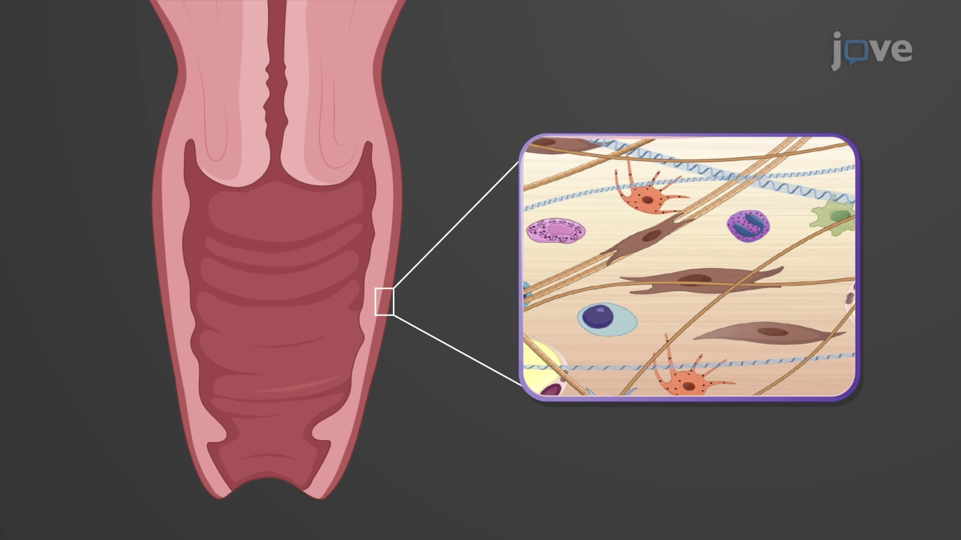
Smooth muscle layers surrounding the mucosa contract in peristaltic waves, similar to intestinal motility but precisely timed for reproductive needs. This muscular activity, combined with ciliary action, ensures proper transport velocity-too fast, and implantation fails; too slow, and ectopic pregnancy risks increase, conditions extensively studied in American reproductive medicine programs.
Understanding what are the uterine tubes in female anatomy proves essential for healthcare professionals diagnosing infertility, ectopic pregnancies, and pelvic inflammatory disease. Students preparing for nursing licensure exams (NCLEX-RN) and medical school entrance tests (MCAT) encounter numerous questions requiring detailed knowledge of uterine tube anatomy and physiology, making this topic crucial for academic success and clinical practice.
Related Micro-courses