Loading subject page…

- Anatomy and Physiology
- The Reproductive System
- Development of the Sexual Organs in the Embryo and Fetus
Micro-course: The Reproductive System
141,004 views
Learn key concepts and earn your certificate.

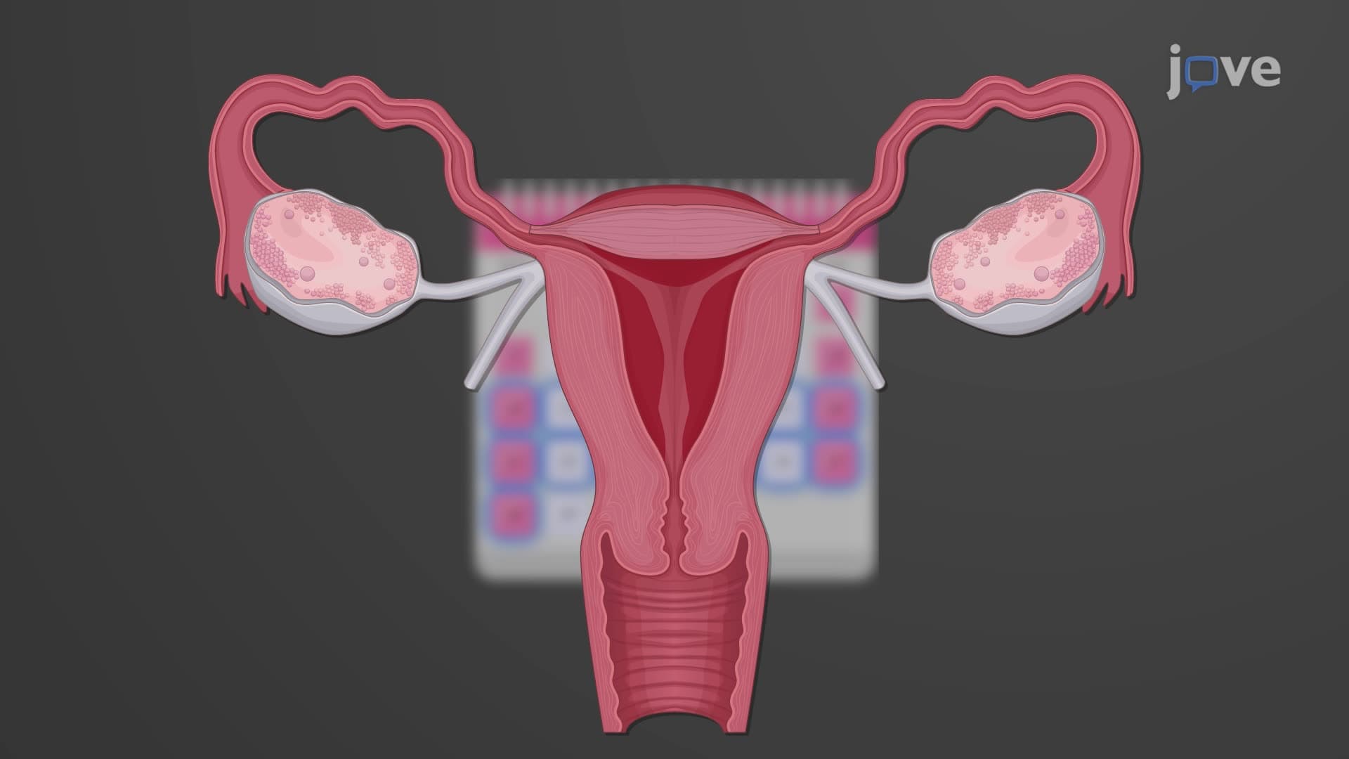
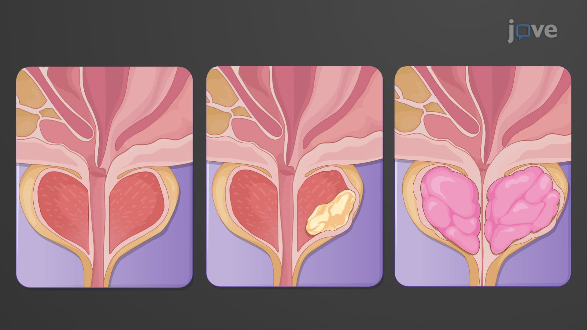
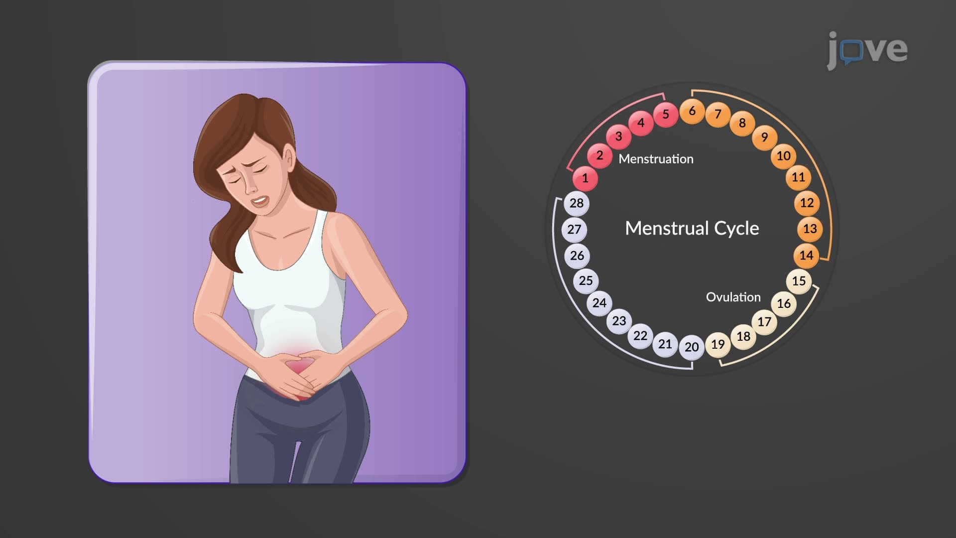
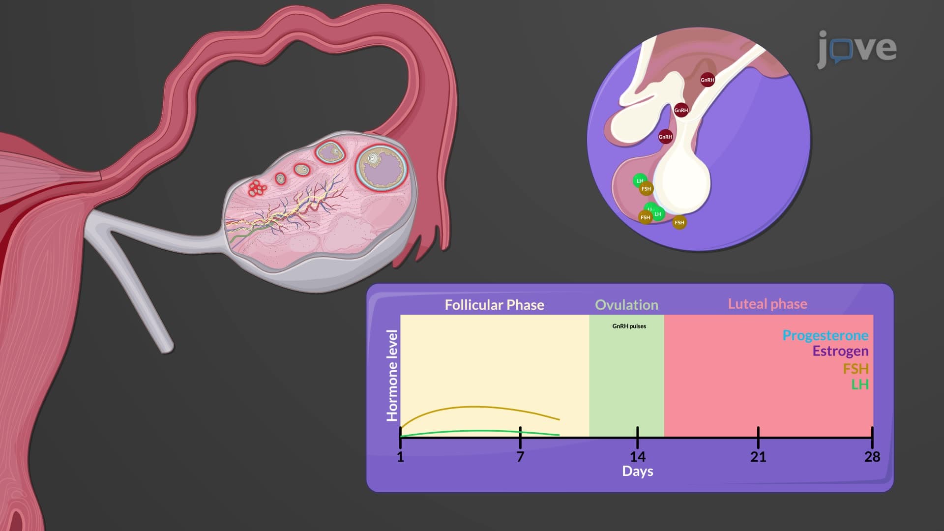

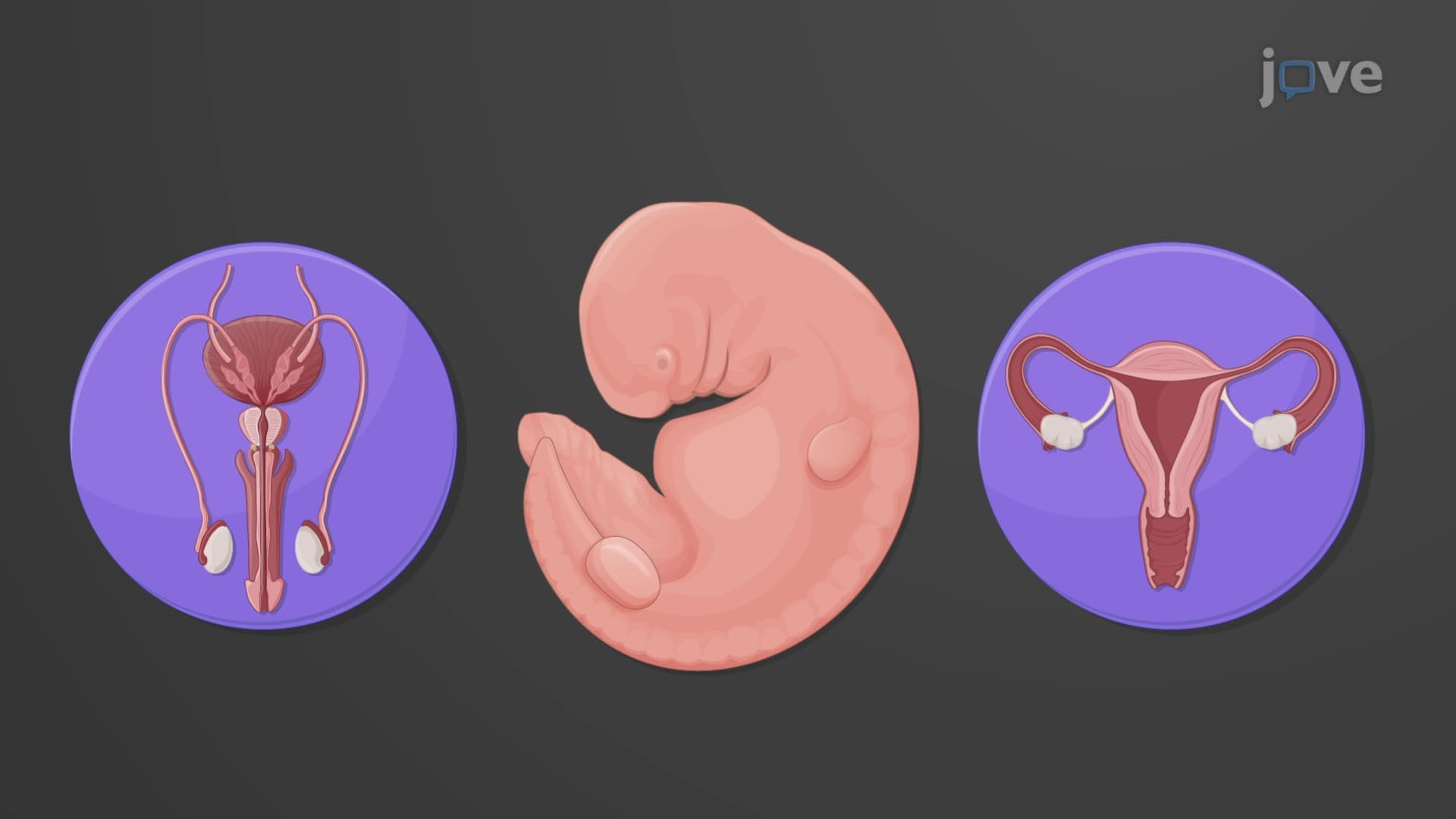
Development of the Sexual Organs in the Embryo and Fetus

00:00
01:15
Video Summary: What Is Development Sexual Organs
Every human embryo begins with the remarkable potential to develop either male or female reproductive systems-a process that determines biological sex through precise genetic and hormonal signals. Sexual organ development biology unfolds through a carefully orchestrated sequence beginning at just five weeks of embryonic life. Consider how genetic counselors at institutions like Johns Hopkins use this knowledge to help families understand conditions affecting reproductive development. What is development sexual organs reveals the intricate interplay between chromosomes, hormones, and tissue differentiation that creates the foundation for human reproduction. Watch the full video on JoVE Coach to master this concept with expert-led visuals and step-by-step explanations.
- Understand what is development sexual organs and how embryonic bipotentiality allows for either male or female reproductive system formation
- Identify the critical role of gonadal ridges, Wolffian ducts, and Müllerian ducts in early sex organ development embryology
- Analyze how the SRY gene on the Y chromosome triggers male reproductive organ differentiation during embryonic development
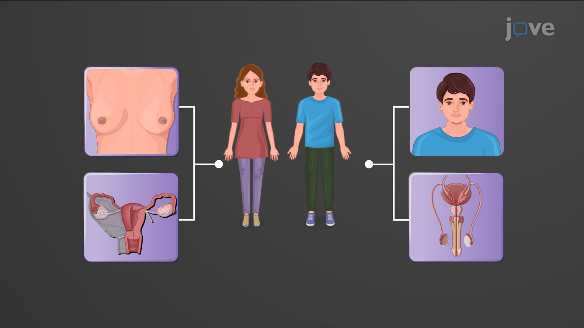
- Compare the developmental pathways that lead to male versus female internal and external genitalia formation
- Evaluate the timeline of sexual organ embryological development from week 5 through week 8 of embryonic life
- Apply knowledge of Müllerian-inhibiting substances and their role in male reproductive tract development
- Recognize the importance of hormonal signals in genital tubercle development and external genitalia formation
- Explore how disruptions in normal sex differentiation embryo processes can lead to differences in sexual development
Understanding What is Development Sexual Organs: The Foundation of Sexual Differentiation
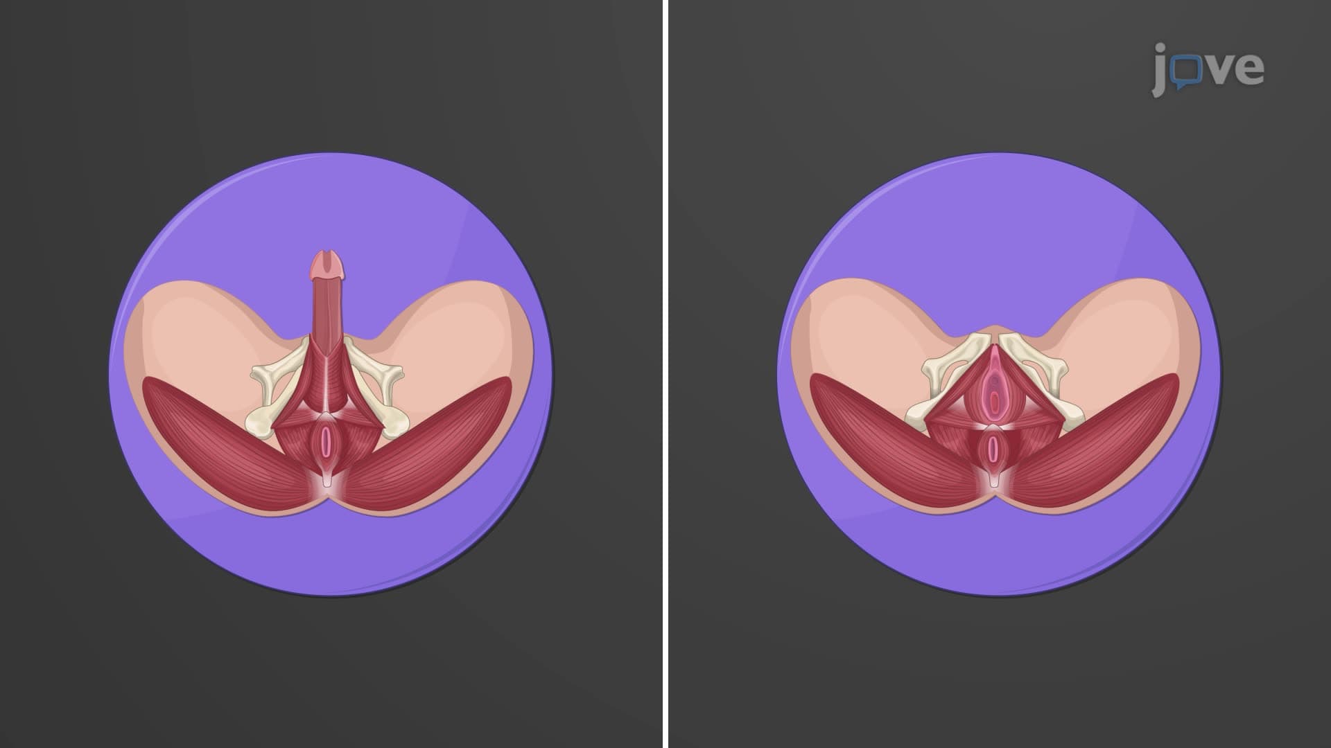
Sexual organ development biology represents one of the most precisely regulated processes in human embryogenesis. Every human embryo possesses bipotential gonads, meaning they have the inherent capacity to develop into either testes or ovaries depending on genetic and hormonal cues. This remarkable developmental flexibility occurs because all embryos initially form the same basic reproductive structures during the first five weeks of development.
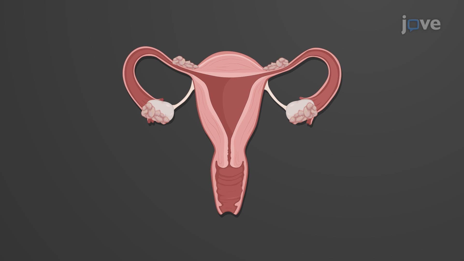
The process begins when specialized tissue called the gonadal ridge development occurs in the intermediate mesoderm. Simultaneously, two critical duct systems emerge: the mesonephric (Wolffian) ducts and the paramesonephric (Müllerian) ducts. These structures serve as the foundation for all future reproductive organs, making early embryonic development a critical period studied extensively in medical schools like Harvard Medical School and Stanford School of Medicine.
The Genetic Switch: SRY Gene and Male Development
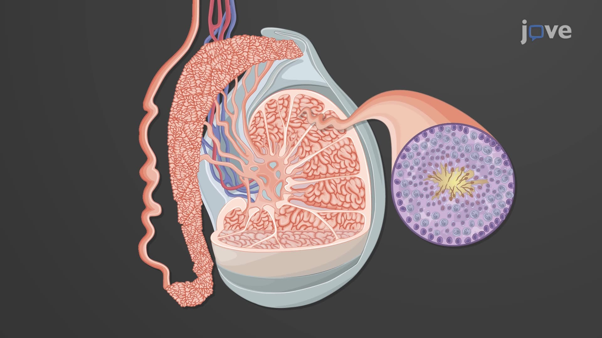
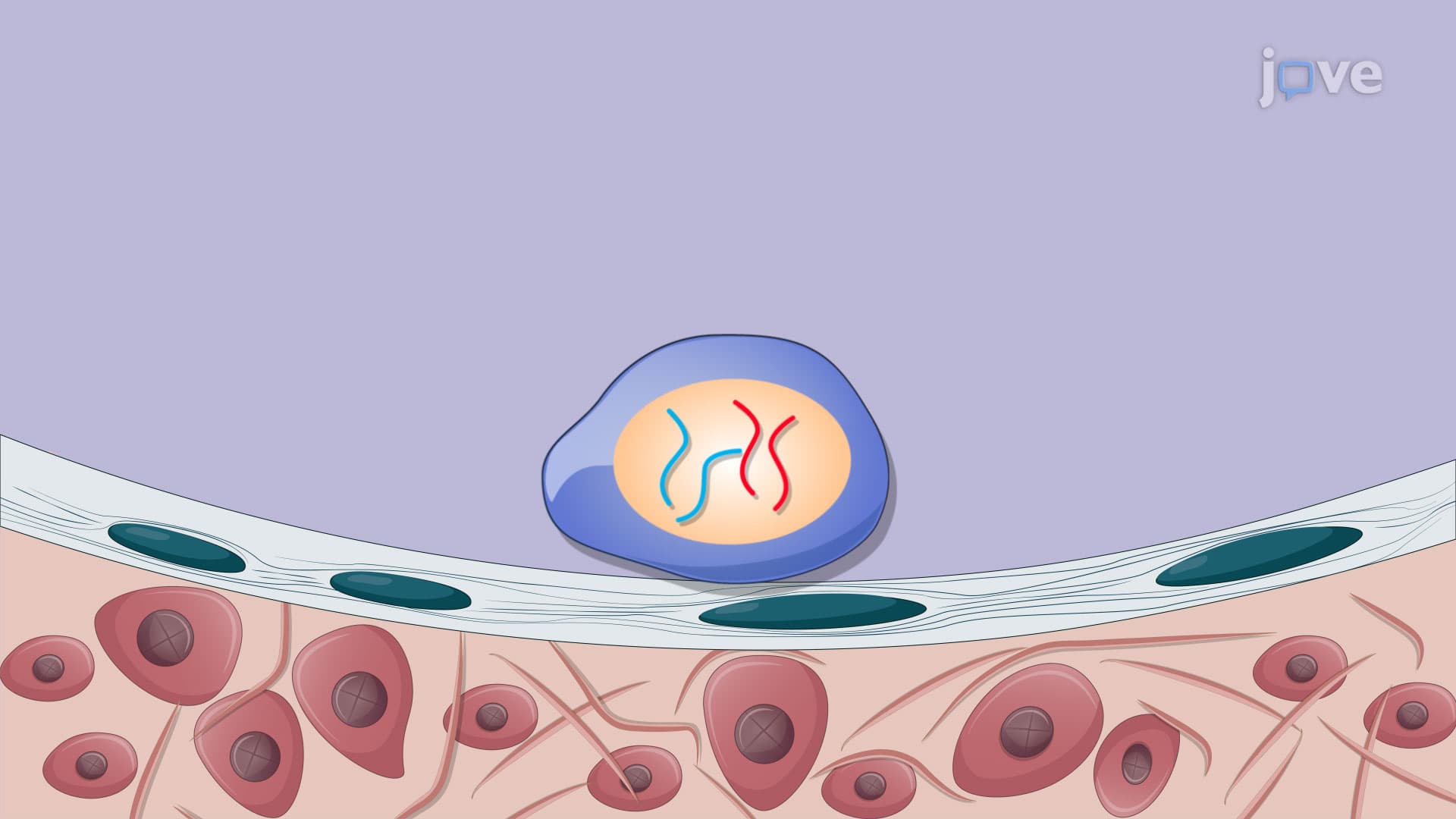
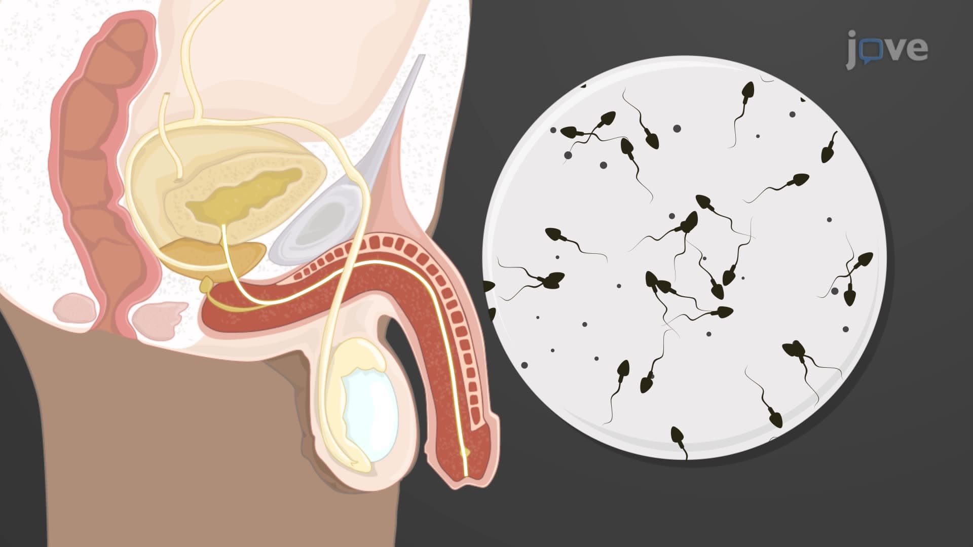
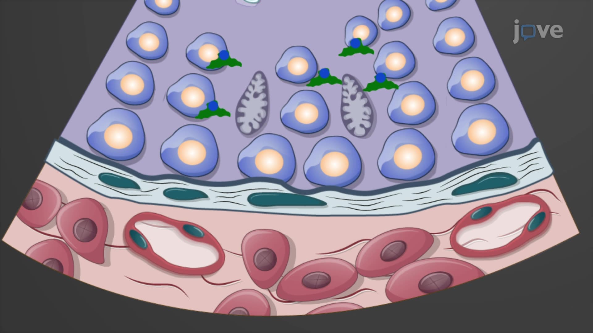
The sex-determining region Y (SRY) gene acts as the master switch in sexual organ embryological development. Located on the Y chromosome, this gene initiates a cascade of molecular events that transform the bipotential embryo into a male phenotype. When SRY activates around week 6-7 of development, it triggers the differentiation of sustentacular cells (Sertoli cells) within the developing testes.
These Sertoli cells then secrete Müllerian-inhibiting hormone (MIH), also called anti-Müllerian hormone (AMH), which causes the Müllerian ducts to regress. Simultaneously, the Wolffian ducts develop into male reproductive structures including the epididymis, vas deferens, and seminal vesicles. This process exemplifies the precision required in sex organ development embryology-a topic frequently tested on the MCAT and featured in AP Biology curricula across US high schools.
Female Development: The Default Pathway
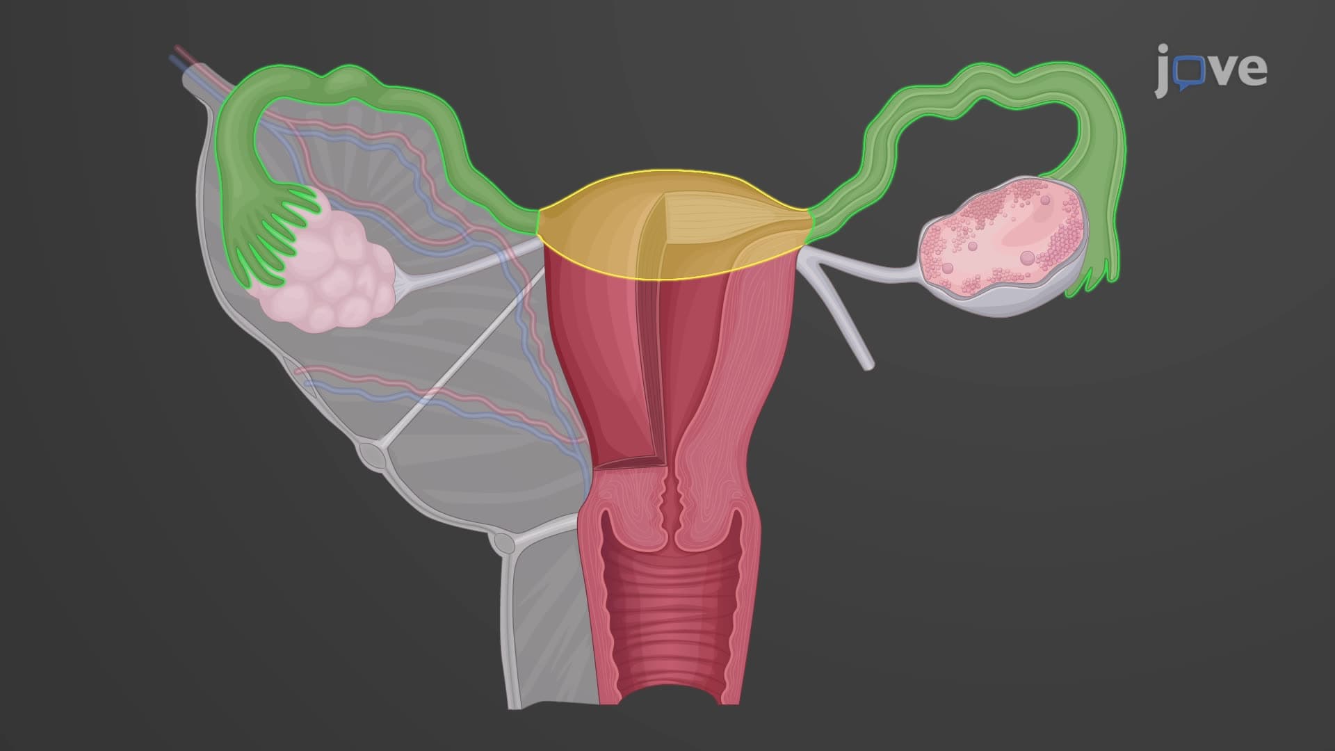
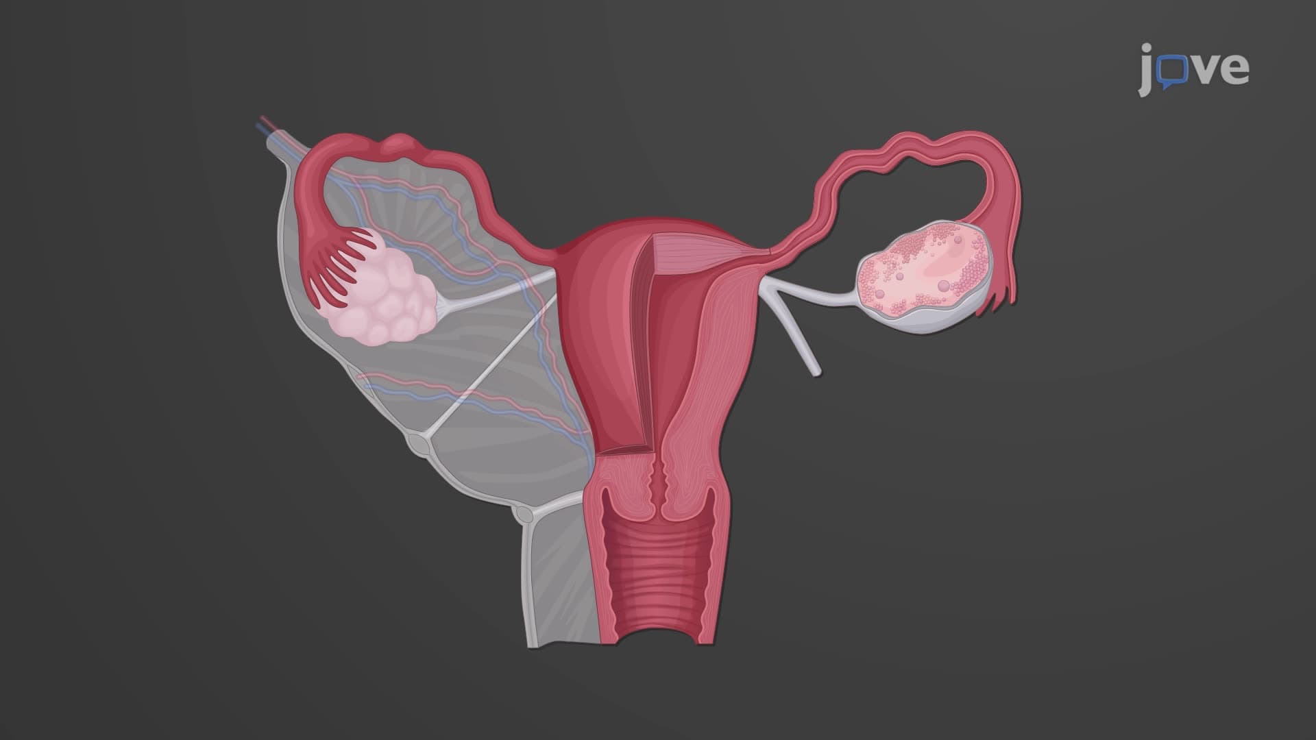
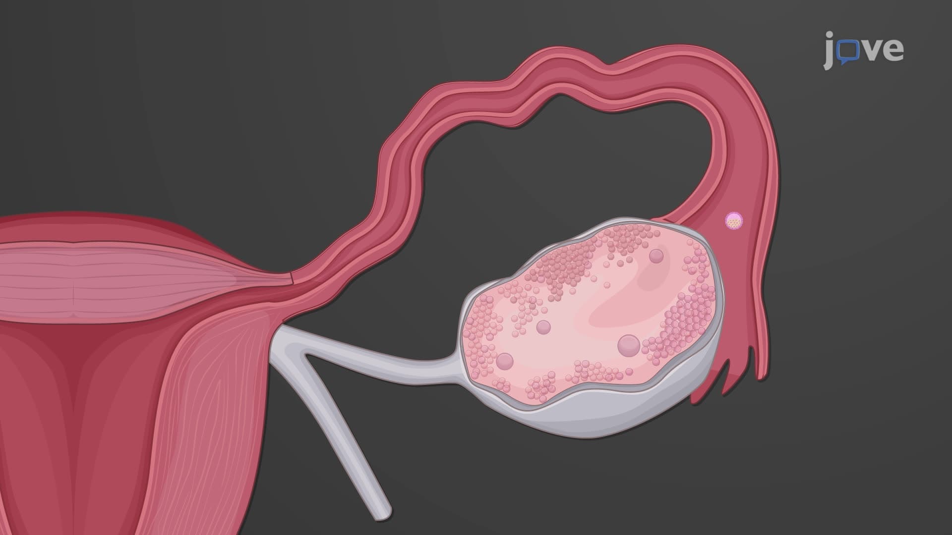
In the absence of the SRY gene, female development proceeds through what was historically called the "default pathway," though modern understanding recognizes female development as an equally active process. The mullerian Wolffian duct system develops differently in genetic females, with Müllerian ducts forming the uterus, fallopian tubes, and upper vagina while Wolffian ducts regress.
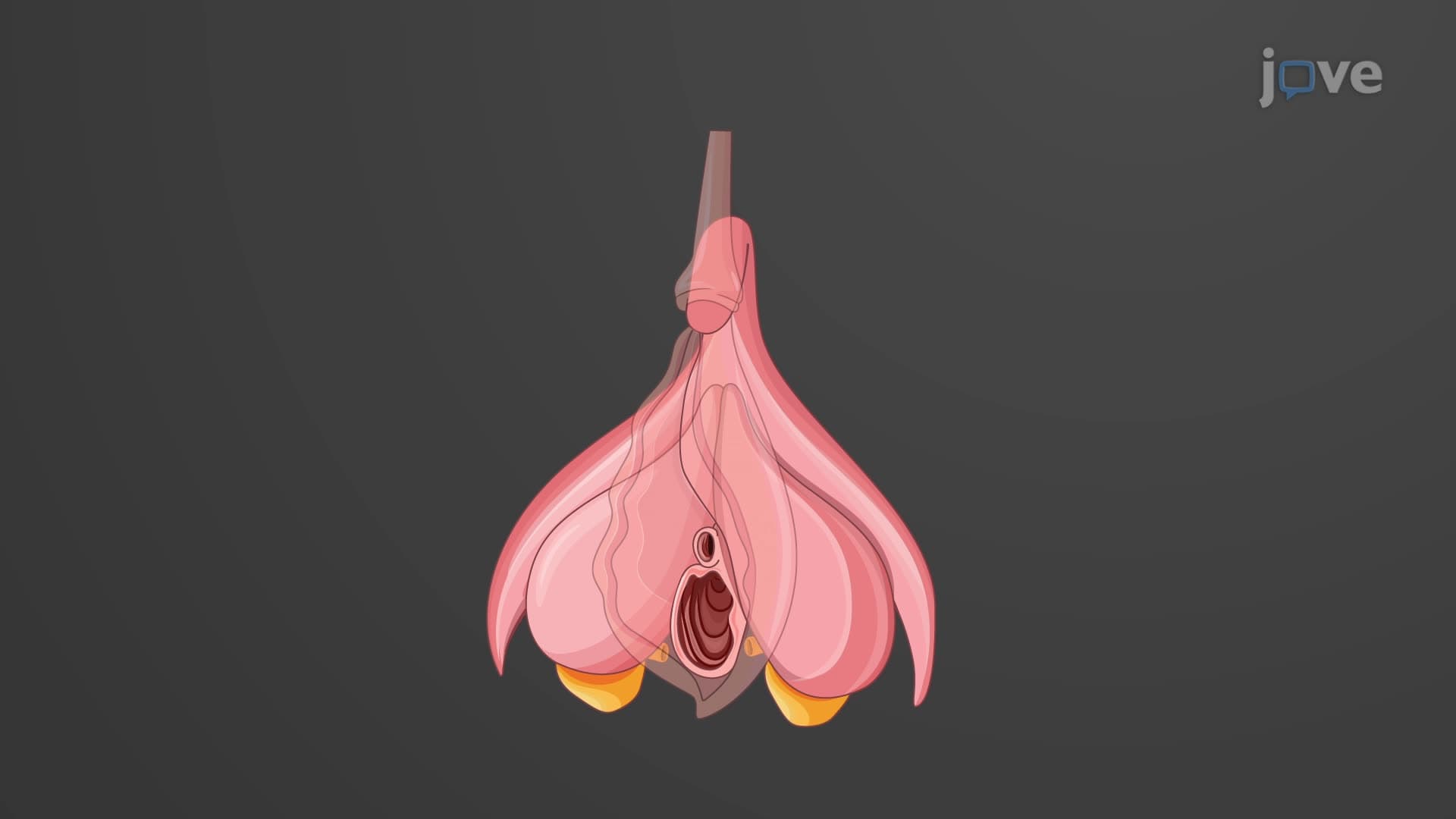
External genital development follows internal organ formation. The genital tubercle development gives rise to the clitoris in females, while labioscrotal swellings become the labia majora and urethral folds form the labia minora. This process typically completes by the eighth week of embryonic development, establishing the foundation for the female reproductive system.
Clinical Significance and Modern Applications
Understanding sex differentiation embryo processes has profound implications for medical practice in the United States. Pediatric endocrinologists at institutions like Children's Hospital of Philadelphia regularly apply this knowledge when treating patients with differences of sexual development (DSD). These conditions, affecting approximately 1 in 4,500 births, often result from disruptions in the normal developmental cascade described above.
Frequently Asked Questions
Development of sexual organs is the embryological process by which bipotential reproductive tissues differentiate into male or female reproductive systems. This process begins around the fifth week of embryonic development when gonadal ridges form, with genetic sex determination occurring by week 6-7 and external genitalia completing development by week 8.
The MCAT frequently tests embryological development through questions about hormonal regulation, genetic control mechanisms, and developmental timelines. Students should focus on understanding the SRY gene function, Müllerian-inhibiting hormone effects, and the relationship between Wolffian and Müllerian duct systems. Practice questions often involve analyzing genetic mutations affecting sexual differentiation.
AP Biology covers this topic under gene expression and regulation, developmental biology, and endocrine system units. Key concepts include transcriptional control by SRY, hormone signaling pathways, and cell differentiation mechanisms. Students should understand how genetic information directs developmental processes and cellular specialization.
Healthcare professionals use this knowledge to understand congenital conditions, counsel families about genetic disorders, and provide appropriate medical care for patients with differences in sexual development. Genetic counselors, pediatric endocrinologists, and reproductive specialists regularly apply these principles in clinical practice at major medical centers across the United States.
While the topic involves complex molecular mechanisms, the basic principles follow logical sequences that become easier with practice. Start with understanding the timeline and major structures, then gradually add details about genetic and hormonal control. Many students find creating flowcharts helpful for visualizing the developmental pathways.
Use active recall by drawing the developmental timeline from memory, create comparison charts between male and female pathways, and practice explaining the process aloud. Focus on understanding cause-and-effect relationships rather than memorizing isolated facts. Connect each step to its genetic or hormonal trigger for better retention.
College-level courses expand into detailed molecular mechanisms, clinical correlations, and comparative embryology across species. Students study specific transcription factors, signaling pathways like Wnt and Hedgehog, and examine how developmental disruptions lead to medical conditions. This foundation proves essential for medical school preparation.
Advanced studies include epigenetic regulation of sexual development, endocrine disruption effects on reproductive health, and stem cell research in regenerative reproductive medicine. Students interested in research careers often explore how environmental factors influence sexual differentiation or investigate new treatments for developmental disorders.
Related Micro-courses

14 Concepts

6 Concepts
21 Concepts