Loading video…


 37 Concepts
 141K views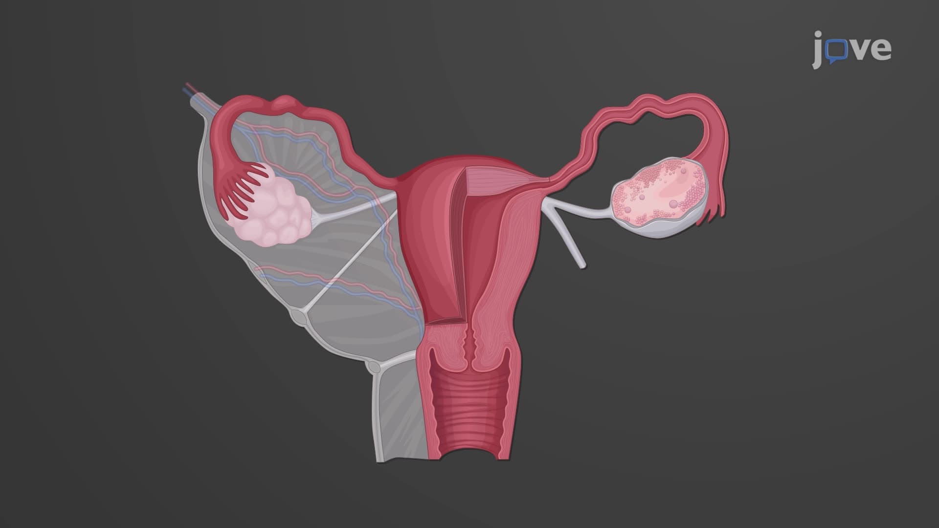

Video Summary: What Is Histology of the Uterus
Did you know that the uterine wall contains specialized tissue layers that can completely regenerate every month? Understanding uterine histology biology reveals how this remarkable organ adapts to hormonal changes throughout a woman's reproductive cycle. Consider how gynecologists at Johns Hopkins Hospital examine endometrial biopsies to diagnose conditions like endometrial hyperplasia-they're analyzing the same histological structures covered in what is the histology of the uterus. Watch the full video on JoVE Coach to master this concept with expert-led visuals and step-by-step explanations.
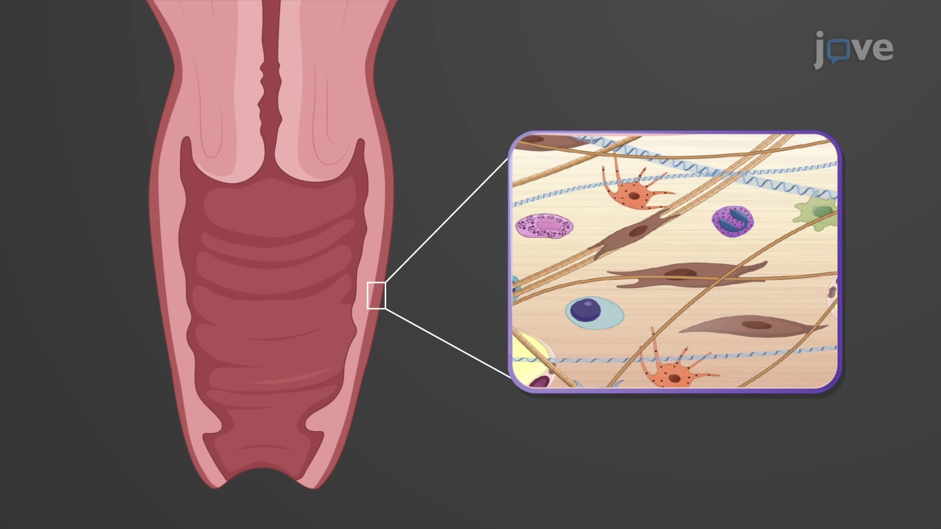
The uterine histology biology represents one of the most dynamic tissue systems in the human body. Unlike static organs, the uterus undergoes continuous structural modifications throughout a woman's reproductive years. Medical students at Harvard Medical School study these uterus histological layers extensively because understanding their organization is crucial for diagnosing reproductive disorders and comprehending normal physiological processes.
The perimetrium outer layer forms the uterus's external coating, consisting of simple squamous epithelium supported by a thin layer of connective tissue. This serous membrane creates a smooth surface that reduces friction as the uterus moves within the pelvic cavity during normal activities. Importantly, this layer connects laterally with the broad ligament, creating anatomical continuity that anchors the uterus in proper position. In AP Biology courses, students learn how serous membranes like the perimetrium prevent organ adhesion through their specialized secretions.
The myometrium smooth muscle comprises the uterus's thickest layer, arranged in three distinct sublayers of smooth muscle bundles. These interlacing fibers create tremendous contractile force during labor-sufficient to generate pressures exceeding 50 mmHg during active contractions, as documented in obstetric units at Mayo Clinic. The myometrium's unique arrangement allows coordinated contractions that facilitate both menstrual flow and parturition. MCAT students frequently encounter questions about how oxytocin receptors in myometrial cells respond to hormonal signals, making this tissue clinically relevant for understanding labor induction protocols.
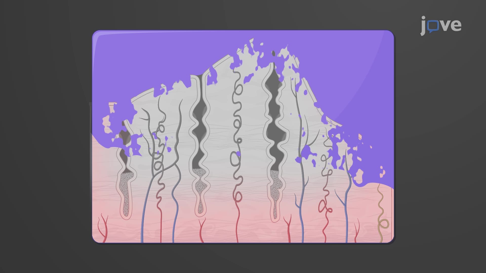
The innermost endometrial layer demonstrates remarkable regenerative capacity through its endometrium functional basal subdivision. The stratum functionalis undergoes complete shedding and regrowth monthly, while the stratum basalis remains stable, serving as the regenerative source. Uterine gland endometrial structures embedded within this layer secrete nutrients that support early embryonic development. These simple tubular glands, lined with columnar epithelium, extend from the surface through the functional layer into the basal region. College anatomy courses emphasize how spiral arterioles in the functional layer respond to hormonal changes, while straight arterioles in the basal layer maintain consistent blood supply for regeneration.
This uterus tissue structure explained becomes particularly relevant when studying endometrial cancer, where pathologists examine cellular changes within these precisely organized layers to determine malignancy staging and treatment approaches used in major cancer centers like MD Anderson.
Related Micro-courses