Loading subject page…

Micro-course: The Reproductive System
141,004 views
Learn key concepts and earn your certificate.


Penis

00:00
01:29
Video Summary: What Is Penis
Did you know that the penis serves dual biological functions that are essential for human reproduction and waste elimination? Understanding penis anatomy biology reveals how this complex organ manages both urination and reproduction through specialized tissue structures. Medical students at Johns Hopkins University study these anatomical details extensively when learning about the male reproductive system. The penis consists of three main regions-the glans, body, and root-each with distinct functions in male genitalia penis explained contexts. Watch the full video on JoVE Coach to master this concept with expert-led visuals and step-by-step explanations.
- Understand what is penis and its three primary anatomical regions: glans, body, and root
- Identify the structural differences between corpora cavernosa and corpus spongiosum in erectile tissue
- Analyze how blood flow creates erection through the filling of cylindrical erectile tissue masses
- Compare the functions of the glans penis versus the penile body in reproduction and urination
- Evaluate the protective role of the prepuce (foreskin) in male genital anatomy
- Apply knowledge of penile root structures (bulb and crura) to understand pelvic anchoring
- Recognize how the corpus spongiosum maintains urethral patency during sexual function
- Explore the relationship between anatomical structure and physiological function in male reproduction
What is Penis: Essential Male Reproductive Anatomy
The penis represents one of the most functionally complex organs in human anatomy, serving dual roles in reproduction and urinary elimination. This external male genital structure demonstrates remarkable engineering through its three distinct anatomical regions, each optimized for specific physiological functions that medical students encounter in courses like Human Anatomy and Physiology at universities across the United States.
Structural Organization and Regional Anatomy
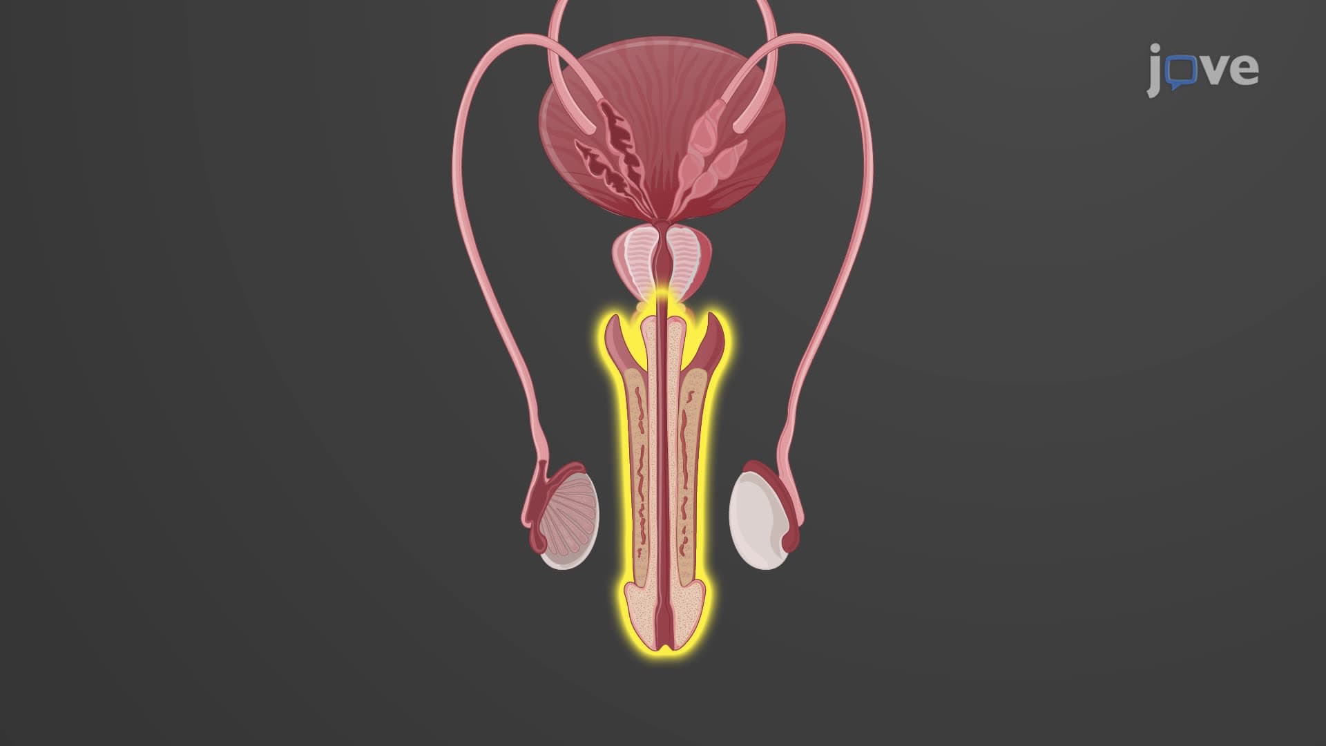
What is the anatomy of the penis becomes clear when examining its three primary regions. The glans penis, or head, contains the external urethral opening and thousands of sensory nerve endings that make it highly sensitive to stimulation. The prepuce, commonly called foreskin, provides protective coverage over the glans, though this structure is frequently removed through circumcision in American medical practice.
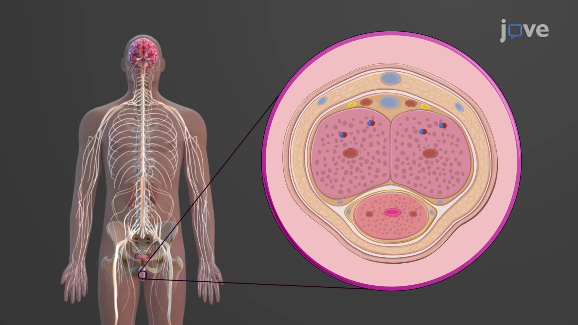
The penile body houses three cylindrical masses of erectile tissue penis components: two corpora cavernosa positioned dorsolaterally and one corpus spongiosum located ventromedially. These structures contain specialized vascular spaces called lacunae that fill with blood during arousal, creating the hydraulic mechanism for erection. Students preparing for the MCAT often focus on understanding how arterial dilation and venous constriction work together in this process.
Erectile Tissue Function and Blood Flow Dynamics
The corpus cavernosum spongiosum terminology reflects the different textures and functions of these tissues. The paired corpora cavernosa provide structural rigidity during erection, while the corpus spongiosum remains softer to maintain urethral patency. This design ensures that semen can pass through the urethra during ejaculation without compression from surrounding erectile tissue.
During sexual arousal, parasympathetic nervous system activation causes smooth muscle relaxation in penile arteries, increasing blood flow into the erectile tissue spaces. This mechanism appears frequently on AP Biology exams when discussing autonomic nervous system control of reproductive functions.
Root Anatomy and Pelvic Attachment
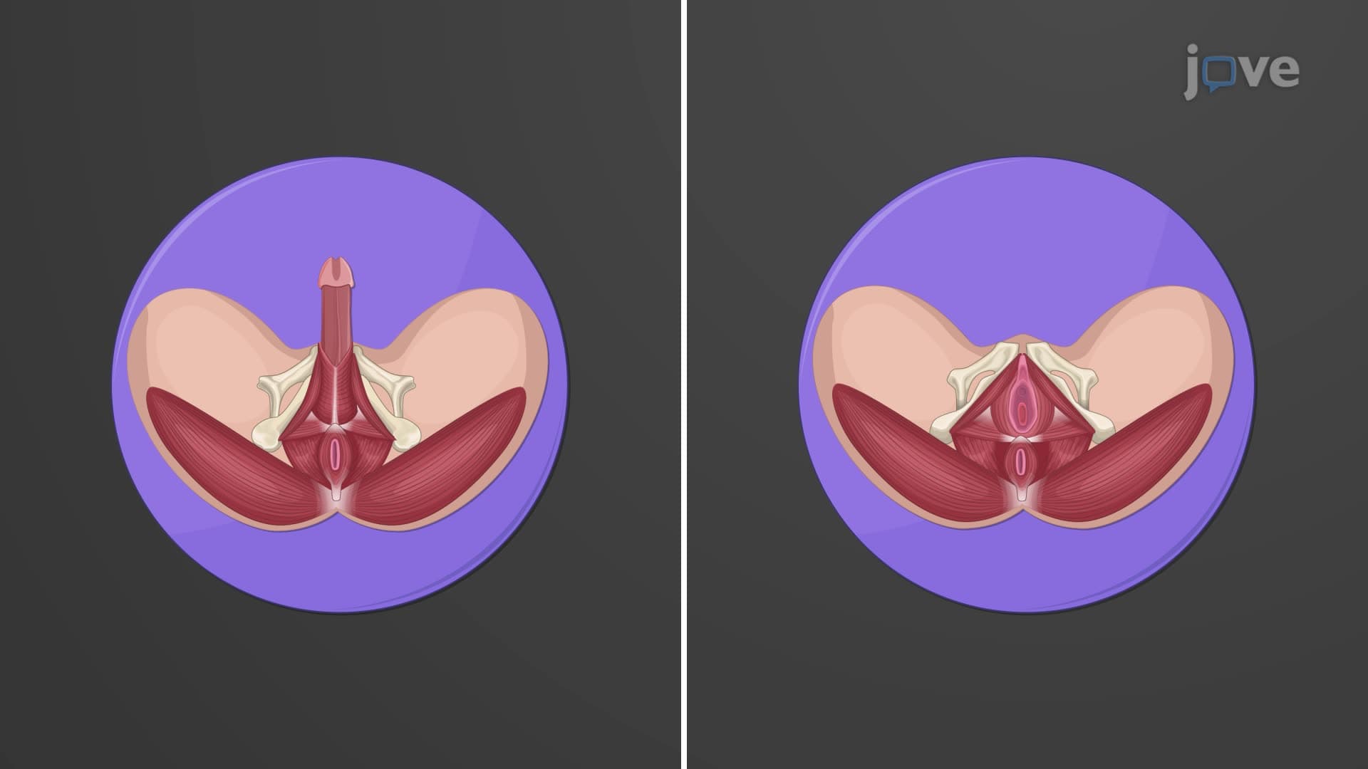
The penile root demonstrates how penis anatomical structure anchors to the pelvis through specialized formations. The corpus spongiosum expands proximally to form the penile bulb, while the corpora cavernosa separate into paired crura (singular: crus) that attach to the ischiopubic rami. These attachments, reinforced by muscles like the ischiocavernosus and bulbospongiosus, provide structural stability during reproductive function.
Understanding glans prepuce penis anatomy and urethra penis anatomy relationships helps students grasp how form follows function in reproductive biology, a concept emphasized in college-level Human Sexuality courses nationwide.
Frequently Asked Questions
The penis is the external male reproductive organ consisting of three main regions: the glans (head), body (shaft), and root. The body contains three cylindrical masses of erectile tissue-two corpora cavernosa and one corpus spongiosum-that enable both reproductive and urinary functions through specialized vascular mechanisms.
The MCAT frequently tests erectile tissue physiology, blood flow regulation, and nervous system control of reproductive functions. AP Biology exams emphasize the relationship between structure and function, particularly how the corpus spongiosum maintains urethral opening during erection while corpora cavernosa provide rigidity.
College exams typically focus on identifying the three erectile tissue masses, understanding the dual innervation (sympathetic and parasympathetic), and explaining how vascular changes create erection. Students must also know the attachments of penile root structures to pelvic bones.
Medical schools teach penile anatomy because urological conditions, erectile dysfunction, and reproductive health issues are common in clinical practice. Understanding normal anatomy is essential for diagnosing conditions like Peyronie's disease, priapism, and congenital abnormalities that physicians encounter regularly.
The NCLEX tests nurses' understanding of male reproductive health, catheter insertion techniques, and patient education about normal anatomy. Knowing the relationship between the corpus spongiosum and urethra is crucial for understanding urological procedures and complications.
Penis anatomy builds on basic knowledge of tissues, blood vessels, and nervous system function typically covered in high school biology. Students who understand how muscles and blood vessels work together can easily grasp erectile tissue function and the organ's dual roles.
Create diagrams showing the three tissue masses and their positions, use mnemonics for remembering "two corpora cavernosa, one corpus spongiosum," and practice identifying structures on anatomical models. Focus on connecting structure to function rather than just memorizing names.
Students should explore male reproductive endocrinology (testosterone effects), the physiology of erection and ejaculation, comparative anatomy across mammals, and clinical conditions affecting penile function. These topics frequently appear in advanced biology and pre-med coursework.
Related Micro-courses

14 Concepts
6 Concepts
21 Concepts