Loading subject page…


141,004 views

Video Summary: What Is Testes Histology
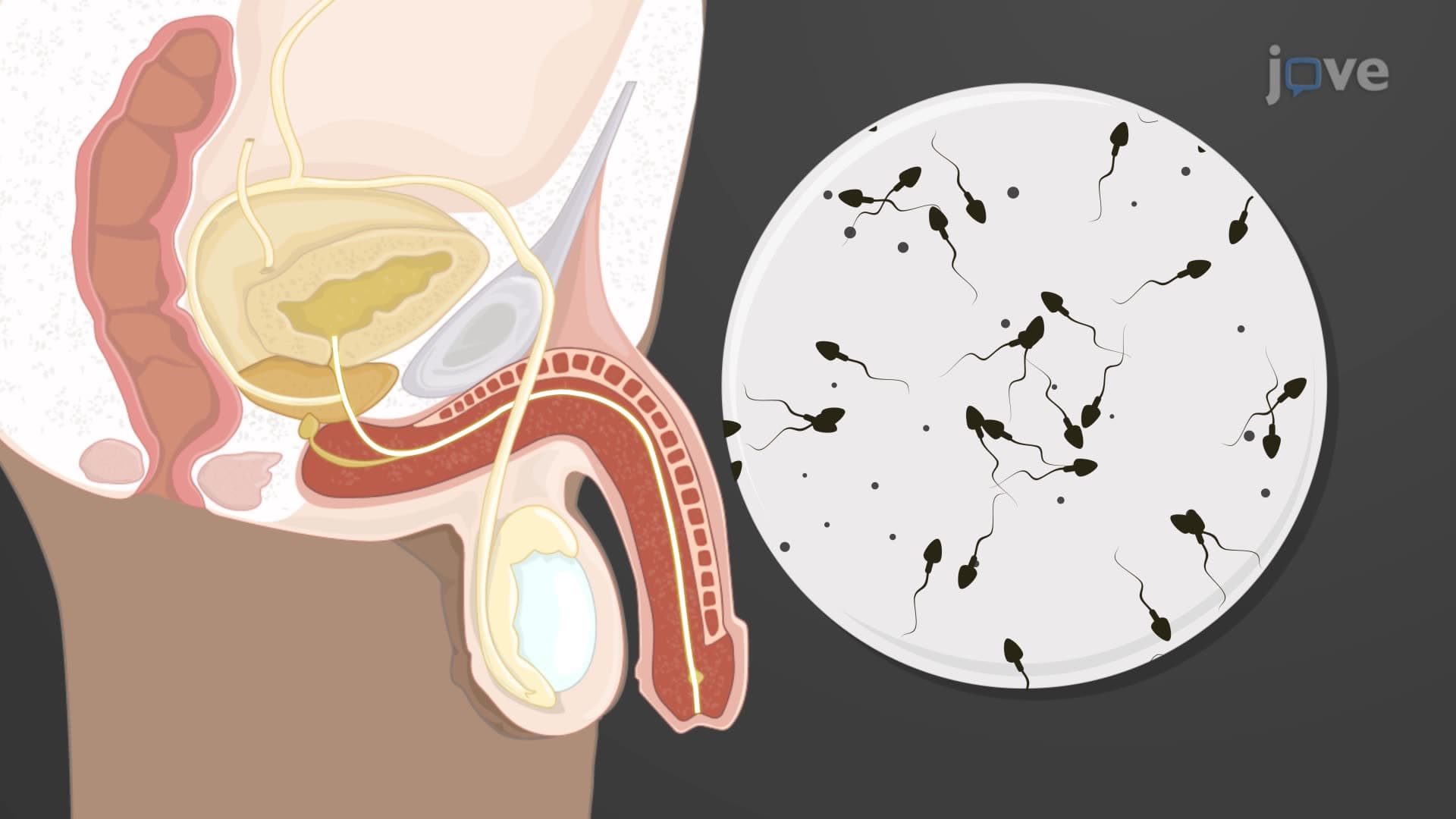
Did you know that a single human testis contains over 800 feet of microscopic tubules packed into an organ smaller than a golf ball? Testes histology biology reveals the intricate cellular architecture that enables male fertility through specialized compartments and cell types. From the protective tunica albuginea to the sperm-producing seminiferous tubules, what is testes histology encompasses the structural organization essential for reproductive function. Medical students at institutions like Johns Hopkins University study these microscopic details to understand male infertility cases. Watch the full video on JoVE Coach to master this concept with expert-led visuals and step-by-step explanations.
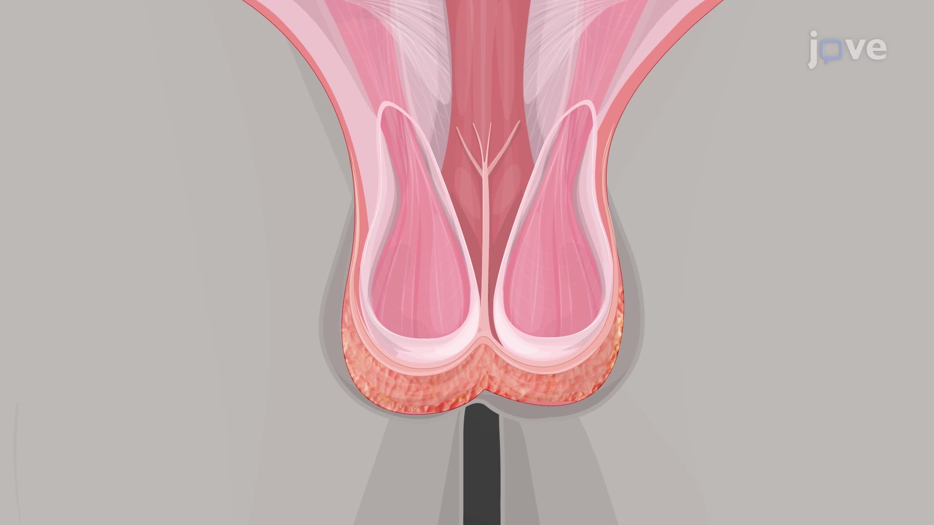
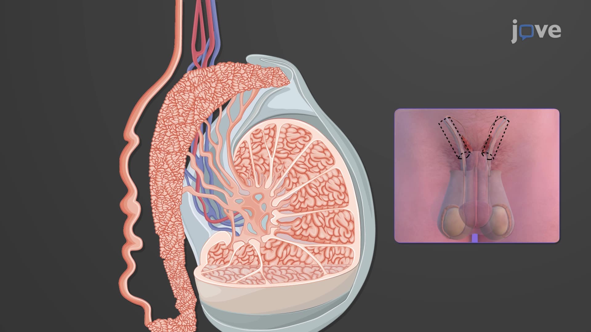
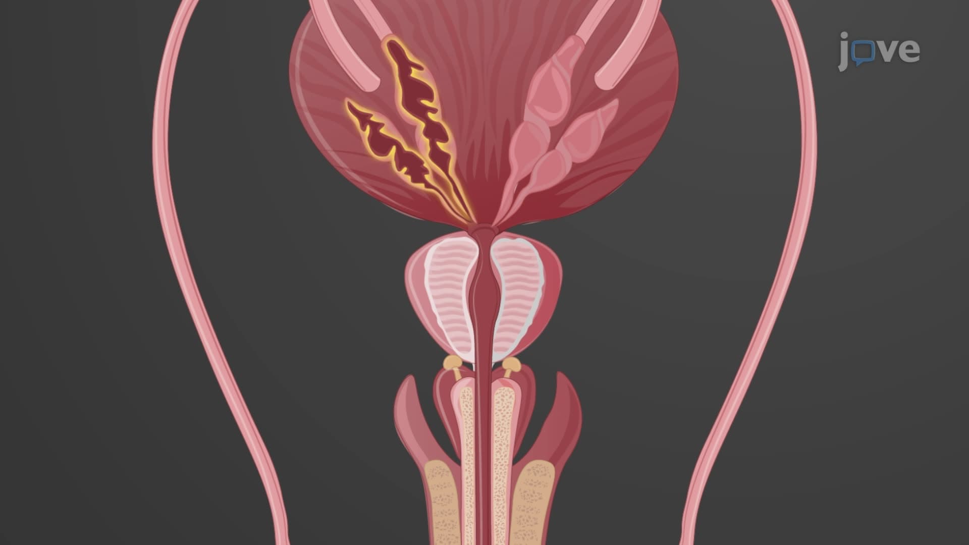
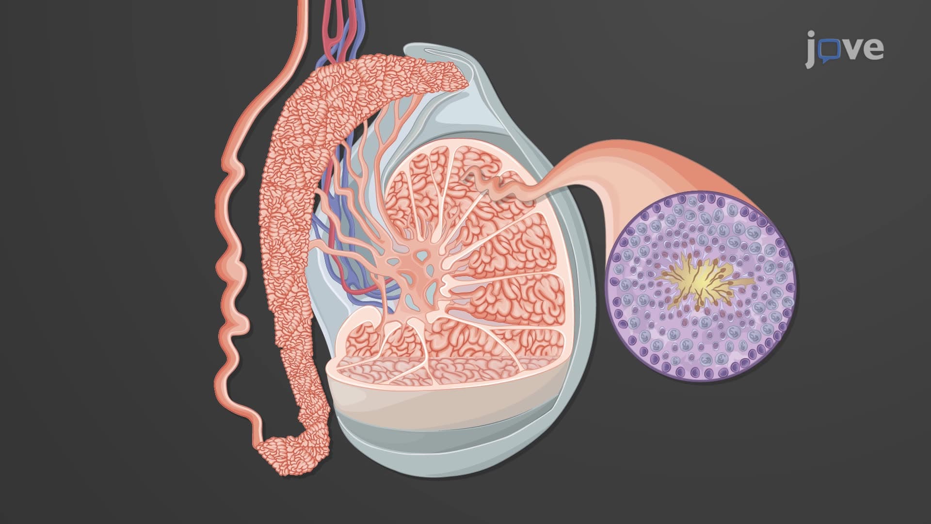
Testes histology biology examines the microscopic organization of male reproductive organs, revealing how structure enables function at the cellular level. The testis represents one of the most compartmentalized organs in the human body, with each structural element serving specific roles in sperm production and hormone synthesis. Students preparing for the MCAT or AP Biology exams must understand how the fibrous tunica albuginea creates internal divisions that organize reproductive processes.
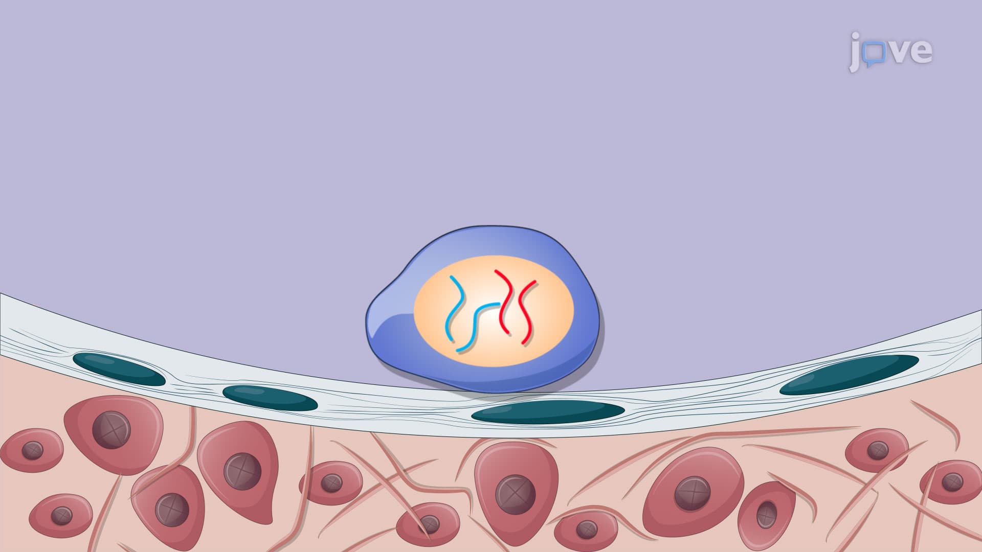
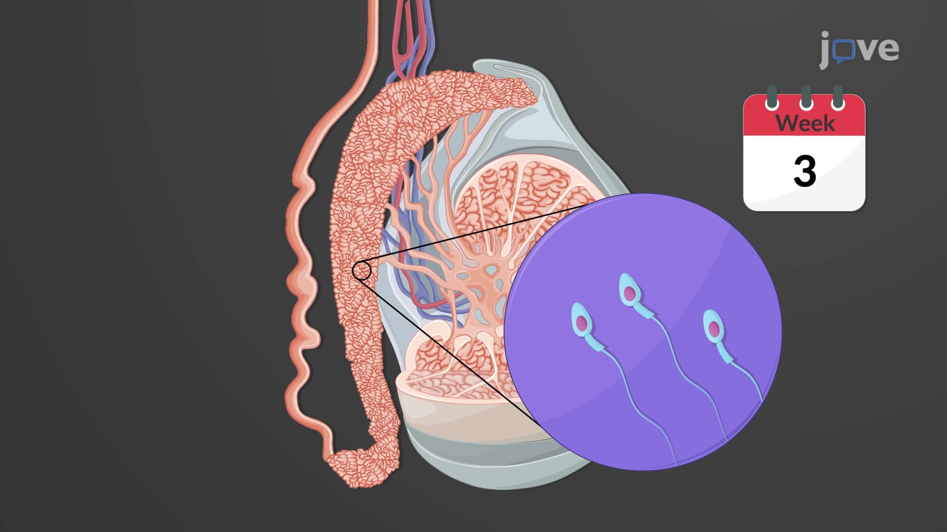
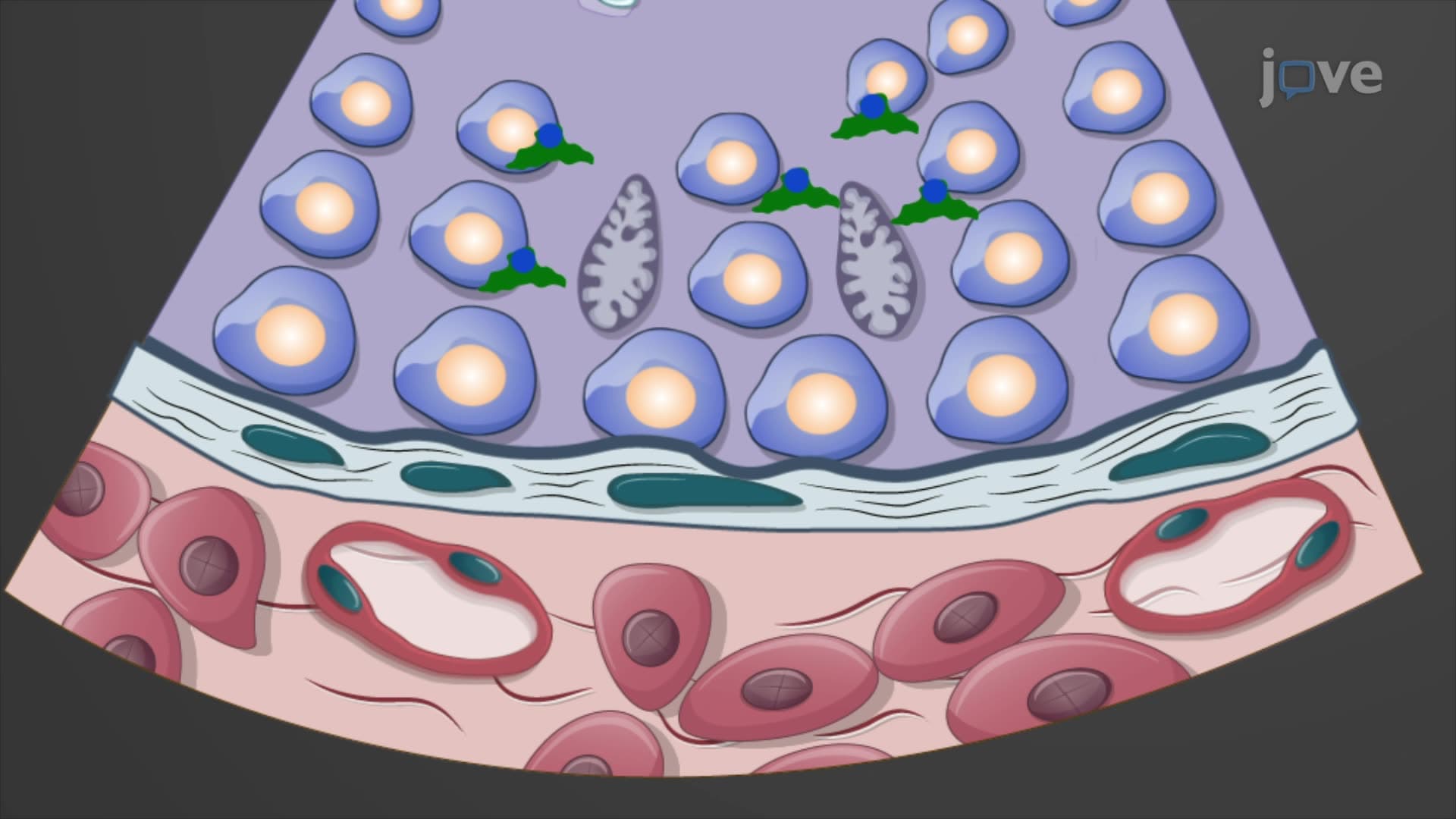
The seminiferous tubule histology forms the cornerstone of male fertility, with each tubule measuring approximately 150-250 micrometers in diameter and containing specialized germinal epithelium. Within this epithelium, Sertoli cell sustentacular function provides both physical support and biochemical nourishment for developing sperm cells. These remarkable cells extend from the basement membrane to the tubule lumen, creating a blood-testis barrier that protects developing gametes from immune system interference. College anatomy courses at universities like UCLA emphasize how Sertoli cells regulate the microenvironment necessary for the 74-day spermatogenesis process.
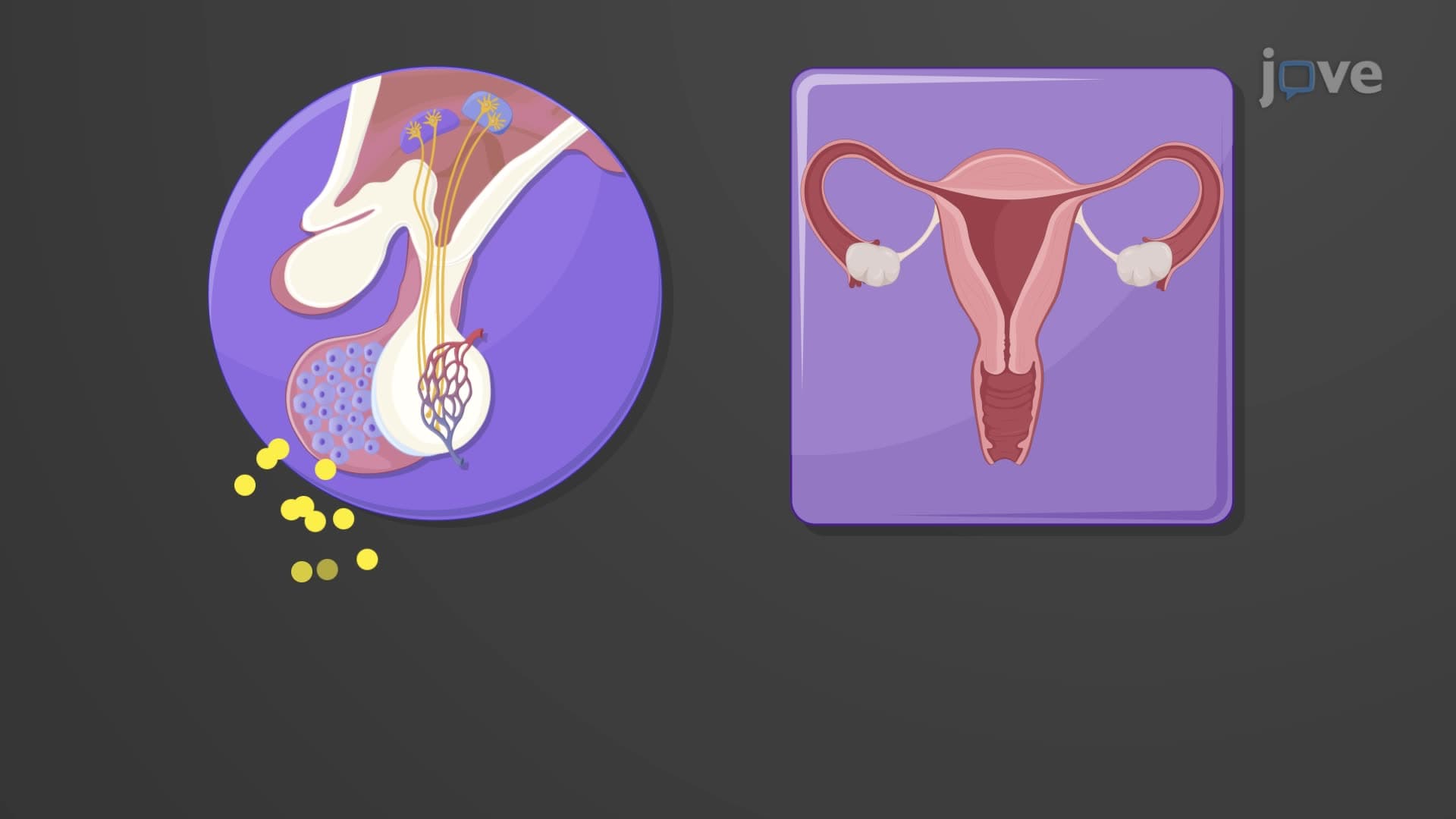
Between the seminiferous tubules lies the interstitial compartment containing Leydig interstitial cells, which produce testosterone and other androgens essential for male sexual development and fertility. These cells respond to luteinizing hormone (LH) signals from the pituitary gland, demonstrating the integration between testicular histology anatomy and endocrine function. Medical students studying for the USMLE Step 1 exam must understand how Leydig cell dysfunction can lead to hypogonadism and fertility issues.
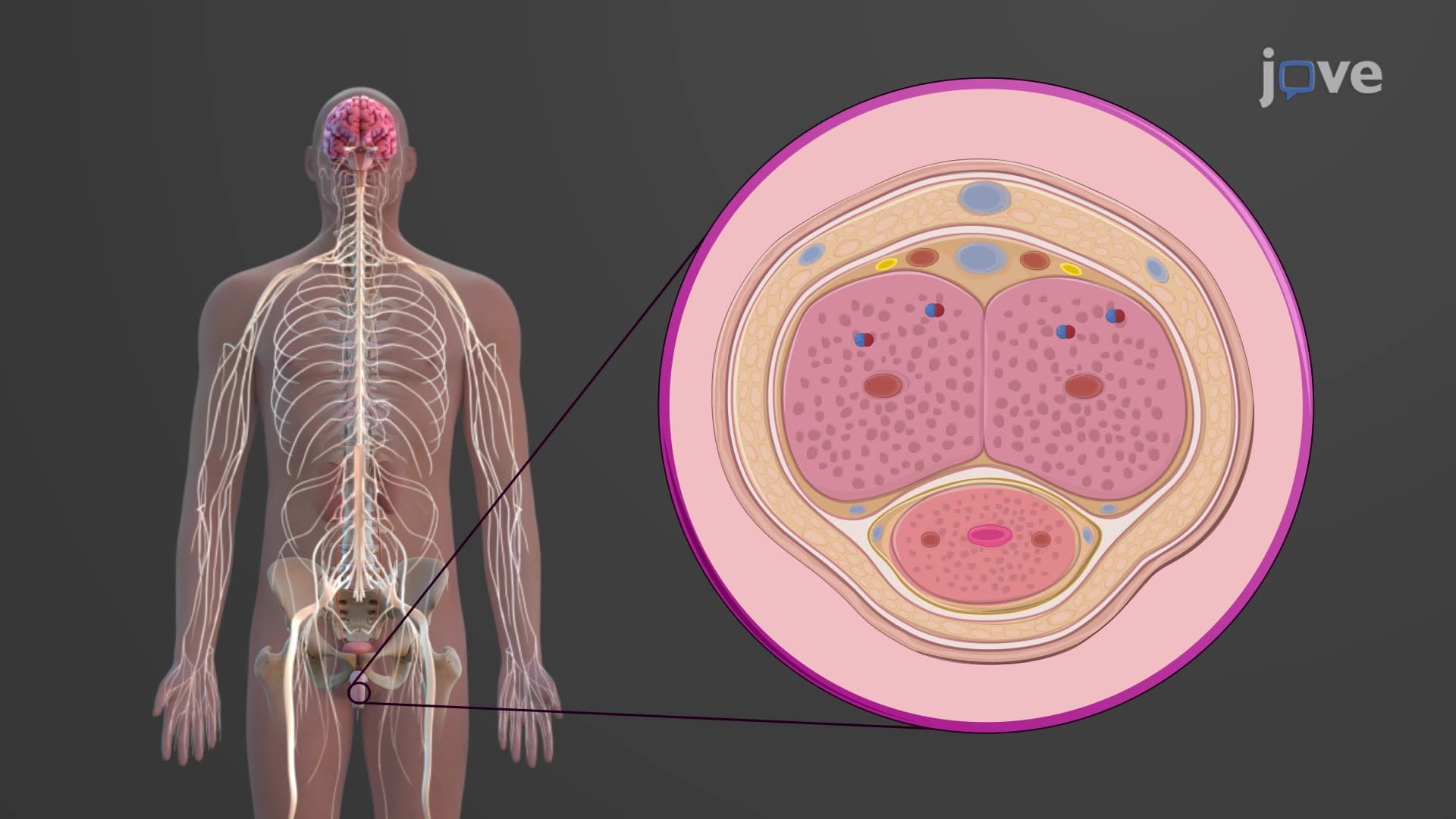
The unique vascular arrangement within testicular tissue reflects the critical importance of temperature control for sperm production. The pampiniform plexus creates a countercurrent heat exchange system that maintains testicular temperature approximately 2-3°C below core body temperature. This testicular histology explained concept appears frequently on nursing exams like the NCLEX, where students must understand how conditions like varicocele can disrupt normal temperature regulation and impact male fertility.
Related Micro-courses