Loading subject page…


141,004 views

Video Summary: What Is Uterus and Cervix
Did you know that the uterus can expand to 500 times its original size during pregnancy? Understanding uterus cervix anatomy biology is fundamental to comprehending female reproductive health. Consider how gynecologists at Johns Hopkins Medical Center examine these structures during routine Pap smears to detect cervical abnormalities. What is Uterus And Cervix explores the intricate pear-shaped organ that serves as the foundation for human development. Watch the full video on JoVE Coach to master this concept with expert-led visuals and step-by-step explanations.
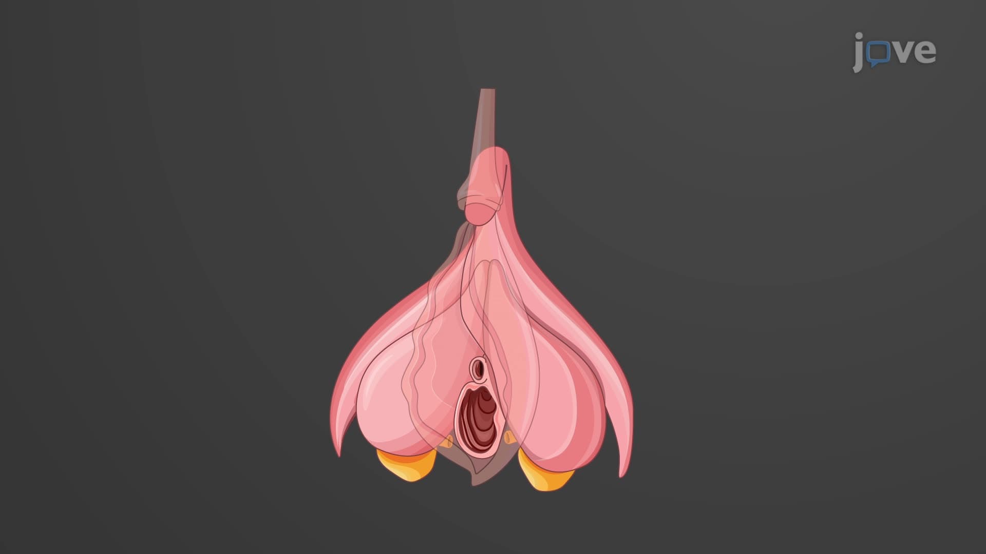
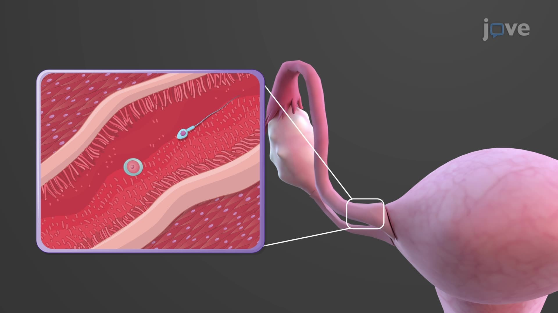
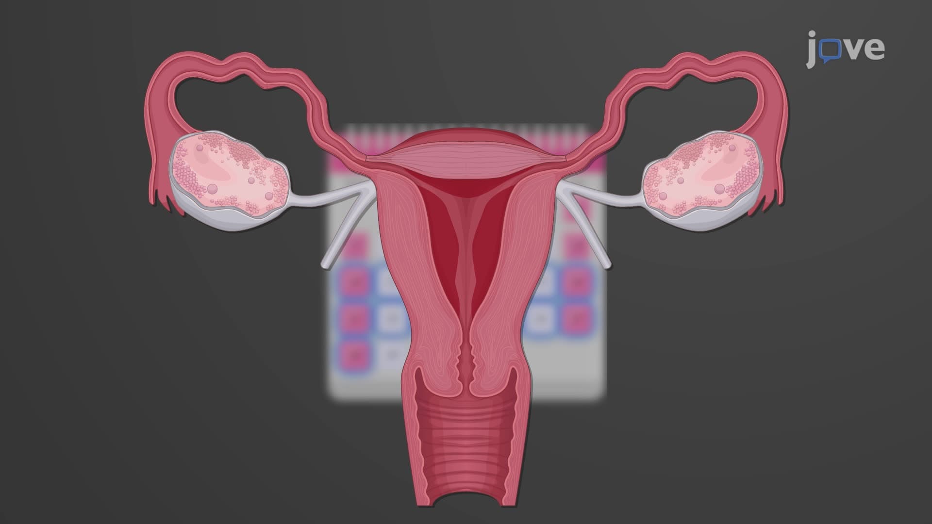
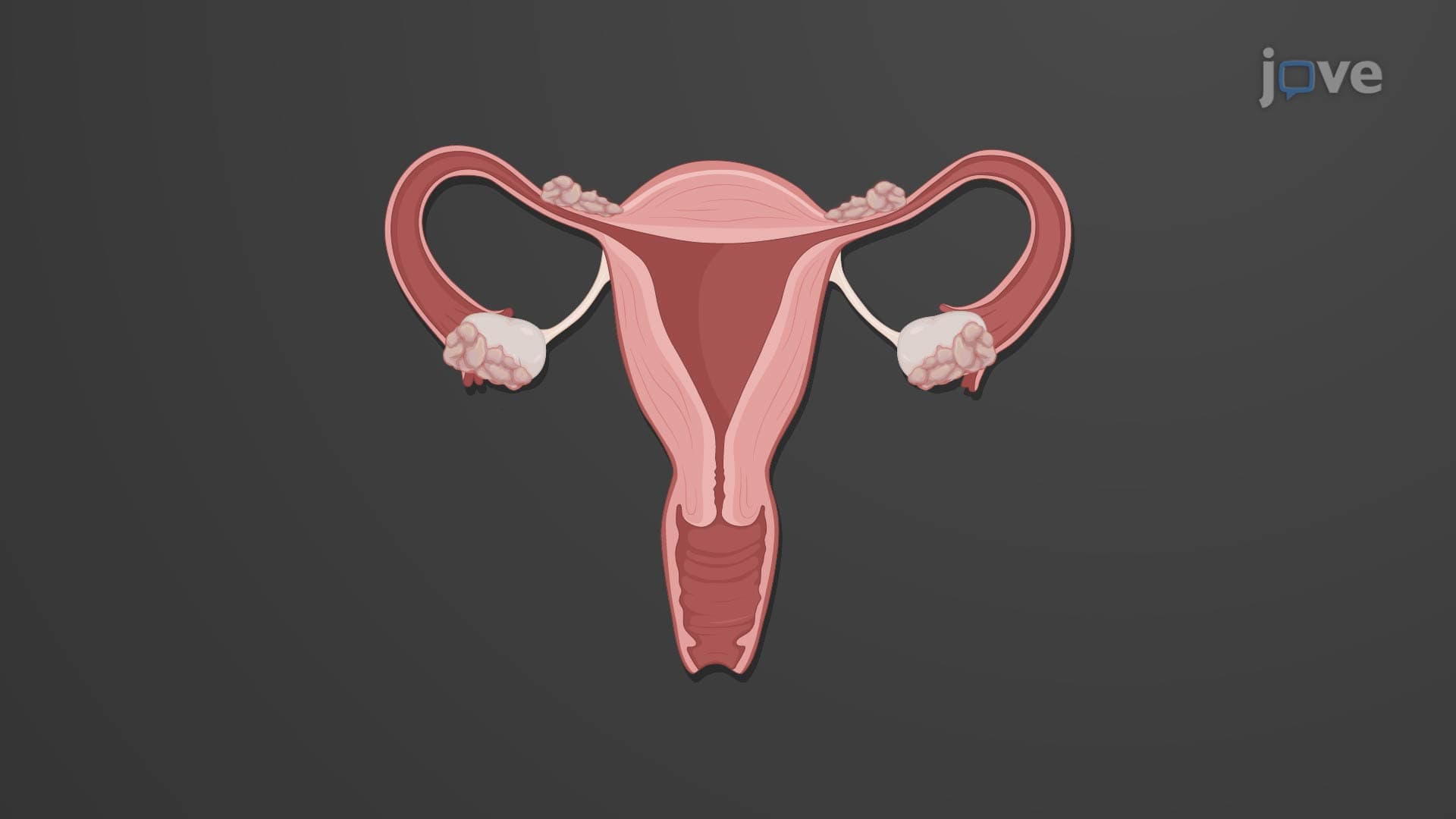
The womb and cervix explained reveals a sophisticated biological system engineered for reproduction. This hollow, muscular organ sits strategically in the female pelvis, positioned between the bladder anteriorly and rectum posteriorly. The typical forward tilt (anteversion) allows the uterus to rest comfortably above the bladder, a positioning that medical students studying for the MCAT must thoroughly understand. At major US medical centers like Mayo Clinic, gynecologists regularly assess this positioning during pelvic examinations.
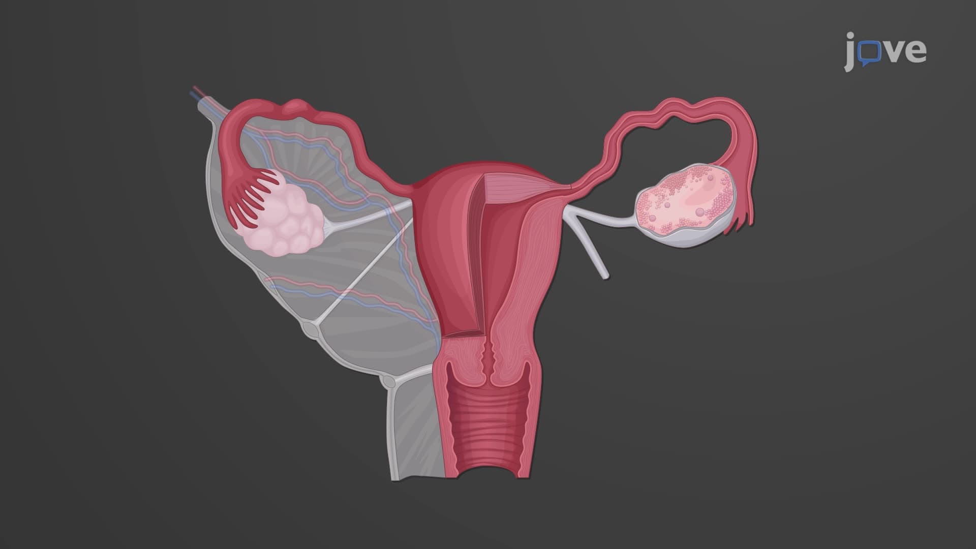
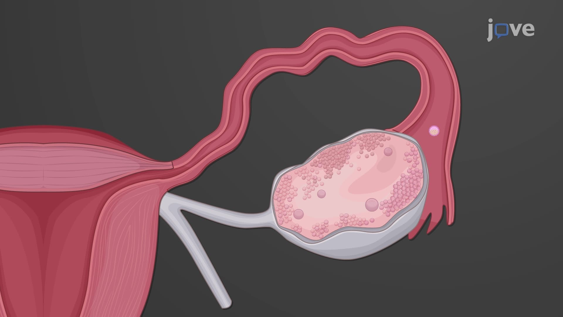
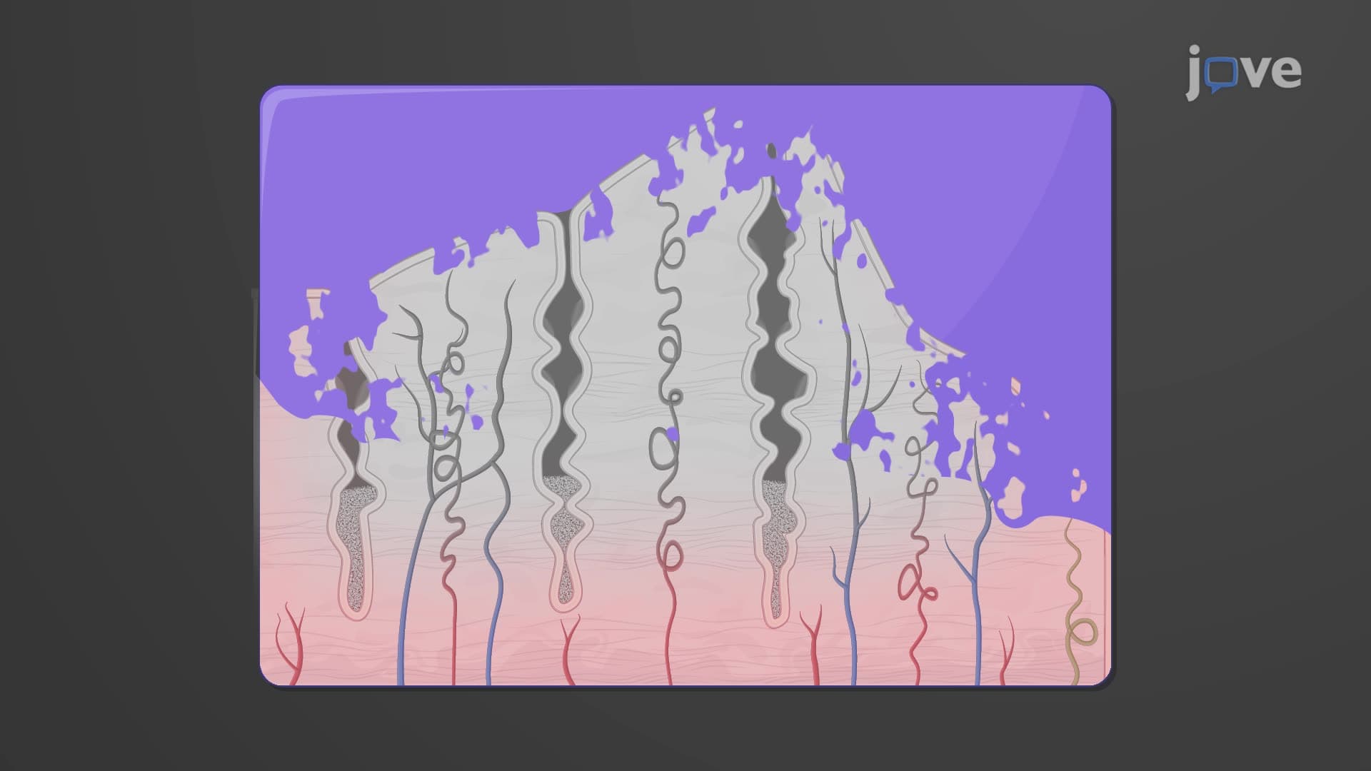
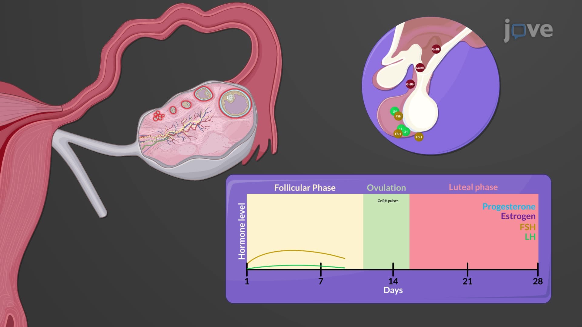
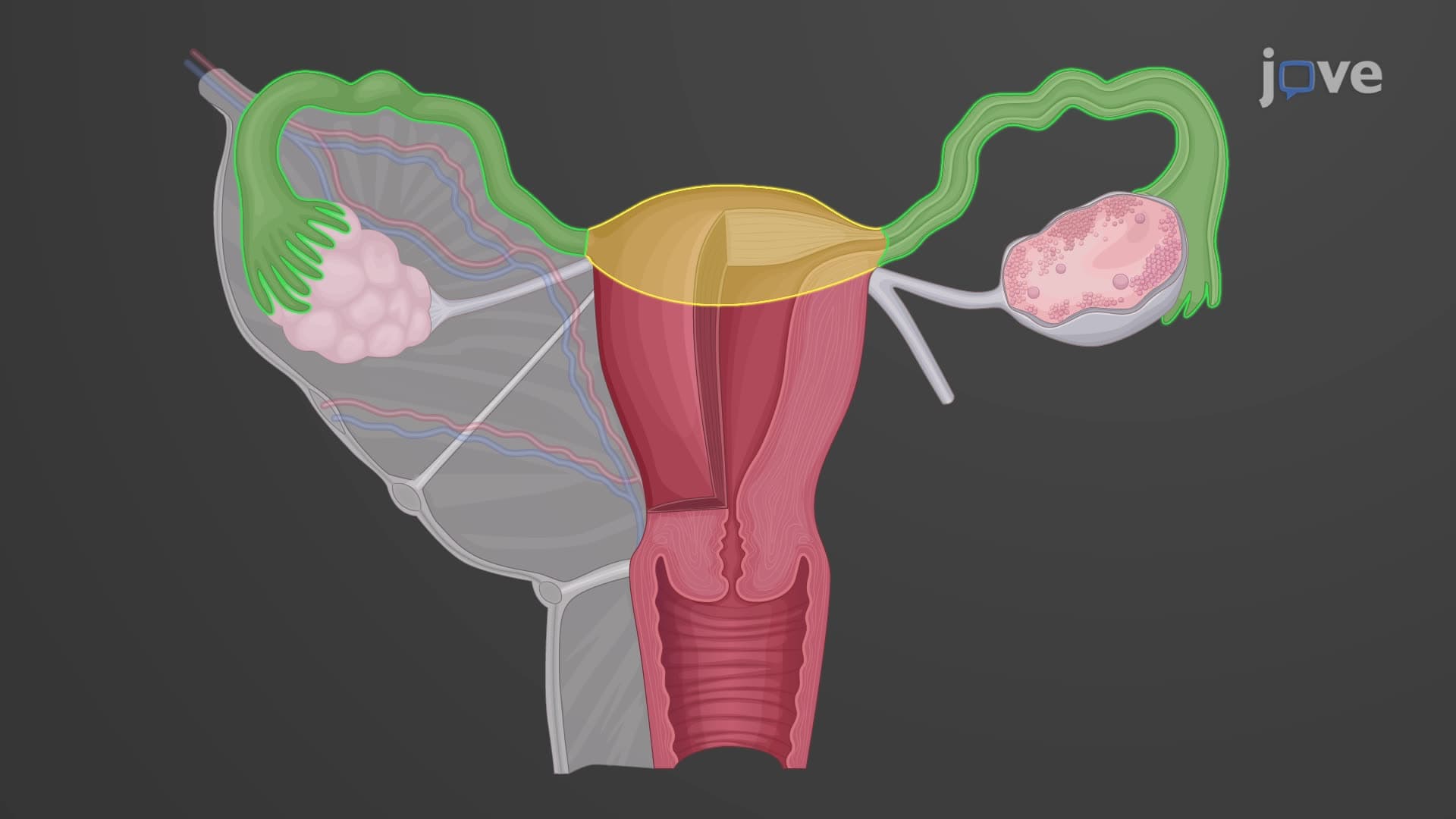
What is the uterus and cervix in female anatomy becomes clearer when examining distinct regions. The fundus represents the rounded upper portion above where fallopian tubes attach, serving as the primary implantation site during early pregnancy. The body contains the main uterine cavity lined with endometrium, which undergoes monthly cyclical changes. AP Biology students frequently encounter questions about endometrial thickness variations throughout the menstrual cycle on college entrance exams.
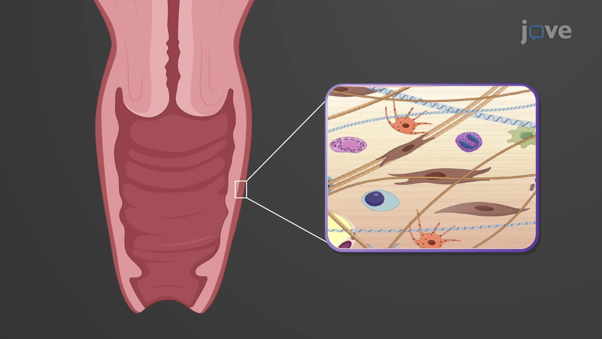
The isthmus creates a narrow transition zone leading to the cervix, a tubular structure extending into the vagina. Understanding cervical canal os cervix anatomy proves crucial for nursing students preparing for NCLEX examinations, as cervical dilation measurements determine labor progress.
The uterus ligament anatomy involves three primary support mechanisms essential for organ stability. Broad ligaments provide lateral support while containing blood vessels and nerves. Round ligaments maintain the forward-tilted position, and uterosacral ligaments offer posterior-inferior support. Weakness in these structures can lead to uterine prolapse, a condition commonly studied in women's health courses at universities like UCLA and Duke.
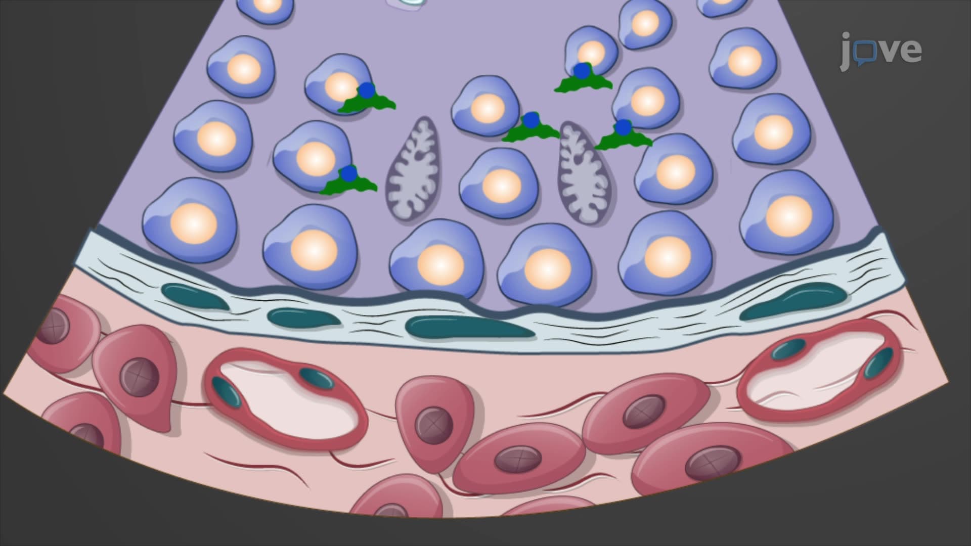
Healthcare professionals at institutions like Cleveland Clinic use standardized measurements when assessing uterine health. The non-pregnant uterus typically measures 7-8 centimeters in length, though these dimensions vary with age and reproductive history. Medical students preparing for USMLE Step 1 must memorize these normal values, as questions frequently test knowledge of anatomical variations. The endometrium myometrium uterus layers serve different functions, with the endometrium providing the nourishing environment for embryonic development while the myometrium contains smooth muscle enabling contractions during childbirth.
Related Micro-courses