Loading subject page…

- Cell Biology
- Cell Cell Interactions
- Adherens Junctions
Micro-course: Cell-Cell Interactions
83,115 views
Learn key concepts and earn your certificate.



Adherens Junctions

00:00
01:24
Video Summary: What are Adherens Junctions
Ever wonder how your skin cells stick together to form a protective barrier, or how intestinal cells maintain tight seals despite constant digestive pressures? Adherens junctions are the cellular "glue" that holds neighboring cells together in tissues throughout your body, from the lining of your stomach to the layers of your epidermis. These complex protein structures act like molecular rivets, anchoring cells while allowing tissues to remain flexible and responsive. Watch the full video on JoVE Coach to master this concept with expert-led visuals and step-by-step explanations.
- Understand what are adherens junctions and their essential role as anchoring structures that maintain tissue integrity
- Identify the three main protein components: cadherins, catenins, and actin cytoskeleton filaments
- Analyze the step-by-step assembly process from initial lamellipodia formation to mature junction development
- Compare the structural differences between branched and unbranched actin arrangements during junction maturation
- Recognize how cadherin clustering and catenin stabilization create strong intercellular connections
- Apply knowledge of adherens junction dysfunction to understand diseases like cancer metastasis and epithelial disorders
- Evaluate the mechanical forces involved in contractile bundle formation and cellular adhesion
- Explore the relationship between adherens junctions and other cell junction types in maintaining tissue architecture
Understanding What Are Adherens Junctions: The Foundation of Tissue Architecture
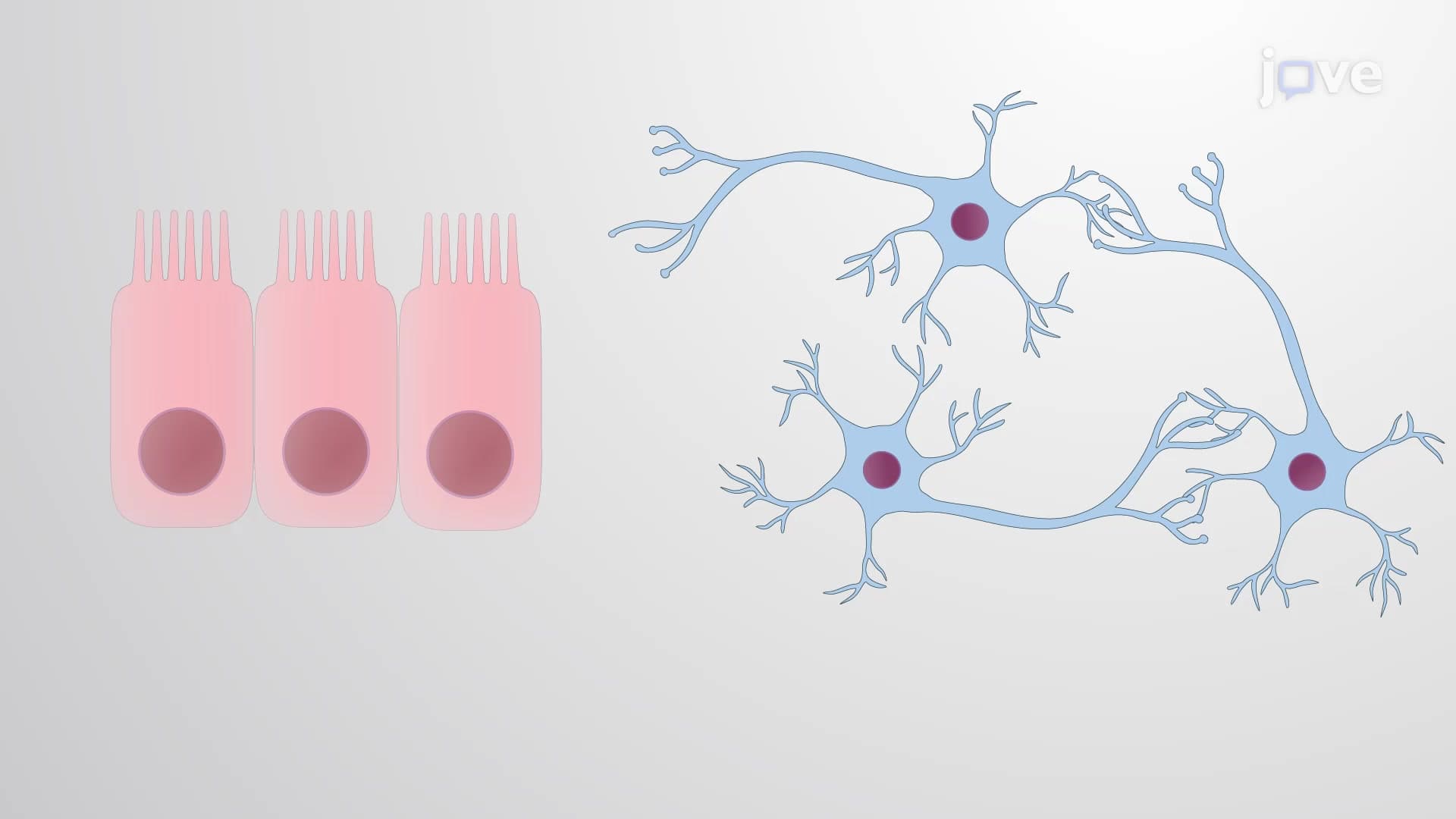
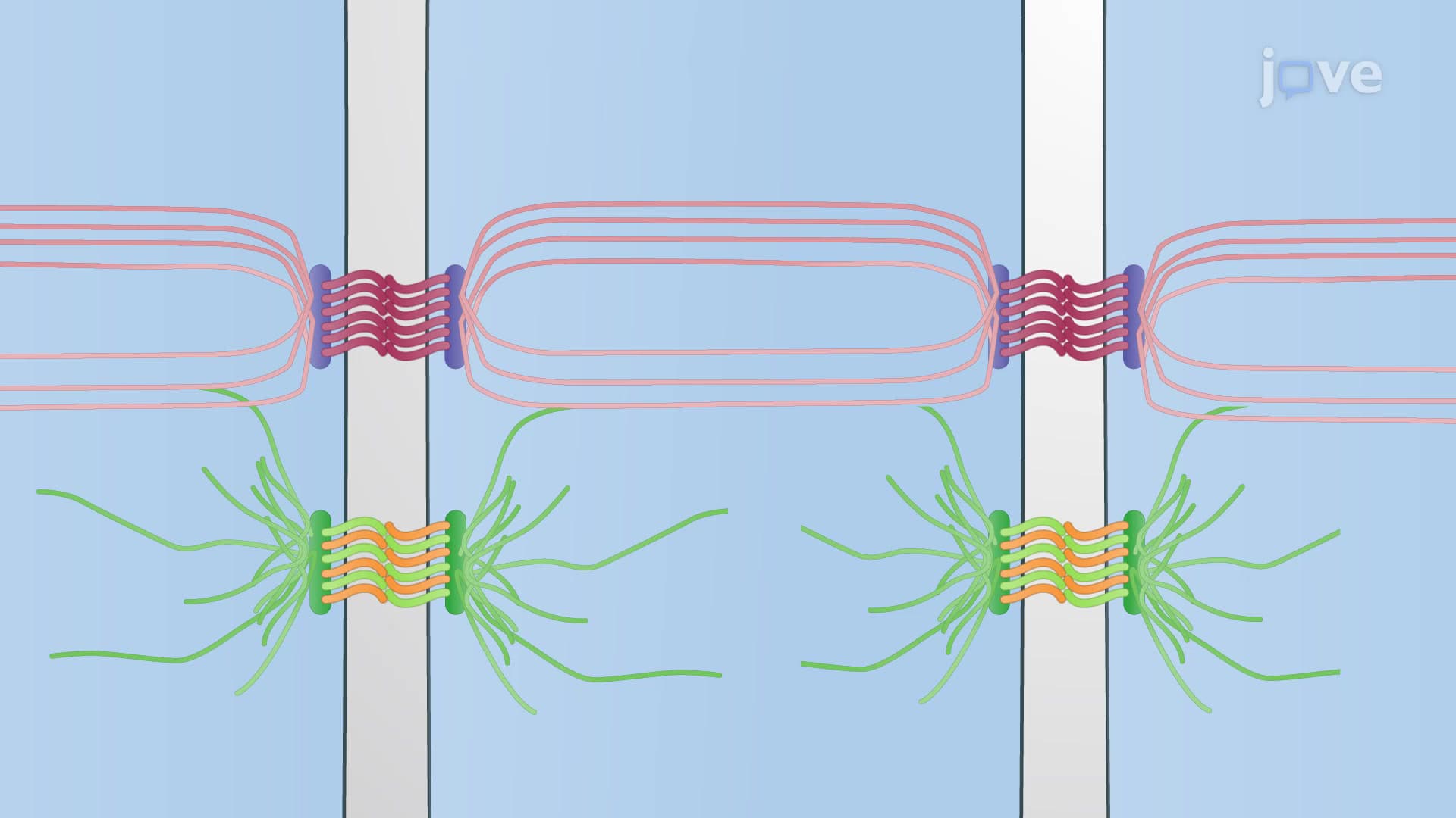
Adherens junctions represent one of nature's most elegant solutions to a fundamental biological challenge: how to hold billions of cells together while maintaining tissue flexibility and function. These sophisticated protein complexes serve as the primary anchoring system in epithelial tissues, which line virtually every surface and cavity in the human body, from your respiratory tract to your digestive system.
The Molecular Architecture of Cellular Adhesion
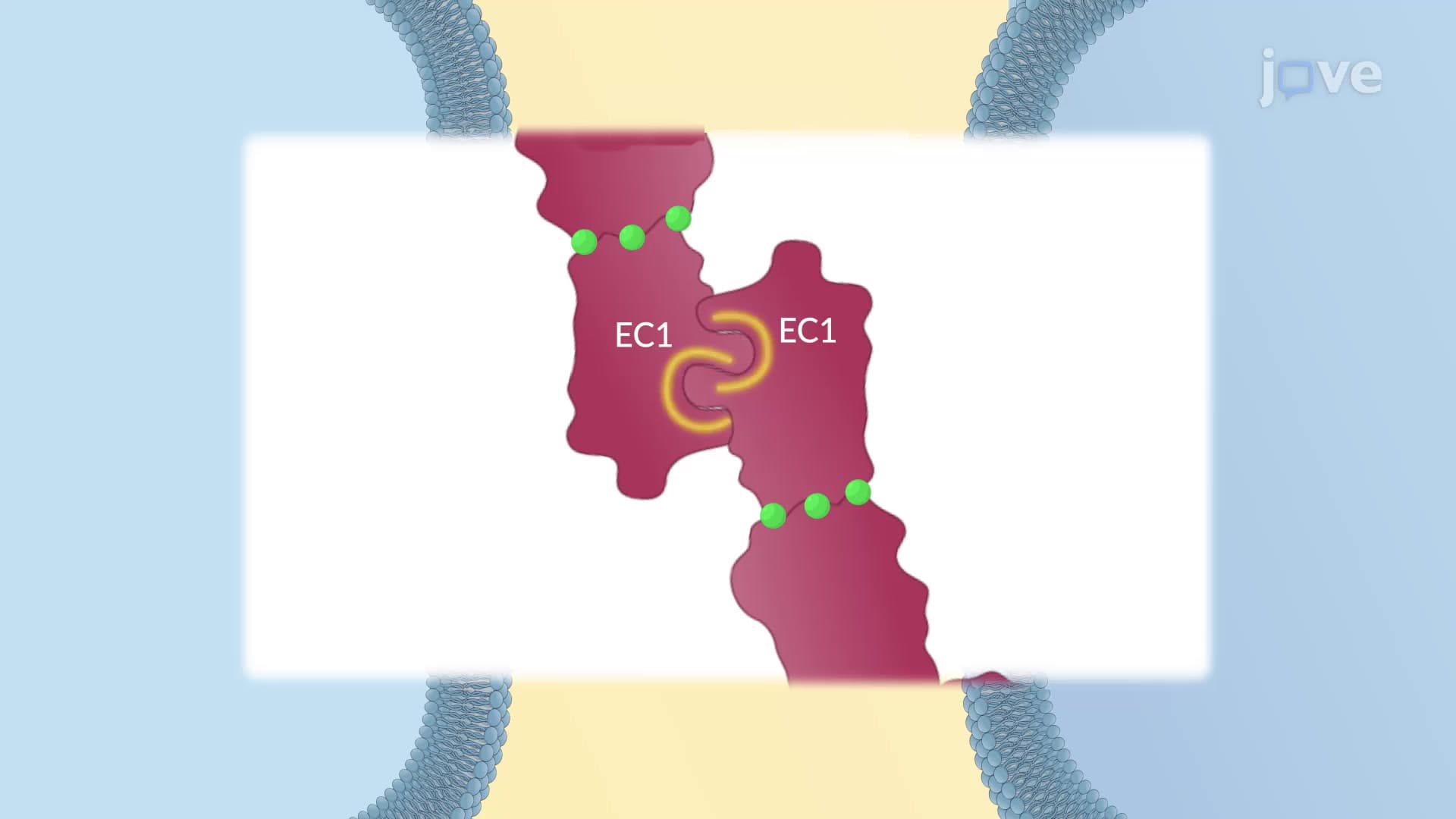
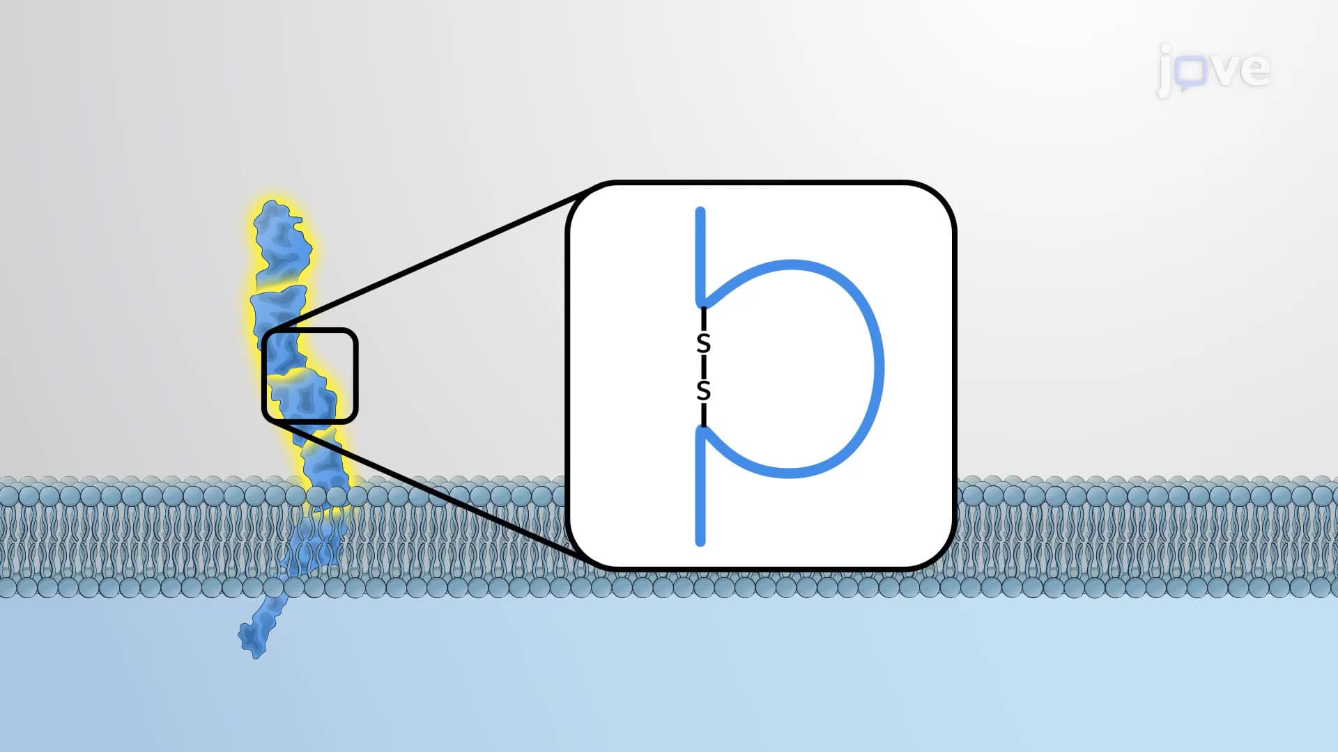
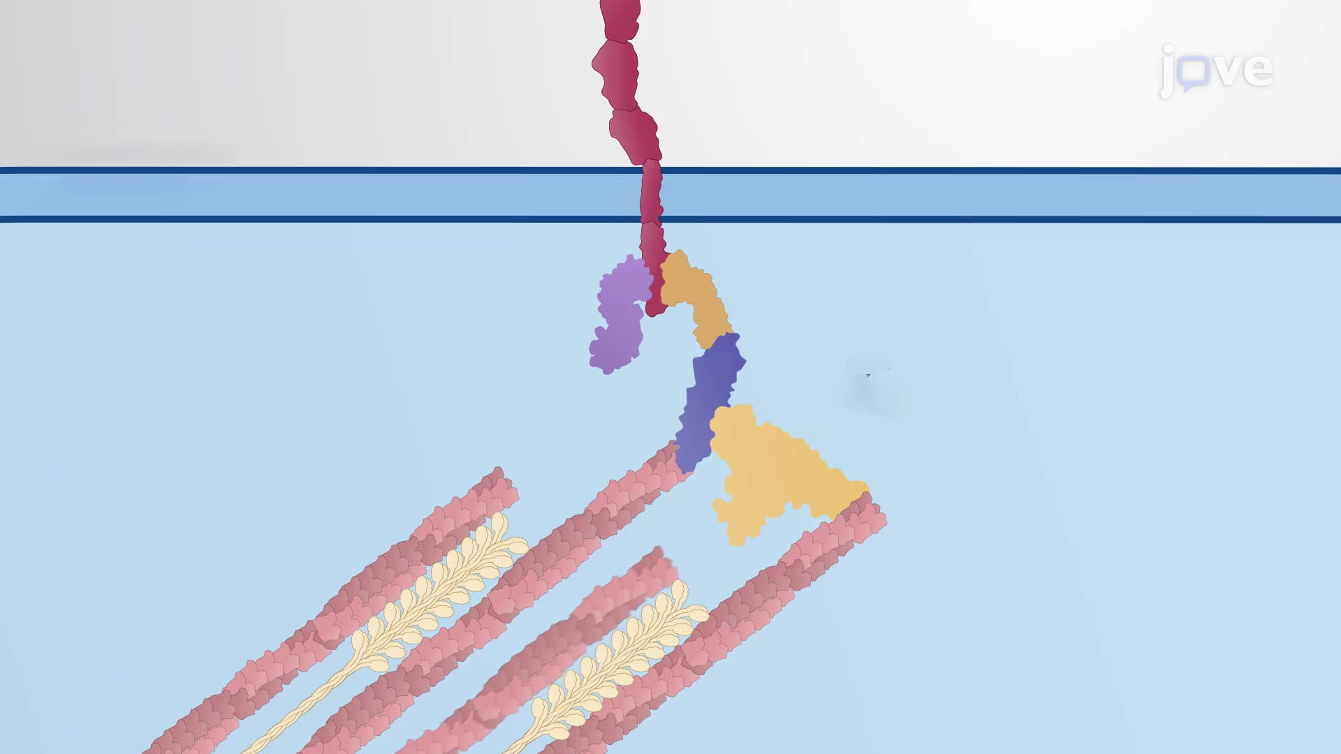
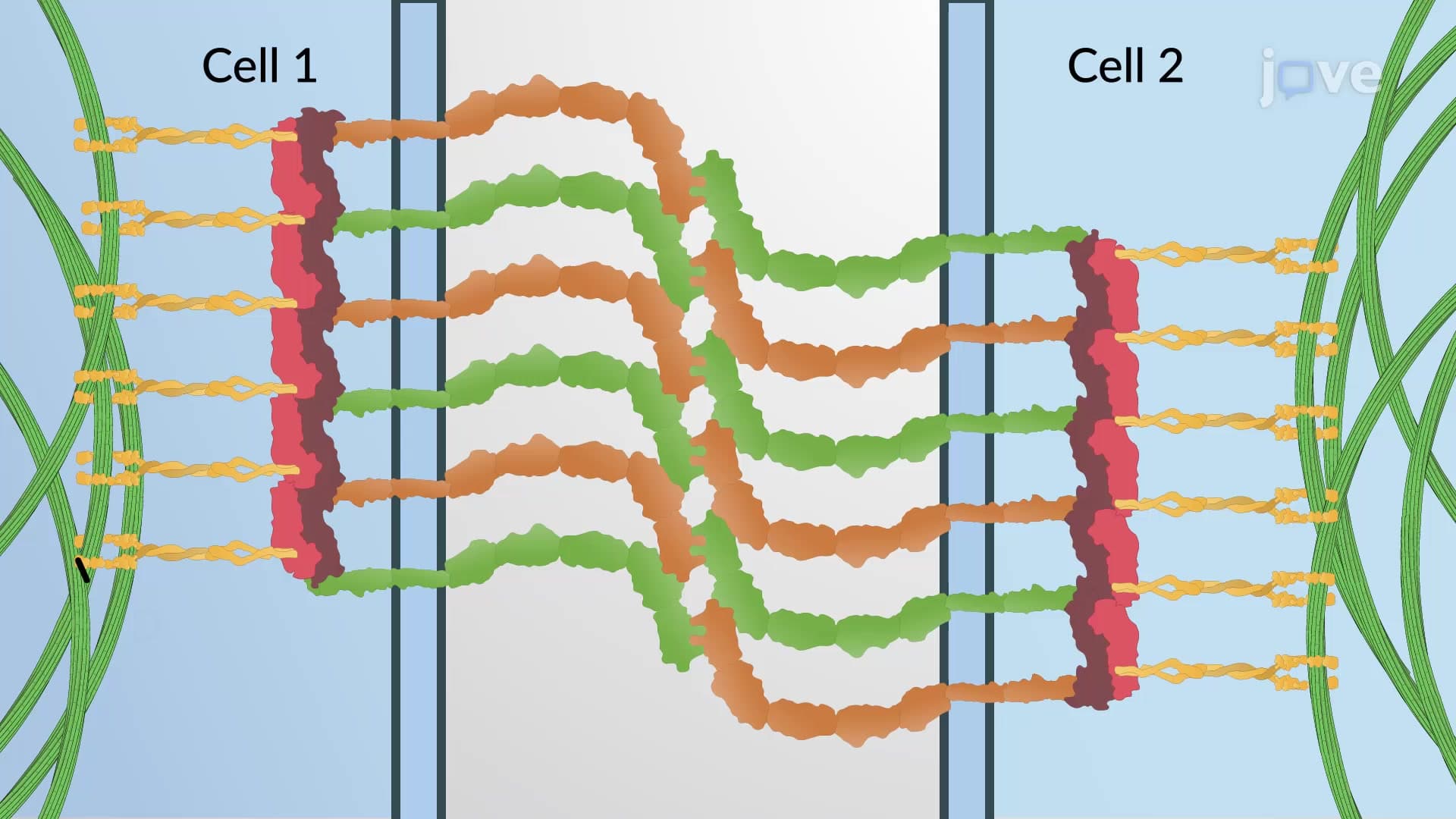
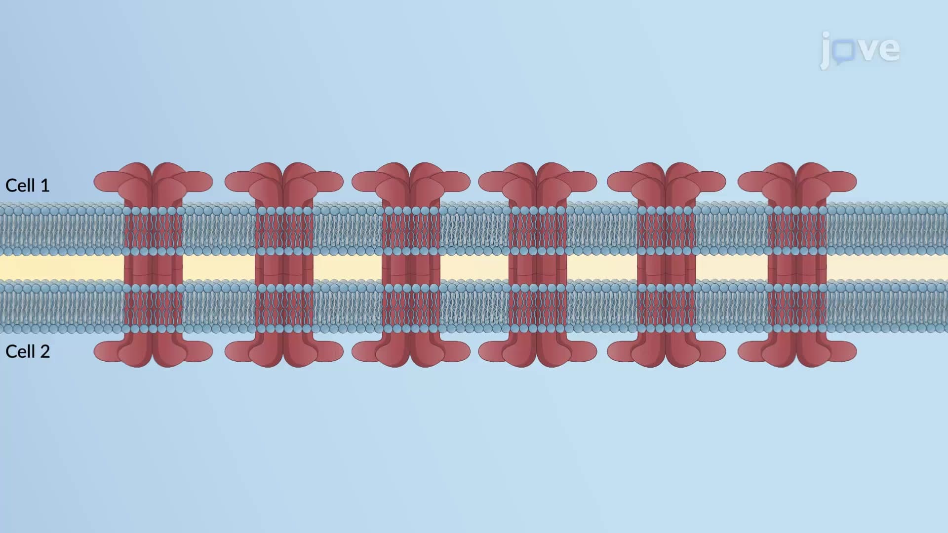
The structural complexity of adherens junctions reflects their critical importance. Three distinct protein families work in concert to create these cellular anchors. Cadherins, the adhesive proteins, extend across the extracellular space like molecular hands reaching out to grasp neighboring cells. These calcium-dependent proteins literally give adherens junctions their name-"adherens" meaning "to stick to."
Within the cell, catenin proteins serve as crucial linkers, connecting the cadherins to the internal cellular machinery. Beta-catenin, alpha-catenin, and p120-catenin each play specialized roles, much like different components in a sophisticated engineering system. The actin cytoskeleton provides the structural foundation, forming contractile bundles that can respond to cellular stresses and maintain tissue integrity under pressure.
Assembly Process: From Contact to Mature Junction
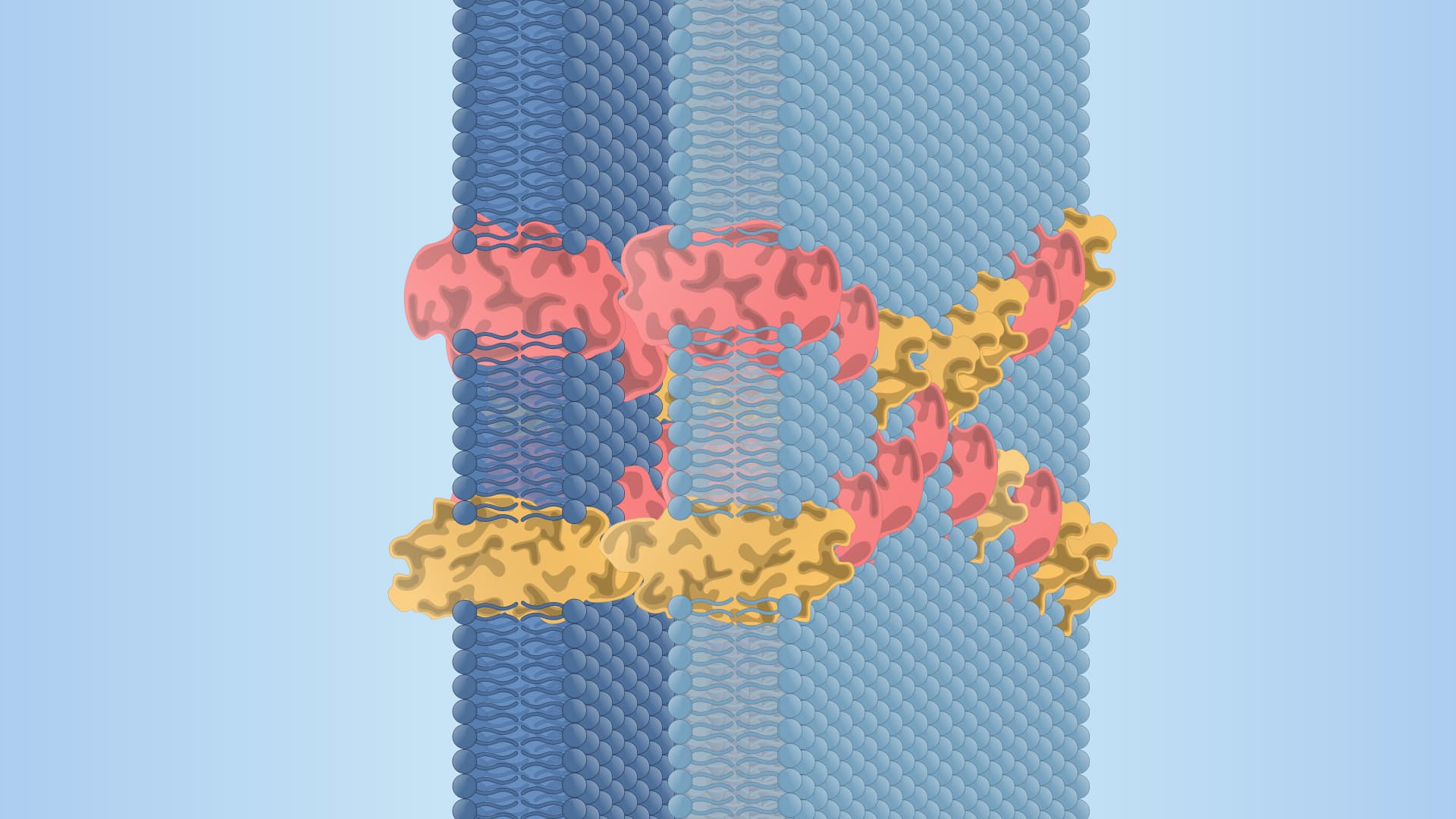
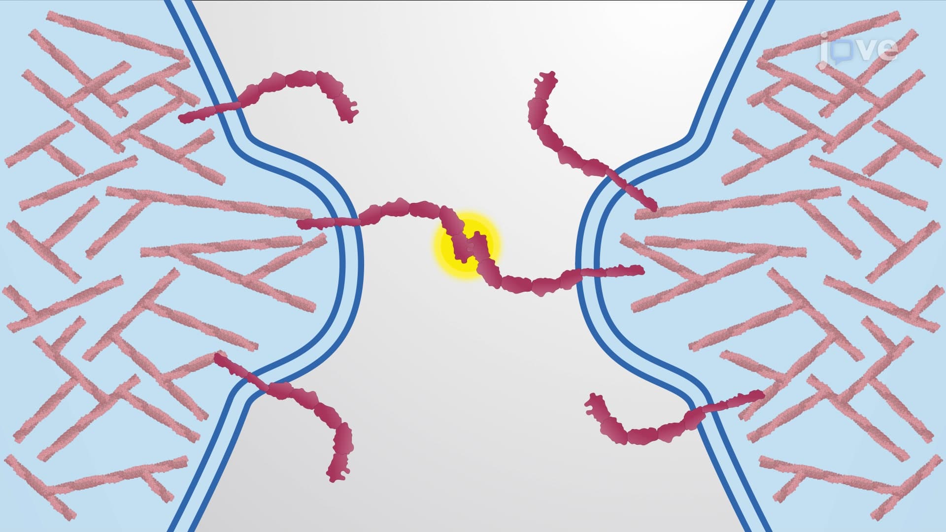
The formation of adherens junctions follows a precisely orchestrated sequence that begins when cells come into contact. Initially, actin polymerization creates lamellipodia-sheet-like protrusions that extend from the cell surface like exploratory fingers. When these structures from adjacent cells encounter each other, cadherins on their surfaces interact, triggering a cascade of molecular events.
This initial contact rapidly amplifies through positive feedback mechanisms. More cadherins cluster at the contact site, while catenins stabilize these growing complexes. As the junction matures, a remarkable transformation occurs: the randomly arranged, branched actin networks reorganize into parallel, unbranched filaments that run alongside the cell membrane. These filaments then associate with myosin motor proteins, creating contractile bundles capable of generating mechanical force.
Clinical and Academic Applications
Understanding adherens junctions proves essential for students pursuing careers in medicine, biomedical research, or related fields. On the MCAT, questions frequently test knowledge of cell junction types and their molecular components. AP Biology courses emphasize these structures when covering cell communication and tissue organization. For pre-med students, adherens junction dysfunction underlies numerous pathological conditions, including cancer metastasis, where tumor cells lose their normal adhesive properties and invade surrounding tissues.
Frequently Asked Questions
Adherens junctions are protein complexes that anchor neighboring cells together, particularly in epithelial tissues like skin and organ linings. They're crucial for maintaining tissue integrity and preventing cells from separating under mechanical stress. When these junctions malfunction, it can lead to serious conditions including cancer metastasis and inflammatory bowel diseases.
While adherens junctions share common structural features, they vary in organization and strength depending on tissue type. Epithelial adherens junctions form belt-like structures around cells, while those in cardiac muscle create intercalated discs for heart contraction. Endothelial adherens junctions in blood vessels regulate permeability and maintain the blood-brain barrier.
MCAT questions often focus on distinguishing adherens junctions from tight junctions and gap junctions, plus their molecular components. AP Biology emphasizes their role in cell communication and tissue organization. Expect diagram interpretation questions showing cadherin-catenin complexes and scenarios involving junction dysfunction in disease states.
Cell Biology and Molecular Biology courses provide the most comprehensive coverage, often including laboratory exercises examining junction proteins. Histology courses emphasize their appearance in different tissues, while Pathology courses explore disease mechanisms. Developmental Biology courses discuss their role in tissue morphogenesis and organ formation.
Adherens junction dysfunction contributes to several common health problems, including inflammatory bowel diseases like Crohn's disease, where intestinal barrier function breaks down. In skin conditions like eczema, compromised junctions allow irritants to penetrate deeper tissue layers. Cancer research increasingly focuses on how tumor cells escape normal junction constraints.
Basic understanding of protein structure and chemical bonding suffices for most high school and introductory college courses. The key concepts involve protein-protein interactions and calcium-dependent binding, which build on fundamental chemistry principles. Focus on the biological functions rather than detailed molecular mechanisms initially.
Use the "CAC" memory device: Cadherins for Adhesion, Catenins for Connection, and aCtin for Contraction. Create concept maps linking each protein family to its specific function. Practice drawing the assembly process step-by-step, as this appears frequently on exams and reinforces the sequential nature of junction formation.
Explore other cell junction types (tight junctions, gap junctions, desmosomes) to understand how cells create diverse connection strategies. Study cytoskeleton dynamics and cell migration mechanisms. Investigate cancer cell biology to see how junction loss enables metastasis, connecting this molecular knowledge to clinical applications.
Current research focuses on targeting adherens junction proteins for cancer therapy, since restoring normal cell adhesion could prevent metastasis. Scientists are also developing treatments for inflammatory diseases by strengthening epithelial barriers. Regenerative medicine research explores how manipulating these junctions could improve tissue engineering and wound healing outcomes.
Related Micro-courses

11 Concepts
7 Concepts
20 Concepts