Loading subject page…


1,346,747 views

Video Summary: What Is DNA Agarose Gel Electrophoresis
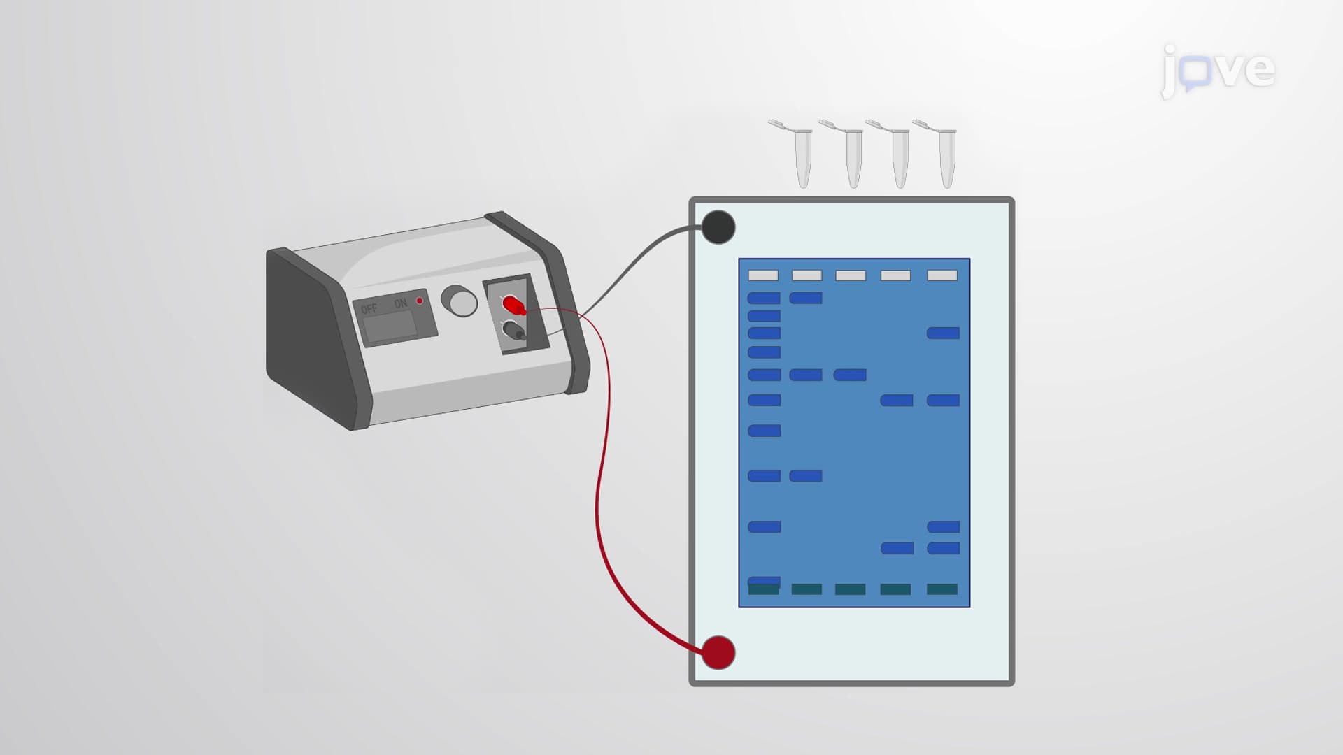
Ever wondered how forensic scientists at the FBI identify suspects from tiny DNA samples found at crime scenes? DNA agarose gel electrophoresis is the foundational laboratory technique that separates DNA fragments by size, enabling everything from criminal investigations to medical diagnostics. This powerful method uses an electric field to pull negatively charged DNA molecules through a gel matrix, with smaller fragments traveling faster than larger ones. What is DNA Agarose Gel Electrophoresis demonstrates this essential molecular biology technique step-by-step. Watch the full video on JoVE Coach to master this concept with expert-led visuals and step-by-step explanations.
DNA agarose gel electrophoresis serves as a cornerstone technique in molecular biology laboratories across American universities and research institutions. This method exploits the uniform negative charge of DNA molecules, which results from phosphate groups in the DNA backbone. When placed in an electric field, these negatively charged molecules migrate toward the positive electrode at rates inversely proportional to their molecular weight.
The technique proves invaluable for students taking AP Biology, college-level molecular biology courses, and pre-medical students preparing for the MCAT. Understanding this concept builds foundation knowledge for advanced topics like PCR analysis, restriction enzyme mapping, and genomic studies.
Agarose, extracted from red seaweed, forms the separation medium through hydrogen bonding between polysaccharide chains. The resulting three-dimensional matrix contains pores whose size depends on agarose concentration. A standard 1% agarose gel (1g agarose per 100mL buffer) creates medium-sized pores suitable for separating DNA fragments ranging from 500 to 10,000 base pairs.
Buffer systems like TAE (Tris-acetate-EDTA) or TBE (Tris-borate-EDTA) maintain stable pH and provide ions necessary for electrical conductivity. These buffers prevent DNA degradation during electrophoresis and ensure consistent migration patterns. Students often encounter questions about buffer selection on college biochemistry exams and laboratory practical assessments.
The separation principle relies on molecular sieving through the agarose matrix. Smaller DNA fragments navigate pores more easily, migrating greater distances in a given time period. Larger fragments encounter more resistance, traveling shorter distances. This size-dependent migration creates distinct bands representing different molecular weights.
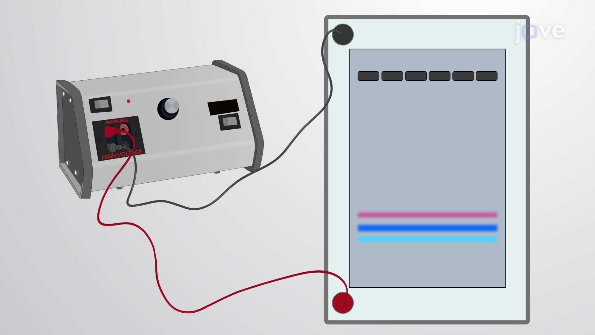
Loading buffers containing glycerol increase sample density, preventing DNA from floating out of wells. Tracking dyes like bromophenol blue and xylene cyanol migrate at predictable rates, allowing researchers to monitor electrophoresis progress without stopping the procedure.
Fluorescent dyes such as ethidium bromide intercalate between DNA base pairs, enabling visualization under UV light. Modern laboratories increasingly use safer alternatives like SYBR Safe or methylene blue. DNA ladders containing fragments of known sizes serve as molecular weight standards, allowing precise size determination of unknown samples.
This technique finds extensive application in American forensic laboratories, medical diagnostic centers, and pharmaceutical companies. Students pursuing careers in biotechnology, medicine, or forensic science frequently encounter gel electrophoresis in both academic coursework and professional settings.
Related Micro-courses